Anatomical dead space
ID: anatomical-dead-space

Anatomical dead space is the volume of the conducting airways that transports gas without providing the pulmonary alveolus surface for gas exchange. It includes passages such as the trachea and bronchi. It is one component of respiratory dead space, distinct from alveolar dead space associated with ineffective exchange in ventilated alveolar regions.[ref]
Pulmonary Function Testing by Warren M. Gold and Laura L. Koth. Single-Breath Nitrogen Test; The Bohr Equation for Respiratory Dead Space
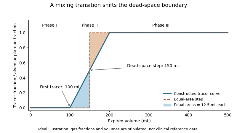
A single-breath tracer experiment makes the conducting volume observable. Following an oxygen inspiration, expired gas initially contains little nitrogen, then passes through a transition as airway and alveolar gas mix, and finally approaches an alveolar plateau. Recording gas fraction against expired volume distinguishes those phases.[ref]
Pulmonary Function Testing by Warren M. Gold and Laura L. Koth. Single-Breath Nitrogen Test, phases I–III
A 1954 primary study explicitly compared single-breath measurements using different gases.[ref]An original idealized calculation uses expired volume and a normalized tracer fraction that rises from zero to an alveolar value of one. Replacing a gradual transition with an equal-area step givesSuppose L, over the first 0.10 L, rises linearly to one over the next 0.10 L, and remains one for the last 0.30 L. The integral is L, giving L. Simply taking the first detectable tracer at 0.10 L would discard the mixing transition.
This is an operational estimate from a gas-transport record, not a direct anatomical scan. Mixing, the selected reference plateau and apparatus volume affect the interpretation. Identifying that measurement convention prevents a tracer-defined boundary from being mistaken for a sharp physical partition in every breath.

New to topics? Read the docs here!