
Papain digestion of IgG 2026-10-18
Limited Papain digestion of IgG cuts each immunoglobulin heavy chain on the N-terminus side of the hinge crosslinks, releasing two Fab fragments and one Fc fragment.[ref]
The structure of a typical antibody molecule. Immunobiology: The Immune System in Health and Disease, 5th edition by Charles A. Janeway, Jr. and others; §3-3 and Figure 3.3: papain cut positions and two Fab plus one Fc
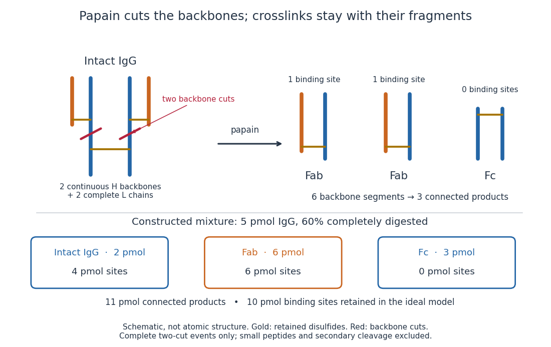
Two backbone cuts give two Fab fragments and one Fc fragment
. Original schematic of ideal limited Papain digestion of IgG, with cuts distinguished from retained protein disulfide bonds. The lower panel counts products for a constructed partial-digestion example, not a measured digest. Small released peptides, incomplete cleavage of one arm and secondary cleavage are excluded from that model. Details.
A connectivity argument explains the three products. Treat each continuous backbone segment as a graph vertex, and retained interchain disulfide bonds as edges. After the two stipulated cuts, six backbone segments remain: two complete immunoglobulin light chains, two immunoglobulin heavy chain arm segments and two immunoglobulin heavy chain stem segments. Each light chain remains attached to its own arm segment, producing two separate Fab fragments. The two stem segments remain attached to each other, producing one Fc fragment. Thus six backbone segments form three connected components of a graph, not six independently released products. Removing crosslinks instead, while keeping each backbone intact, would leave four chain components in this simplified covalent-connectivity model.
For a constructed mixture, start with of intact IgG and stipulate that undergoes the complete two-cut event while the rest remains intact. Count amount of substance of each product:The total amount of separately connected products becomes , although the starting amount was . Each digested IgG replaces one connected assembly with three. The antibody binding site inventory is conserved in this model:The factor two counts sites per intact IgG, not extra immunoglobulin light chains per Fab fragment. Fragment counts, backbone-segment counts and antibody binding site counts must be kept distinct. A real digest's retained binding would additionally depend on preservation of the paratopes.
The historical functional test was more subtle than asking whether a fragment still formed a visible precipitate. Rodney Robert Porter's earlier rabbit anti-ovalbumin preparation was digested with papain powder. The resulting fragment did not precipitate ovalbumin itself, but specifically inhibited ovalbumin precipitation with undigested antiserum. The intact-antiserum reaction supplied the positive comparison: failure to precipitate alone did not mean failure to recognize the antigen.[ref]
Separation and Isolation of Fractions of Rabbit Gamma-Globulin containing the Antibody and Antigenic Combining Sites by Rodney Robert Porter. Rodney R. Porter, Separation and Isolation of Fractions of Rabbit Gamma-Globulin containing the Antibody and Antigenic Combining Sites, 1958, opening paragraph: earlier papain-powder digest and inhibition of precipitation
The inference is that binding and assembly of a precipitating network are different abilities. A one-site Fab fragment can occupy an epitope without providing the two-arm bridge available to intact IgG. The inhibition readout supports retained recognition under the tested conditions; it does not locate individual contacts or show that every papain-generated product binds. This is the conceptual benefit of fragmenting an antibody: one can ask which functions survive loss of a structural connection.