Topics (318k) Articles (329k) Users (390) Discussions (237) Comments (383) Files (10k) New article

Radiochromic film is a type of dosimetric film used for measuring radiation exposure. It is primarily used in medical physics, radiation therapy, and radiation safety because of its ability to visually indicate radiation dose through changes in color. ### Key Characteristics of Radiochromic Film: 1. **Composition**: Radiochromic films are typically made from polymers that contain special dyes that change color when exposed to ionizing radiation.
Radiation treatment planning is a crucial process in radiation therapy, which is a common treatment for cancer and some other diseases. This planning involves several steps to ensure that the radiation is delivered accurately and effectively while minimizing exposure to surrounding healthy tissues. The primary objectives of radiation treatment planning include: 1. **Patient Simulation**: This involves positioning the patient in a way that reflects how they will be treated during radiation therapy.
Radiation therapy, also known as radiotherapy, is a medical treatment that uses high doses of radiation to kill or damage cancer cells. It is a common treatment for various types of cancer and can be employed either alone or in conjunction with other treatments, such as surgery and chemotherapy. ### Key Points about Radiation Therapy: 1. **Mechanism:** Radiation works by damaging the DNA within cells. Cancer cells are generally more sensitive to radiation because they are dividing more rapidly than most normal cells.
Proton therapy is a type of radiation therapy used primarily to treat cancer. Unlike conventional X-ray radiation therapy, which uses high-energy X-rays to kill cancer cells, proton therapy uses protons—positively charged particles that are part of the atomic nucleus. ### Key Features of Proton Therapy: 1. **Precision Targeting**: Protons have unique physical properties that allow them to deliver high doses of radiation directly to the tumor while sparing surrounding healthy tissues.
Proton computed tomography (pCT) is an advanced imaging technique that utilizes protons instead of traditional X-rays to create detailed cross-sectional images of objects, particularly in the context of medical imaging. This method leverages the interactions between protons and matter, providing unique advantages when it comes to imaging tissues, especially for cancer treatment and radiation therapy planning. Key characteristics of proton computed tomography include: 1. **Proton Sources**: pCT typically employs proton beams generated by particle accelerators.
Prostate brachytherapy is a form of radiation therapy used to treat prostate cancer. It involves placing radioactive seeds directly into or near the prostate gland to deliver targeted radiation to cancerous tissues while minimizing exposure to surrounding healthy tissues. ### Key characteristics of prostate brachytherapy include: 1. **Minimally Invasive**: The procedure is typically performed under local anesthesia and sedatives, and can often be done on an outpatient basis.
Preclinical imaging refers to a set of imaging techniques used to visualize biological processes in animal models (usually small animals like mice or rats) prior to human clinical trials. This field is crucial in biomedical research as it allows scientists to study disease mechanisms, evaluate therapeutic interventions, and monitor treatment responses in vivo. Preclinical imaging helps bridge the gap between basic science and clinical application by providing insights into the efficacy and safety of new drugs and therapies.
Positron Emission Tomography (PET) is a medical imaging technique that helps visualize and measure metabolic processes in the body. It is often used in clinical and research settings to assess conditions such as cancer, neurological disorders, and cardiovascular diseases. Here's how PET works: 1. **Radiotracer Injection**: A small amount of a radioactive substance, called a radiotracer, is introduced into the body, usually via injection.
Photothermal therapy (PTT) is a minimally invasive treatment method that utilizes light-absorbing agents, often in the form of nanoparticles, to convert light energy into heat. This heat can selectively destroy targeted cells, such as cancer cells, while minimizing damage to surrounding healthy tissue. The therapy is typically induced by shining near-infrared (NIR) light onto the treatment area, which penetrates the tissue and is absorbed by the nanoparticles.
Photomedicine is a field of medicine that involves the use of light to diagnose, treat, and prevent various medical conditions. It encompasses a range of therapies that utilize different types of light, including visible light, lasers, and other forms of electromagnetic radiation. Key applications of photomedicine include: 1. **Phototherapy**: This includes treatments like light therapy for skin conditions such as psoriasis, eczema, and acne. In this context, ultraviolet (UV) light is often used.
Photodynamic therapy (PDT) is a medical treatment that utilizes light-activated compounds to treat various conditions, including certain types of cancer, skin disorders, and age-related macular degeneration. The therapy involves three key components: 1. **Photosensitizer**: This is a special drug that is administered to the patient and accumulates in the target tissue. Photosensitizers are typically non-toxic before they are activated by light.
Pertechnetate, specifically known as sodium pertechnetate, is a chemical compound with the formula NaTcO₄. It is the sodium salt of pertechnetic acid (H₄TcO₄) and contains the technetium isotope Tc-99m, which is widely used in nuclear medicine.
Perfusion scanning is a medical imaging technique used to assess blood flow to various tissues and organs in the body. It helps in evaluating the perfusion (the passage of fluid through the circulatory system) in areas like the heart, brain, and other vital organs. ### Key Aspects of Perfusion Scanning: 1. **Purpose**: The primary purpose is to detect abnormalities in blood flow that may indicate conditions such as ischemia (reduced blood flow), tumors, or other vascular disorders.
The Percentage Depth Dose (PDD) curve is an important concept in radiation therapy that describes how the dose of radiation delivered by a therapy beam decreases with increasing depth in a given medium, such as tissue. This curve is essential for understanding how radiation penetrates and interacts with the tissues of the body.
Peptide receptor radionuclide therapy (PRRT) is a form of targeted cancer treatment that utilizes radioactive substances attached to peptides—short chains of amino acids that can bind to specific receptors on the surface of certain cancer cells. This therapy is primarily used to treat neuroendocrine tumors (NETs), which are tumors that arise from neuroendocrine cells and often express specific receptors such as somatostatin receptors.
Pencil-beam scanning is a technique used in radiation therapy, particularly in proton therapy, for the precise delivery of radiation to tumors while minimizing exposure to surrounding healthy tissues. This method involves using a narrow, focused beam of protons (or other charged particles) that can be accurately directed to specific locations within a tumor. ### Key Aspects of Pencil-Beam Scanning: 1. **Precision Targeting**: The pencil-beam is small in diameter, allowing for highly precise targeting of tumors.
A passive dual coil resonator is a type of resonant circuit that comprises two inductive coils (or inductors) arranged in a specific configuration to create resonance at a particular frequency. These circuits are commonly used in various applications involving resonance, such as in radio frequency (RF) systems, wireless power transfer, and electromagnetic field sensing.
Particle therapy, also known as charged particle therapy, is an advanced form of radiation therapy used primarily in cancer treatment. Unlike conventional radiation therapy that uses X-rays (photons), particle therapy utilizes charged particles, such as protons or heavier ions like carbon, to deliver radiation to tumors. ### Key Features of Particle Therapy: 1. **Precision**: Particle therapy provides more precise targeting of tumors while sparing surrounding healthy tissues.
Positron Emission Tomography (PET) for bone imaging is a diagnostic imaging technique that uses radioactive tracers to visualize metabolic processes in the body. Specifically, in the context of bone imaging, PET is often used to assess bone health, detect tumors, evaluate infection, and monitor the metabolism of bone tissue.
PET-CT, or Positron Emission Tomography-Computed Tomography, is a medical imaging technique that combines two different imaging modalities: PET and CT. 1. **Positron Emission Tomography (PET)**: This technique uses a small amount of radioactive material (radiotracer) that is injected into the body. The radiotracer emits positrons, which are detected by the PET scanner to produce images that reflect the metabolic activity of tissues.
Pinned article: Introduction to the OurBigBook Project
Welcome to the OurBigBook Project! Our goal is to create the perfect publishing platform for STEM subjects, and get university-level students to write the best free STEM tutorials ever.
Everyone is welcome to create an account and play with the site: ourbigbook.com/go/register. We belive that students themselves can write amazing tutorials, but teachers are welcome too. You can write about anything you want, it doesn't have to be STEM or even educational. Silly test content is very welcome and you won't be penalized in any way. Just keep it legal!
Intro to OurBigBook
. Source. We have two killer features:
- topics: topics group articles by different users with the same title, e.g. here is the topic for the "Fundamental Theorem of Calculus" ourbigbook.com/go/topic/fundamental-theorem-of-calculusArticles of different users are sorted by upvote within each article page. This feature is a bit like:
- a Wikipedia where each user can have their own version of each article
- a Q&A website like Stack Overflow, where multiple people can give their views on a given topic, and the best ones are sorted by upvote. Except you don't need to wait for someone to ask first, and any topic goes, no matter how narrow or broad
This feature makes it possible for readers to find better explanations of any topic created by other writers. And it allows writers to create an explanation in a place that readers might actually find it.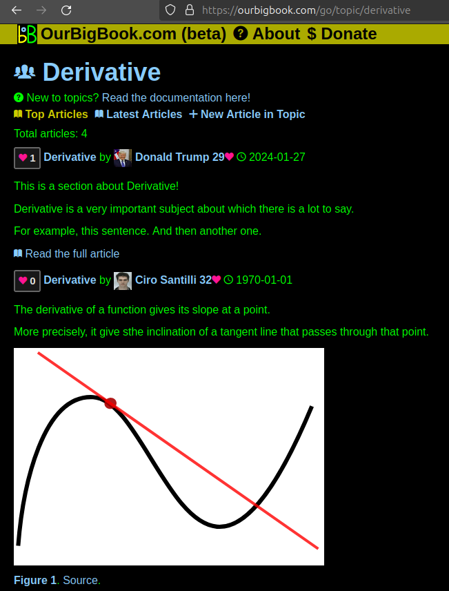
Figure 1. Screenshot of the "Derivative" topic page. View it live at: ourbigbook.com/go/topic/derivativeVideo 2. OurBigBook Web topics demo. Source. - local editing: you can store all your personal knowledge base content locally in a plaintext markup format that can be edited locally and published either:This way you can be sure that even if OurBigBook.com were to go down one day (which we have no plans to do as it is quite cheap to host!), your content will still be perfectly readable as a static site.
- to OurBigBook.com to get awesome multi-user features like topics and likes
- as HTML files to a static website, which you can host yourself for free on many external providers like GitHub Pages, and remain in full control

Figure 2. You can publish local OurBigBook lightweight markup files to either OurBigBook.com or as a static website.
Figure 3. Visual Studio Code extension installation.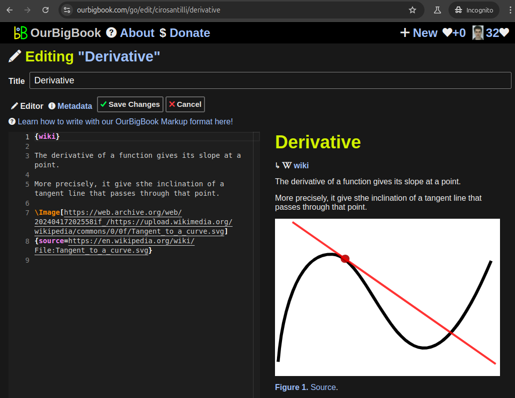
Figure 5. . You can also edit articles on the Web editor without installing anything locally. Video 3. Edit locally and publish demo. Source. This shows editing OurBigBook Markup and publishing it using the Visual Studio Code extension. 
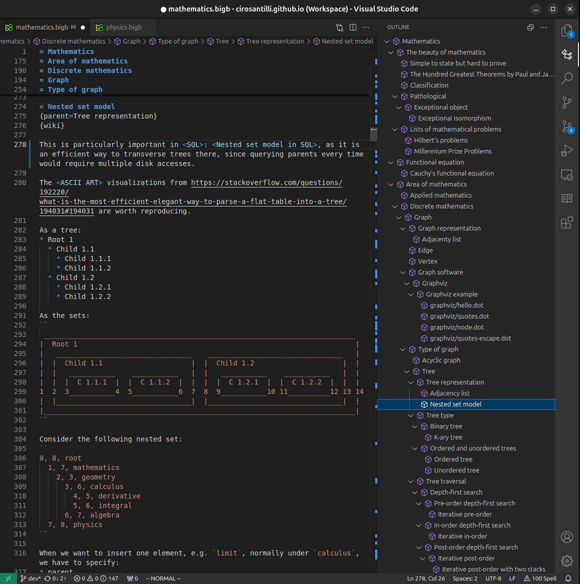
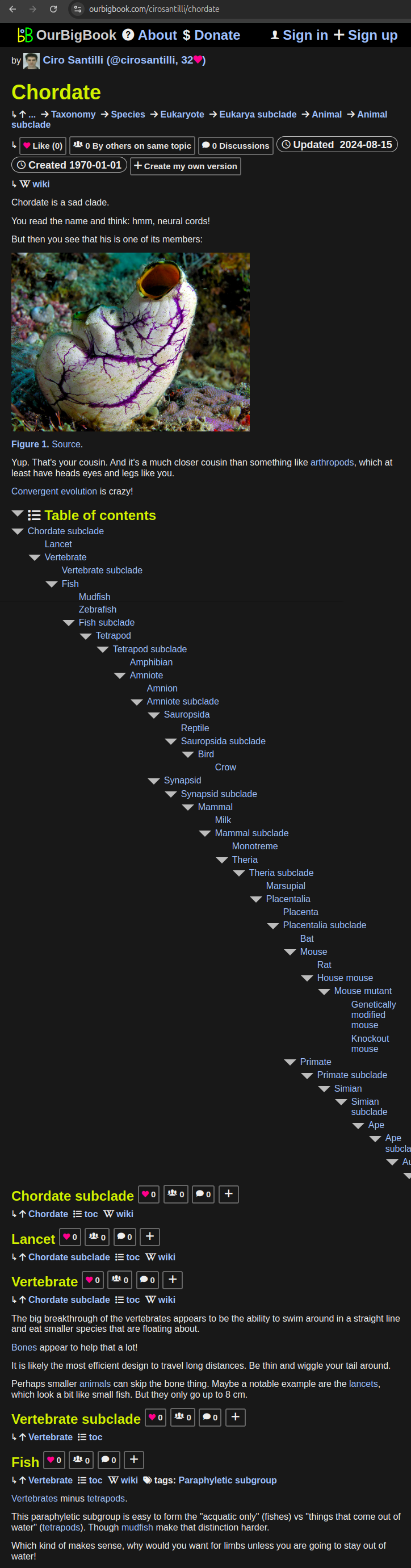
- Infinitely deep tables of contents:
All our software is open source and hosted at: github.com/ourbigbook/ourbigbook
Further documentation can be found at: docs.ourbigbook.com
Feel free to reach our to us for any help or suggestions: docs.ourbigbook.com/#contact