Ultraviolet (UV) refers to a type of electromagnetic radiation that has a wavelength shorter than visible light but longer than X-rays. Specifically, UV radiation falls within the wavelength range of approximately 10 nanometers (nm) to 400 nm. It is categorized into three main types based on its wavelength: 1. **UVA (320-400 nm)**: This type of UV radiation penetrates the skin more deeply and is primarily responsible for skin aging and long-term skin damage.
Articles by others on the same topic

Ultraviolet is electromagnetic radiation with wavelengths shorter than the violet end of ordinarily visible light. The CIE convention uses –, subdivided into UV-A at –, UV-B at – and UV-C at –. These divisions name intervals; they do not mark universal changes in every material's response. The boundary with visible light is not sharp, and other fields use near-, far-, vacuum- and extreme-ultraviolet divisions. [ref]
https://cie.co.at/eilvterm/17-21-008. CIE, International Lighting Vocabulary (2020), ultraviolet radiation: ranges and boundary caveats
. For example, NASA's Solar Dynamics Observatory observes extreme-ultraviolet bands below ; the CIE interval is therefore not the full extent of astronomical ultraviolet usage. [ref]https://sdo.gsfc.nasa.gov/data/channels.php. NASA, Solar Dynamics Observatory HMI and AIA channels: the Atmospheric Imaging Assembly's ultraviolet and extreme-ultraviolet bands
.The CIE ultraviolet interval corresponds to frequencies about – and photon energies –, using vacuum wavelength and with Planck constant and speed of light . Shorter wavelength means larger energy per photon; larger irradiance means more incident power per area. These are different comparisons. At the same incident power, a ultraviolet beam has fewer photons per second than a infrared beam, even though each ultraviolet photon has more energy.
A common ultraviolet mechanism is photon absorption promoting an Electron from a ground state to an excited state of a chemical atom or molecule. By contrast, many infrared absorption bands involve stretching or bending chemical bonds. For either mechanism, both the energy difference and the coupling to the incident electromagnetic field matter. One concrete example is the absorption discussed for butadiene: an Electron moves between molecular orbitals, and the position of the absorption reveals their energy separation. Ultraviolet absorption can therefore probe molecular orbitals, while infrared spectroscopy probes many molecular motions. [ref]
https://openstax.org/books/organic-chemistry/pages/14-7-ultraviolet-spectroscopy. John McMurry, Organic Chemistry, §14.7: ultraviolet excitation and the 217 nm butadiene example
.Subsequent chemical reactions depend on the absorbing chemical substance and its environment; the label ultraviolet alone does not predict a reaction rate. At sufficiently short wavelength, a photon can cause photoionization if its energy exceeds the relevant ionization energy. There is no single wavelength that separates all ionizing from all nonionizing behavior for every chemical substance. Likewise, eventual heating after photon absorption is possible in both ultraviolet and infrared. The distinction is useful because different optical spectra expose different processes, not because the two ranges obey different laws of electromagnetism.
Ritter's 1801 observation used a chemical readout rather than a thermometer. In Caltech/IPAC's account, sunlight passed through a glass optical prism and silver chloride was placed in different parts of the optical spectrum. Darkening was weak toward red, stronger toward violet, and pronounced beyond the visible violet edge. The measured evidence was a change in the chemical substance's appearance at a spatial location with no visible color, establishing radiation beyond the eye's ordinary range. It was not a calibrated measurement of a universal ultraviolet optical spectrum. [ref]
https://coolcosmos.ipac.caltech.edu/page/lesson_ritter_experiment. Caltech/IPAC Cool Cosmos, The Ritter Experiment (2022): the historical silver-chloride observation
.A controlled modern interpretation of such a chemical test would compare equal exposure durations and matched fresh samples, keep an unexposed sample in darkness, monitor temperature, and distinguish scattered visible light from the intended short-wavelength beam with characterized filtering. These are proposed controls, not an assertion that Ritter performed them. Unequal sample preparation or heating can change a reaction rate. The optical prism, terrestrial atmosphere and sample response restrict which wavelengths the test detects. Darkening is a detector-weighted response, so its spatial maximum is not automatically the maximum of incoming radiant flux or of photon energy.
Thomas Larason and Jeanne Houston's NIST report documents a quantitative ultraviolet detector comparison. An argon source feeds a double-diffraction grating spectrometer; a shutter and mirrors deliver the selected band to a working-standard or test photodiode, successively at the same position. A beam splitter supplies a monitor photodiode. Amplifiers and voltmeters record detector and monitor simultaneously; shutter-closed readings provide dark baselines. The – scan, typically with bandpass, measures spectral responsivity. Monitor normalization suppresses source drift; known spectral lines calibrate wavelength. The report assesses temperature effects and unwanted light outside the band, weighted by detector response. The latter need not cancel between different spectral responsivities. [ref].
This determines electrical response without visual perception. Bandpass and spatial nonuniformity limit the calibration; aging and changed temperature can affect later use. The scan includes visible light and ultraviolet; no response below follows from it.
For a narrow beam, the idealized substitution calculation isHere is spectral responsivity; and are dark-subtracted detector and monitor electric currents. This follows by writing for each detector and using the monitor to track changes in radiant flux. It assumes linear response, unchanged beam splitting and collection geometry, and a sufficiently narrow band. For an illustrative standard with , ratios and give . These invented arithmetic inputs illustrate the method; they are not NIST measurements.
An independent photon-counting model connects spectral responsivity to quantum efficiency. If is the number of collected Electrons per incident photon, there is no internal multiplication, and is the elementary charge, thenAt illustrative constant and , this gives about at and at . The ultraviolet readout is smaller in this model because there are fewer incident photons, not because their individual energies are smaller. Real quantum efficiency depends on wavelength, windows, absorption and carrier collection; a detector's band gap and construction determine its usable range. Constant across these two wavelengths is an analytical comparison, not a claimed specification of a real photodiode.
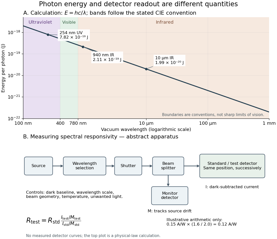
Photon energy across conventional ultraviolet and infrared ranges, with a schematic detector-substitution experiment
. Details. The curve is the calculated relation , and the shaded ranges use the stated CIE conventions. The lower diagram abstracts the detector comparison rather than reproducing an instrument drawing. Together with the infrared heat-balance illustration, it shows why a calibrated detector response, rather than what looks bright to an observer, is the bridge between invisible radiation and a quantitative measurement.