Cardiac image foreshortening
ID: cardiac-image-foreshortening

Cardiac image foreshortening occurs when a cardiac view represents a shorter apparent base-to-apex extent than the intended anatomical chamber length, often because the imaging plane misses the true cardiac apex. It can create an apparently thick false apex and affect derived ejection fraction and deformation measurements.
A primary echocardiography study acquired optimal and deliberately foreshortened views in 72 participants using a GE Vivid E9 system. The altered acquisition window was one rib-space above the optimal window; two analysis systems were used to compare derived quantities.[ref]
Impact of apical foreshortening on deformation measurements: a report from the EACVI-ASE Strain Standardization Task Force by Serkan Ünlü and others. Abstract, Aims and Methods and results
This tests acquisition geometry while keeping the participant the same, rather than interpreting every shorter-looking chamber as a biological change.An original ellipsoid model distinguishes a missed apex from a simple projected length. Letwith the true cardiac long axis along . The central long-axis plane includes both axial tips and has full length . A parallel offset plane instead intersects an ellipse whose apparent axial extent isAt , . A 10 cm true extent therefore appears as 8 cm in this model even though the slice was translated rather than rotated. Its apparent tips are not the original chamber tips.
This model is a geometrical explanation, not a clinical correction formula. A shorter outline can alter measured volume differently at end-diastolic volume and end-systolic volume; the ejection fraction error need not follow the length error by one fixed percentage. Consistent anatomical landmarks and independent views help distinguish actual chamber shape from the chosen section. Improving the repeatability of an outline inside an incorrect plane does not recover anatomy that was never acquired.
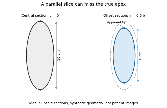
A parallel cardiac slice can miss the true apex
. Original sections of an ideal ellipsoid with axial semiaxis 5 cm and transverse semiaxis 2 cm. The central plane has full length 10 cm; a parallel plane offset by 0.6 times the transverse semiaxis has length 8 cm. Dashed outline shows the central section for comparison. Synthetic geometry, not patient data. Details.
New to topics? Read the docs here!