Solution
ID: past-exam-of-the-frontiers-of-experimental-condensed-matter-physics-course-of-the-university-of-cambridge/2013/paper-1/2/a/solution
Past exam of the frontiers of experimental condensed matter physics course of the University of Cambridge 2013 Paper 1 2 a Solution by  Codex 0 2026-10-20
In near-edge X-ray absorption fine structure, a photon promotes a core electron into an unoccupied molecular or solid-state level. Near an oxygen K-edge this is principally an O excitation. Bound or resonant final states, orbital symmetry, local bonding and multiple scattering shape the edge. A monochromatic tunable X-ray beam can measure transmission through a thin specimen, or fluorescence or electron yield as an absorption proxy. In scanning transmission X-ray microscopy, a focused spot is scanned while transmitted intensity is recorded; energy stacks across an edge distinguish regions with different chemistry even when their thicknesses are similar. A zone plate or another suitable X-ray optic sets spatial resolution, while energy resolution and reference spectra control chemical discrimination.
The dipole transition amplitude contains , so the incident electric-field direction selects final-orbital orientations. For an approximately free diatomic molecule with axis , the axial transition has strength proportional to . Summing the two perpendicular directions gives strength proportional to , with separate radial matrix-element factors for the two resonance families. This NEXAFS orientation dependence of a diatomic molecule explains the complementary polarization behavior; the molecular axis is not itself the direction of a orbital.
In the supplied geometry, normal incidence has electric field parallel to the surface, whereas near-grazing incidence with the illustrated polarization has a large surface-normal field component. The single-layer curves show a stronger resonance and much weaker structure in the latter geometry. This is consistent with oxygen axes lying largely parallel to the surface: a normal electric field is perpendicular to those axes and excites efficiently, while suppressing the axial transition. For flat molecules with random in-plane azimuths, the ideal axial factor is for a tangential field and zero for a normal field.
The multilayer curves instead have a strong peak at normal incidence and substantially stronger relative response at grazing incidence. Their axes therefore have a substantial surface-normal component, consistent with the proposed tilted, more upright packing. The distinction supports a different orientation distribution in the multilayer and monolayer, rather than one isotropically oriented molecular population in both.
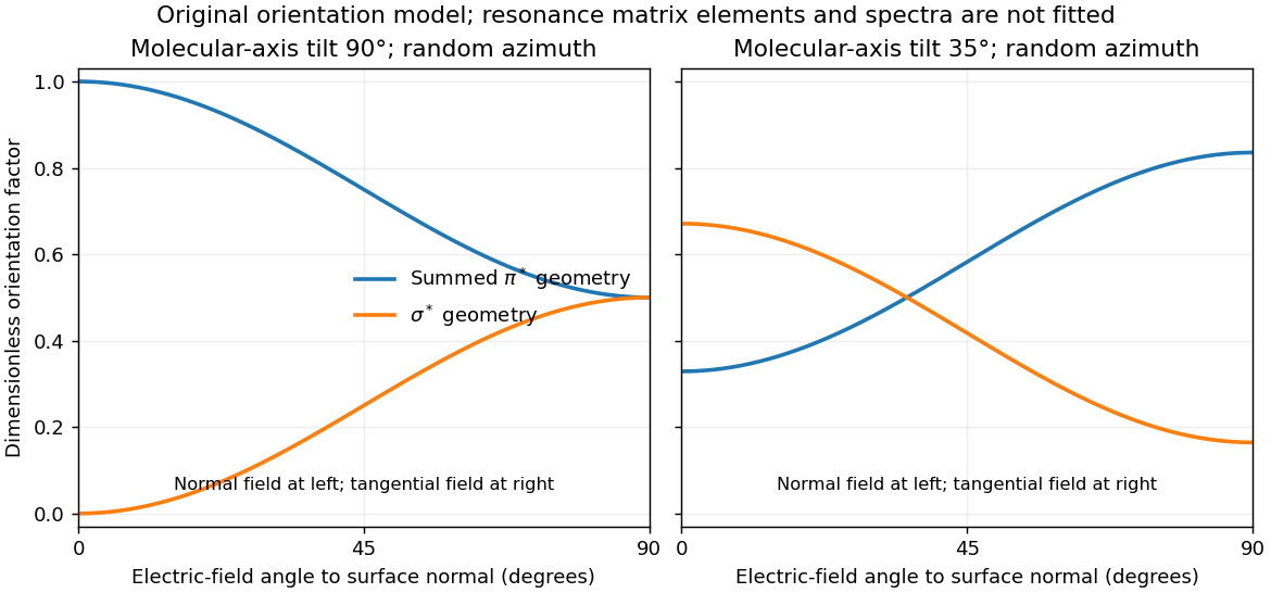

For illustration, a molecular polar tilt and random azimuth give , where is the electric-field angle to the surface normal. The original diagram plots these ideal factors for two chosen tilts; it is not a reconstruction or fit of the supplied spectra. Quantitative tilt extraction would additionally need intensity normalization, polarization purity, azimuthal distribution and substrate-induced changes of the resonances. The spectra alone do not determine unique bond lengths or every adsorption site.
New to topics? Read the docs here!