Coaptation area is the area of the contact region formed when opposing valve leaflet surfaces meet during coaptation. The measurement must specify the valve, phase and surface convention. Counting the same interface once is different from summing the apposed faces of both leaflets, and a projected image area is different from the area measured along a curved surface.
A primary investigation examined quantitative assessment of mitral coaptation using three-dimensional transesophageal echocardiography.[ref]
Quantitative assessment of mitral valve coaptation using three-dimensional transesophageal echocardiography by Frederick C. Cobey and others. Title and indexed Abstract, three-dimensional coaptation-zone assessment
This provides an experimental context for defining the zone before assigning it one scalar area. The observed or reconstructed leaflet geometry remains part of the measurement.For a specified contact patch on one reference face,If a planar patch is viewed at angle between its normal and the viewing direction, its projected area is . A true area of 20 mm viewed at therefore projects to 10 mm. This is an original geometrical example, not an echo calibration rule for all curved leaflets.
Area also omits the arrangement of the contact. In an ideal 10 mm by 10 mm gap map, a 10-by-2 mm contact strip spanning the full width has the same area as a 2-by-10 mm strip running along its height. If flow is constrained to travel from the bottom edge to the top edge within this two-dimensional map, the first strip blocks every path while the second leaves open side paths. Both contact areas are 20 mm.
Real valves have three-dimensional fluid gaps, deformation and changing pressure. The map is a counterexample to inferring closure from area alone, not a prediction of leakage magnitude. The location of contact, any remaining connected gaps and the surrounding heart valve mechanics matter alongside the reported coaptation area.
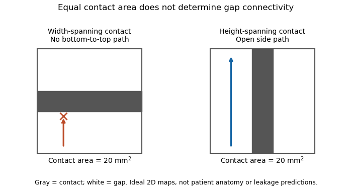
Equal coaptation area need not determine gap connectivity
. Original ideal two-dimensional contact maps. Each 10 mm square has a 20 square-millimetre contact strip. With paths confined inside the map, the horizontal strip blocks bottom-to-top passage, while the vertical strip leaves side paths. This illustrates geometric information lost by a scalar area, not measured valve leakage or patient anatomy. Details.
The mitral coaptation index is a dimensionless normalization of the mitral valve's coaptation area. In the primary method of Frederick C. Cobey and colleagues, it is the three-dimensional end-systolic coaptation-zone area divided by the three-dimensional mitral-valve area used in that protocol:The denominator and surface-reconstruction convention are part of the definition.[ref]
The study used intraoperative transesophageal echocardiography in 25 patients with functional mitral regurgitation, followed by offline analysis of the end-systolic geometry.[ref]
Quantitative assessment of mitral valve coaptation using three-dimensional transesophageal echocardiography by Frederick C. Cobey and others. Abstract, Methods
This supplies an experimental acquisition context for the ratio; it does not make every area called annular area interchangeable with the protocol's valve-area measurement.An original normalization example uses cm and cm, giving . A second constructed valve with areas 3 and 20 cm has the same index despite twice the absolute contact area. Conversely, keeping contact area 1.5 cm while changing the denominator to 15 cm gives . Absolute area and normalized coverage answer different questions.
Uniformly scaling every length by a positive factor multiplies both areas by , leaving unchanged. Inconsistent surface conventions do not cancel: counting two apposed faces in the numerator while counting their interface once in another reconstruction can change the ratio without a biological change.
Finally, the index does not describe the connectivity of any remaining gaps. Two contact patches with equal coaptation area and equal denominator can have different shapes and positions, as illustrated under coaptation area. The mitral coaptation index is a geometric descriptor to interpret alongside the reconstructed valve and blood flow, rather than a complete description of valve sealing.
Articles by others on the same topic
There are currently no matching articles.