A lateral ventricle is one of the two large cerebrospinal fluid-filled cavities within the cerebral hemispheres. Each connects with the third ventricle through an interventricular foramen. Its location inside a hemisphere makes it a useful landmark when interpreting sections or images of the brain.[ref]
Lateral Ventricles in MeSH. Definition, paired cerebral cavities and communication with the third ventricle
The choroid plexus projects into ventricular cavities and contributes to cerebrospinal fluid production; the ependyma lines the ventricular wall. The resulting environment joins a secretory interface, a fluid compartment and neighbouring neural tissue. These components have different roles even though they appear close together in an anatomical section.[ref]
Anatomy and Physiology 2e by J. Gordon Betts and others. §12.2, ependymal cells; §13.3, cerebrospinal fluid and choroid plexuses
The connections below provide a simplified route through the ventricular system. Actual transport also has a time dependence: phase-contrast magnetic resonance imaging has resolved cerebrospinal fluid movement over the cardiac cycle, while work on the third ventricle has revealed structured local flows driven by motile cilia.[ref]
Normal flow patterns of intracranial and spinal cerebrospinal fluid defined with phase-contrast cine MR imaging by D. R. Enzmann and others. abstract, cardiac-cycle-dependent flow encoding
[ref]Cilia-based flow network in the brain ventricles by Regina Faubel and others. abstract, ciliary modules and ventricular flow networks
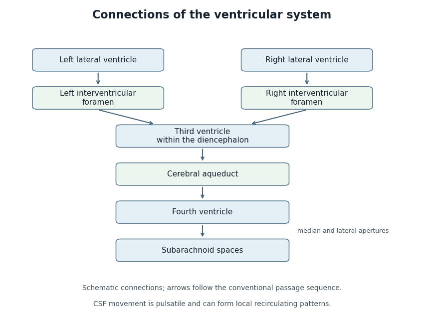
Anatomical connections of the ventricular system
. Original schematic showing paired lateral ventricles, the interventricular foramina, third ventricle, cerebral aqueduct, fourth ventricle and subarachnoid spaces. Arrows follow the conventional passage sequence; cerebrospinal fluid also undergoes pulsatile movement and local recirculation.[ref]Neuroscience, 2nd edition edited by D. Purves and others. The Ventricular System, Figure 1.17, anatomical connections
Details.
Articles by others on the same topic
There are currently no matching articles.