The anterior central nervous system of a vertebrate, developing principally from the neural tube rather than directly from neural crest.
The forebrain, also called the prosencephalon in embryological terminology, is the anterior major division of the developing vertebrate brain. It subdivides into the telencephalon and diencephalon. The name describes a developmental and anatomical grouping, rather than one nucleus with a single task.[ref]
Prosencephalon in NLM MeSH. Scope note and Forebrain entry term: anterior primitive cerebral vesicle derived from the neural tube and its two subdivisions.
The telencephalon includes the cerebral-hemisphere system, while the diencephalon includes regions such as the thalamus, hypothalamus and epithalamus. Thus the visible cerebral cortex is part of the forebrain but does not exhaust it. Deep nuclei and connecting pathways also belong to the region.[ref]
Neuroscience, 2nd edition edited by D. Purves and others. Gross organization of the brain and ventricular-system anatomical landmarks.
The ventricular spaces provide an illuminating map. The lateral ventricles lie within the cerebral hemispheres, while the third ventricle is associated with the diencephalic region. A fluid-filled cavity and its surrounding neural tissue are different structures: locating the cavity helps orient the tissue, but does not make the cavity itself a neuron-processing center.[ref]
This developmental grouping is useful when comparing brains whose proportions differ greatly. Expansion of one forebrain component does not erase the distinction among its components. Likewise, observing a response in the lateral habenula identifies activity in one forebrain circuit, not a computation performed uniformly by the entire forebrain.
The telencephalon is the anterior subdivision of the embryonic forebrain and the corresponding territory of the mature brain. Its derivatives include the cerebral hemispheres, with cerebral cortex, deeper basal ganglia and associated structures, including the olfactory bulbs. It is an anatomical and developmental division, rather than a name for one behavior or one type of neuron.[ref]
Telencephalon in NLM MeSH. D013687 scope note and anatomy hierarchy: anterior prosencephalic subdivision; cerebral and associated structures.
The forebrain divides into telencephalon and diencephalon. These names should not be treated as interchangeable: the thalamus belongs to the diencephalon, whereas cerebral cortex belongs to the telencephalon. The large cerebral hemispheres can cover deeper regions in a surface view, so apparent position in a photograph alone is a poor guide to developmental membership.[ref]
Prosencephalon in NLM MeSH. D016548 scope note: subdivision into diencephalon and telencephalon.
A practical example is classifying structures in a section through the brain. A region of cerebral cortex and a deep basal ganglia nucleus can both be telencephalic even though one is superficial and the other deep. Conversely, an adjacent thalamus is diencephalic. This distinction makes the telencephalon useful as a map of related structures without assuming that everything within that map performs the same computation.
The diencephalon is a division of the forebrain surrounding the third ventricle, deep beneath the cerebral hemispheres. It includes the thalamus, hypothalamus and epithalamus. The anatomical name groups several interacting regions: circuits of the thalamus connect with the cerebral cortex, while the hypothalamus coordinates aspects of homeostasis and regulates signals to the pituitary gland.[ref]
Anatomy and Physiology 2e by J. Gordon Betts and others. §13.2, The Diencephalon, Thalamus and Hypothalamus
The relation to the third ventricle gives a useful way to read a section of the brain. The paired thalami lie beside the narrow midline fluid space; the surrounding neural tissue belongs to the diencephalic region, while cerebrospinal fluid occupies the cavity. Distinguishing the cavity from its walls connects the ventricular system to the organization of the surrounding brain.[ref]
Neuroscience, 2nd edition edited by D. Purves and others. The Ventricular System, Figure 1.17; ventricular spaces as anatomical landmarks
The epithalamus is a dorsal posterior subdivision of the diencephalon, associated with the roof of the third ventricle. Its usual anatomical scope includes the habenula and associated fiber systems, the pineal gland and the epithelial ventricular roof. It is a region containing distinct structures, not merely another name for the pineal gland.[ref]
Epithalamus in NLM MeSH. Scope note: dorsal posterior position and usual components; classification convention for paraventricular thalamic nuclei.
One component supplies an experimentally clear example. Masayuki Matsumoto and Okihide Hikosaka recorded from the lateral habenula during a reward-associated eye-movement task in rhesus monkeys. Many neurons increased activity for an unrewarded target and decreased activity for a rewarded target, while recorded dopamine-neuron responses showed the opposite pattern.[ref]
Lateral habenula as a source of negative reward signals in dopamine neurons by Masayuki Matsumoto and Okihide Hikosaka. 2007 abstract: visually guided task and opposite reward-related response signs.
Reversing which target location earned reward also reversed the responses. This is an illuminating control: a response following the reward assignment is harder to explain as a fixed preference for one screen position. Habenular excitation preceded dopamine-neuron inhibition on unrewarded trials, and weak electrical stimulation of the lateral habenula inhibited dopamine neurons.[ref]
Lateral habenula as a source of negative reward signals in dopamine neurons by Masayuki Matsumoto and Okihide Hikosaka. Reward-location reversal, temporal order and stimulation results.
Recording and intervention provide different evidence. Opposite responses and timing motivate a circuit hypothesis; stimulation tests influence on another population. Neither establishes that every epithalamic structure performs reward evaluation, nor that the influence is necessarily a direct monosynaptic inhibitory connection. Anatomical membership should not be confused with uniform physiological function.
The pineal gland is a small neuroendocrine organ of the epithalamus, attached to the roof of the third ventricle. It secretes melatonin, linking a neural timing system to a chemical signal distributed beyond the brain. Its anatomical position is distinct from that of the nearby habenula.[ref]
Pineal Gland in NLM MeSH. D010870 scope note and anatomical hierarchy.
A classic experiment located a strong daily rhythm at an enzymatic step in pineal metabolism. In 1970, D. C. Klein and J. L. Weller reported that rat pineal serotonin N-acetyltransferase activity was more than 15 times higher at night than during the day. The rhythm persisted in complete darkness and in blinded animals, whereas constant illumination suppressed it. Their experiments in isolated tissue also implicated norepinephrine in regulation of the enzyme activity.[ref]
Indole metabolism in the pineal gland: a circadian rhythm in N-acetyltransferase by D. C. Klein and J. L. Weller. Science169,1093–1095(1970), abstract: lighting conditions, enzyme rhythm and in-vitro regulation.
The darkness control is particularly illuminating. A mechanism that merely switched on whenever light was absent would predict a continuously high output in constant darkness. Continued rhythmic variation instead supports an internally generated timing signal. It does not show that the pineal gland alone generates the organism's entire circadian rhythm: the measurement concerns one gland and one metabolic activity in an intact timing system. Likewise, the reported activity ratio is an enzyme-assay result, not a universal ratio of human blood melatonin concentrations.
The habenula is a small nuclear complex of the epithalamus at the dorsal posterior wall of the third ventricle, adjacent to the dorsal thalamus and pineal gland. In mammals, its medial habenula and lateral habenula are distinct divisions with different circuits. The name denotes neural tissue, rather than a hormone-secreting gland.[ref]
Habenula in NLM MeSH. D019262 scope note: location, habenular nuclei and epithalamic membership.
The distinction between divisions matters when assigning a measured function. In a reward-location reversal task, recordings from the lateral habenula of monkeys showed responses opposing those of dopamine neurons: many lateral habenular cells were more active when an outcome was worse than expected. Weak electrical stimulation of the lateral habenula inhibited recorded dopamine neurons.[ref]
Lateral habenula as a source of negative reward signals in dopamine neurons by Masayuki Matsumoto and Okihide Hikosaka. Nature447,1111–1115(2007), abstract and reward-reversal/stimulation experiments.
For example, if a cue that formerly predicted a reward ceases to do so, the experiment asks whether neural activity follows the new outcome relationship rather than merely the cue's appearance. A change after reversal supports an outcome-related signal. It does not establish that every neuron in the habenula encodes disappointment, nor does electrical stimulation establish a direct single-synapse pathway. Keeping the whole complex, its divisions and the recorded cell populations distinct makes the anatomical term useful when comparing experiments.
The medial habenula is the medial division of the habenula, a nuclear complex of the epithalamus. It participates in a circuit projecting to the interpeduncular nucleus. The medial habenula and lateral habenula are neighboring anatomical divisions with different circuitry; results from one should not automatically be assigned to the other.[ref]
Habenular α5 nicotinic receptor subunit signalling controls nicotine intake by Christie D. Fowler and others. Originating 2011 article, medial-habenula/interpeduncular circuit and region-specific receptor manipulations.
A motivating experiment tested an inhibitory contribution to nicotine intake. Christie D. Fowler and colleagues found increased nicotine intake in mice lacking the α5 nicotinic acetylcholine receptor subunit. Re-expression of that subunit in the medial habenula rescued the altered intake phenotype; knockdown in the medial habenula of rats reproduced increased intake. The work distinguished nicotine's rewarding effects from an inhibitory effect appearing at higher tested doses.[ref]
Habenular α5 nicotinic receptor subunit signalling controls nicotine intake by Christie D. Fowler and others. Abstract: knockout, local rescue, rat knockdown and reward/inhibitory-signal distinction.
The rescue is particularly informative. A whole-animal mutation can alter many tissues and developmental processes. Restoring the relevant component in a specified region tests whether that regional circuit can account for an important part of the phenotype. Conversely, the comparison does not show that every medial habenula neuron has the same function or that the entire region is merely a nicotine-detector. The measured intake change is a result in specified rodent preparations, not a direct measurement of human smoking behavior or a clinical intervention.
The lateral habenula is the lateral division of the habenula, an epithalamic nuclear complex. Its circuits participate in linking information about outcomes and aversive events to other brain systems. It is an anatomical structure containing varied neuronal activity, rather than a uniform label for every negative emotion.[ref]
Lateral habenula as a source of negative reward signals in dopamine neurons by Masayuki Matsumoto and Okihide Hikosaka. Abstract: epithalamic location and reward-related recordings.
In a rhesus-monkey eye-movement task, many lateral-habenular neurons responded more strongly to targets predicting no reward and less strongly to rewarded targets. Reversing which location earned reward reversed these responses. That control distinguishes outcome association from an unchanging preference for a screen location. Habenular excitation preceded dopamine-neuron inhibition on unrewarded trials, and weak stimulation inhibited dopamine-neuron activity.[ref]
Lateral habenula as a source of negative reward signals in dopamine neurons by Masayuki Matsumoto and Okihide Hikosaka. Reward-location reversal, temporal ordering and stimulation experiment.
Temporal organization is another experimentally important property. A 2018 study of rat and mouse models examined increased burst firing and its suppression by ketamine. Pharmacology and modeling implicated NMDA receptors and T-type calcium channels, and controlled current changes could switch recorded neurons between bursting and tonic firing modes.[ref]
An invented comparison makes the distinction clear: ten evenly spaced spikes and two tight bursts of five spikes over the same observation period have the same total count and mean rate, but different timing. A mean firing rate alone can miss the property a burst experiment tests.
The studies support specific circuit and cellular hypotheses. They do not establish a direct monosynaptic inhibitory connection from the stimulation result alone, or show that rodent behavioral assays explain every aspect of human depression or ketamine action. Anatomy, response polarity and firing pattern answer related but different questions.
A set of interacting nuclei with diverse cortical and subcortical connections; anterior and mediodorsal nuclei have distinct anatomical relations and should not be treated as one undifferentiated memory relay.
A thalamic visual structure receiving retinal input and participating in pathways to visual cortex. Chromatic nulling experiments constrain how particular cells combine cone signals, while recurrent and descending interactions complicate a pure feed-forward relay description.
Thalamic region with prominent prefrontal connections, implicated in aspects of cognition and memory. It is anatomically distinct from the anterior thalamic nuclei.
Thalamic nuclei connected with the mammillary bodies, hippocampal formation and cingulate regions. Their dysfunction can compromise spatial and event-related memory.
Intracranial pressure is the pressure within the cranial compartment, commonly assessed through a specified fluid or tissue measurement. Brain tissue, blood and cerebrospinal fluid share a constrained space. Changes in their volumes and drainage therefore interact rather than acting as isolated pressures.
The brainstem connects the spinal cord with more rostral parts of the brain and includes the medulla oblongata, pons and midbrain. Its pathways and nuclei participate in sensory, motor and autonomic functions. A compact region can therefore support several interacting systems.
A cranial nerve nucleus is a central collection of neuronal cell bodies participating in a cranial-nerve pathway. Motor nuclei contain projecting neurons, while sensory nuclei receive and process input. A central nucleus should be distinguished from a peripheral ganglion.
The pons is a brainstem region between the medulla oblongata and midbrain. It contains long pathways, cranial-nerve nuclei and connections with the cerebellum. Its visible bulge does not correspond to one uniform functional circuit.
The locus coeruleus is a pontine nucleus containing many noradrenaline-releasing neurons with widespread projections. It participates in state-dependent regulation of neural activity. Activity in this population is one component of a distributed system rather than a complete scalar measure of arousal.
A vestibular nucleus is one of the brainstem nuclei processing input from the vestibular system and connected pathways. Outputs contribute to eye movements, posture and further central processing. These nuclei combine incoming signals rather than merely reproducing one peripheral measurement.
The facial motor nucleus is a pontine motor nucleus whose axons travel in the facial nerve to facial-expression muscles. Its fibers curve around the abducens nucleus, contributing to the facial colliculus. The surface landmark therefore reflects more than one nerve system.
The abducens nucleus lies in the dorsal pons and contains neurons involved in horizontal eye-movement control. Its motor output reaches the lateral rectus through the abducens nerve. A nuclear lesion can affect coordinated gaze more broadly than an isolated peripheral nerve lesion.
The medulla oblongata is the Caudal brainstem region adjoining the spinal cord. It contains ascending and descending pathways and nuclei involved in sensory, motor and autonomic regulation. Its structural compactness connects local lesions with combinations of otherwise distinct functions.
The solitary nucleus is a medullary sensory complex receiving visceral and taste-related inputs through several cranial nerves. Its connections contribute to autonomic and behavioral responses. Nucleus tractus solitarius and nucleus of the solitary tract are alternate names for this complex.
Nucleus tractus solitarius is the Latin-derived name commonly used for the solitary nucleus. The name refers to a central sensory complex associated with the solitary tract, rather than to a separate peripheral ganglion.
The hypoglossal nucleus contains motor neurons whose axons travel through the hypoglossal nerve to most muscles of the tongue. It lies near the floor of the fourth ventricle, beneath the hypoglossal trigone. Surface position and motor connections describe different aspects of the same nucleus.
The spinal trigeminal nucleus is an elongated brainstem sensory complex involved particularly in pain and temperature information from the face and associated territories. Its Caudal extension and incoming tract distinguish it from a single compact relay at the entry of the trigeminal nerve.
The nucleus of the solitary tract is another name for the solitary nucleus. It receives visceral sensory input and contributes to distributed reflex circuits, including those regulating cardiorespiratory and digestive functions.
The dorsal motor nucleus of the vagus contains preganglionic parasympathetic neurons whose axons enter the vagus nerve. Many outputs regulate thoracic and abdominal organs. It is distinct from sensory vagal nuclei and from the branchial motor populations of the nucleus ambiguus.
The nucleus ambiguus is a medullary neuronal group contributing branchial motor pathways to pharyngeal and laryngeal muscles and containing cardiac vagal populations. Different cell groups have different targets. The anatomical label therefore spans more than one physiological role.
The inferior olive is a medullary nuclear complex providing climbing-fiber input to the cerebellum. Its connections participate in cerebellar timing and plasticity. A teaching-related role in one task should be distinguished from a claim that every olivary signal encodes the same error variable.
The ventricular system is the connected set of fluid-filled cavities within the brain, continuous caudally with the central canal of the spinal cord. Cerebrospinal fluid passes between ventricular compartments and the surrounding subarachnoid spaces through defined openings.
The interventricular foramina are the paired openings through which the lateral ventricles communicate with the third ventricle. Each opening connects its own side's lateral ventricle to the shared midline cavity. This arrangement provides a route for cerebrospinal fluid to reach the rest of the ventricular system.[ref]
Lateral Ventricles in MeSH. Definition, each lateral cavity communicating with the third ventricle
Both sides reach the shared third ventricle, which joins the fourth ventricle through the cerebral aqueduct.
Measurements add a temporal picture to this connection map. In a 1991 study, phase-contrast magnetic resonance imaging encoded the direction of cerebrospinal fluid movement across the cardiac cycle. The ventricular measurements contained systolic and diastolic flow components, with essentially simultaneous timing at the interventricular opening and cerebral aqueduct. Anatomical continuity and measured pulsatile transport describe complementary features of the same pathway.[ref]
Normal flow patterns of intracranial and spinal cerebrospinal fluid defined with phase-contrast cine MR imaging by D. R. Enzmann and others. abstract, flow encoding and timing at ventricular passages
A lateral ventricle is one of the two large cerebrospinal fluid-filled cavities within the cerebral hemispheres. Each connects with the third ventricle through an interventricular foramen. Its location inside a hemisphere makes it a useful landmark when interpreting sections or images of the brain.[ref]
Lateral Ventricles in MeSH. Definition, paired cerebral cavities and communication with the third ventricle
The choroid plexus projects into ventricular cavities and contributes to cerebrospinal fluid production; the ependyma lines the ventricular wall. The resulting environment joins a secretory interface, a fluid compartment and neighbouring neural tissue. These components have different roles even though they appear close together in an anatomical section.[ref]
Anatomy and Physiology 2e by J. Gordon Betts and others. §12.2, ependymal cells; §13.3, cerebrospinal fluid and choroid plexuses
The connections below provide a simplified route through the ventricular system. Actual transport also has a time dependence: phase-contrast magnetic resonance imaging has resolved cerebrospinal fluid movement over the cardiac cycle, while work on the third ventricle has revealed structured local flows driven by motile cilia.[ref]
Normal flow patterns of intracranial and spinal cerebrospinal fluid defined with phase-contrast cine MR imaging by D. R. Enzmann and others. abstract, cardiac-cycle-dependent flow encoding
[ref]Cilia-based flow network in the brain ventricles by Regina Faubel and others. abstract, ciliary modules and ventricular flow networks
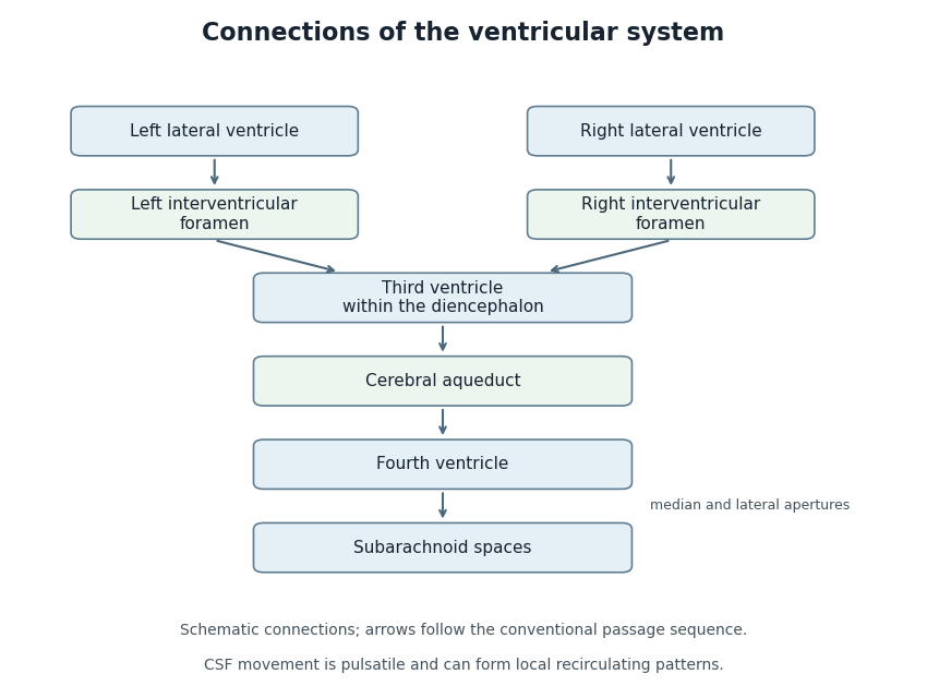
Anatomical connections of the ventricular system
. Original schematic showing paired lateral ventricles, the interventricular foramina, third ventricle, cerebral aqueduct, fourth ventricle and subarachnoid spaces. Arrows follow the conventional passage sequence; cerebrospinal fluid also undergoes pulsatile movement and local recirculation.[ref]Neuroscience, 2nd edition edited by D. Purves and others. The Ventricular System, Figure 1.17, anatomical connections
Details.The third ventricle is the narrow midline cavity of the diencephalon, between the paired thalami and adjoining the hypothalamus. It contains cerebrospinal fluid and forms part of the ventricular system. The lateral ventricles communicate with it through the interventricular foramina; the cerebral aqueduct connects it to the fourth ventricle.[ref]
This cavity is more than a passive container. A 2016 study of mouse third ventricles found organized groups of cilia producing a network of cerebrospinal fluid flows, including a periodically changing pattern. Related complex flows were observed in rat and pig third ventricles.[ref]
Cilia-based flow network in the brain ventricles by Regina Faubel and others. Abstract; ciliary modules and time-dependent flow
An opening specifies a possible route between cavities; local cilia-driven transport specifies how material moves within a cavity. A single well-mixed compartment model can therefore conceal spatial differences in the delivery of signaling molecules.
The central canal of the spinal cord is the narrow cavity along the central spinal region, developmentally continuous with the ventricular system. In adults its patency varies. Anatomical continuity does not imply that it carries most cerebrospinal-fluid outflow from the fourth ventricle.
The cerebral aqueduct is the narrow midbrain passage connecting the third and fourth ventricles. Its small caliber makes the relation between geometry and fluid passage important. It is a conduit rather than the principal site of cerebrospinal-fluid production.
A choroid plexus is a vascular and epithelial structure projecting into a brain ventricle and contributing to cerebrospinal fluid production. Its epithelial barrier separates blood-associated and ventricular compartments. Secretion is a regulated transport process rather than simple unfiltered leakage from vessels.
The choroid plexus of the fourth ventricle is the vascular secretory structure associated with the tela choroidea of that ventricle. Its position near the roof distinguishes it from neural nuclei underlying the ventricular floor.
The cellular lining of the ventricular system and central canal of the spinal cord.
An ependymal cell is a cell of the ependyma, the lining of the brain ventricular system and central canal of the spinal cord. Many ventricular ependymal cells carry multiple motile cilia facing the cerebrospinal fluid. Their coordinated beating can move nearby fluid; the cells are distinct from the neurons whose signaling circuits surround the cavity.[ref]
Anatomy and Physiology 2e by J. Gordon Betts and others. Nervous tissue: ependymal cells and cerebrospinal-fluid circulation
In a 2016 study, organized cilia modules in the mouse third ventricle generated structured fluid-flow networks and a periodically switching flow pattern.[ref]
Cilia-based flow network in the brain ventricles by Regina Faubel and others. Abstract; collective ciliary transport
The result illustrates how an epithelial lining can actively organize its fluid environment rather than simply delimit it. A cilium on one cell acts locally; tissue-level organization determines the collective transport pattern.Tanycytes are specialized ependymal-related cells with long processes. Their geometry and functions should not be assigned indiscriminately to every ependymal cell.
Cerebrospinal fluid is the fluid occupying the brain ventricles and surrounding subarachnoid spaces. Production, movement and removal connect its composition and pressure to the function of several interfaces. It is not interchangeable with either blood or fluid inside neurons.
The fourth ventricle is the ventricular cavity between the dorsal pons and medulla oblongata anteriorly and the cerebellum posteriorly. It communicates with the cerebral aqueduct and with subarachnoid spaces through median and lateral apertures.
A lateral aperture of the fourth ventricle is an opening near the end of a lateral recess of the fourth ventricle. The paired apertures allow communication between ventricular and subarachnoid fluid spaces. They are distinct from the single median aperture of the fourth ventricle.
The paired lateral aperture of the fourth ventricle openings.
The median aperture of the fourth ventricle is the midline opening connecting the ventricular cavity with the cisterna magna region. It is a principal route into the subarachnoid space, together with the paired lateral apertures.
The tela choroidea is a thin vascular membrane associated with the roof of a ventricular cavity and its choroid plexus. In the fourth ventricle it contributes to the thin inferior roof. A vascular membrane and the neighboring neural sheet are different structural components.
The inferior medullary velum is a thin neural sheet associated with the inferior roof of the fourth ventricle, extending from the nodular region toward the floccular attachments. It is distinct from the adjacent vascular tela choroidea.
The superior medullary velum is a thin neural sheet spanning between the superior cerebellar peduncles and contributing to the upper roof of the fourth ventricle. Its ventricular surface and cerebellar attachment provide landmarks for neighboring pathways.
A lateral recess of the fourth ventricle is a lateral extension of the ventricular cavity toward a lateral aperture of the fourth ventricle. Its direction differs from the main cranial-to-caudal ventricular axis, making three-dimensional relations important in interpreting a section.
Plural name of the lateral recess of the fourth ventricle.
The fastigium is the apex of the tent-like roof of the fourth ventricle. It is a geometric landmark of the cavity, distinct from a neural nucleus or a fluid outlet. Its name helps orient descriptions of the superior and inferior roof.
The diamond-shaped floor of the fourth ventricle, formed by the dorsal pons and upper medulla oblongata.
The funiculus separans is a narrow ridge in the Caudal floor of the fourth ventricle, separating the vagal trigone from the area postrema. It is a surface landmark, not a complete boundary between all underlying neural pathways.
The area postrema is a specialized vascular region near the Caudal fourth ventricle, involved in detecting circulating signals and contributing to emetic responses. Its close relation to vagal sensory circuits connects blood-borne information with coordinated behavior.
The vagal trigone is a surface region of the floor of the fourth ventricle associated with the underlying dorsal motor nucleus of the vagus. Surface shape and the full extent of the nucleus are different anatomical descriptions.
The hypoglossal trigone is a triangular surface region near the Caudal midline of the floor of the fourth ventricle. It overlies the region of the hypoglossal nucleus, connecting an external landmark with a deeper motor structure.
The vestibular area is a lateral region of the floor of the fourth ventricle associated with vestibular nuclear structures. It helps orient the sensory side of the floor relative to more medial motor landmarks.
The superior fovea is a small depression associated with the sulcus limitans in the pontine part of the floor of the fourth ventricle. It is a regional surface landmark rather than an isolated functional center.
The facial colliculus is a dorsal pontine elevation in the floor of the fourth ventricle. It reflects the underlying abducens nucleus and looping fibers of the facial nerve. Its name therefore should not be read as indicating the location of the facial motor nucleus itself.
The striae medullares of the fourth ventricle are transversely running fiber markings across the ventricular floor, associated with auditory pathways. Their visibility and arrangement vary, so a surface specimen need not display one invariant pattern.
The sulcus limitans is a longitudinal groove associated with the developmental division between sensory and motor regions of the neural tube. In the floor of the fourth ventricle, it helps orient the medial eminence relative to lateral sensory regions.
The medial eminence is a longitudinal elevation on each side of the midline in the floor of the fourth ventricle. The sulcus limitans marks its lateral relation. Local elevations within it include the facial colliculus.
The obex is a landmark at the Caudal tip of the fourth ventricle, near its transition toward the central canal of the spinal cord. It locates the inferior end of the ventricular floor rather than serving as a separate large cerebrospinal-fluid outlet.
One of the two major divisions of the cerebrum. A functional difference between the left and right cerebral hemispheres is distinct from a claim that a process occurs exclusively on one side.
Interconnected subcortical nuclei participating in circuits with cortical and thalamic systems. Contributions to action, learning and motivation should be distinguished rather than described as one general movement or reward center.
A component of the basal ganglia. Bilateral pallidal injury has been associated with severe disturbances of spontaneous activity, though clinical brain lesions and toxic exposures are not anatomically selective experiments.
A midbrain region containing dopamine neurons and other cell types. Identifying the region does not establish the transmitter identity of every recorded unit.
An anatomical and functional grouping including central amygdala and bed nucleus of the stria terminalis circuits. It contains multiple cell types and pathways rather than one homogeneous output.
A set of nuclei participating in extended-amygdala circuits, including sustained threat responses in particular paradigms. A duration-related dissociation in one assay is not a universal division between all fear and anxiety.
A collection of nuclei involved in learning, valuation and defensive regulation among other functions. Different nuclei and projections can have different or opposing effects; the anatomical name is not a unitary fear centre.
An amygdala region involved in organizing selected defensive and autonomic outputs, with heterogeneous neuronal populations and projection targets.
Amygdala nuclei with major sensory, cortical and other inputs and outputs, involved in learning about biologically significant events.
A brain region containing structures involved in sensory processing and action, including the superior colliculus.
A layered midbrain structure involved in orienting. Its deeper layers include neurons with visual, auditory or somatosensory influences; their spatial and temporal interactions provide an experimental model for multisensory processing. Its role in orienting does not make it a complete explanation of conscious multisensory perception.
A brainstem nucleus projecting to the mammillary bodies, named after Bernhard von Gudden. It is distinct from the commonly discussed dopaminergic ventral tegmental area.
A brainstem nucleus containing many serotonin-releasing neurons. In stress-controllability experiments its activity depends on network regulation, illustrating why global transmitter amount alone is an inadequate account of behaviour.
The projection from the mammillary bodies to the anterior thalamic nuclei. A pathway lesion can impair network function while leaving the connected nuclei anatomically present.
A major fibre bundle carrying multiple hippocampal connections. Selectively interrupting its descending postcommissural component is anatomically different from destroying the entire fornix or hippocampus.
A set of anatomically related structures including the hippocampus, entorhinal cortex and perirhinal cortex. Membership in the same anatomical region does not imply identical cognitive function.
Cortex adjoining the hippocampus, implicated in processing contextual and spatial information. Its relation to rodent postrhinal cortex does not justify treating every cross-species task or anatomical boundary as identical.
Rodent cortex adjoining the perirhinal cortex, important in contextual and spatial processing. A lesion including both regions is not evidence about the perirhinal region alone.
Cortex adjoining the rhinal sulcus, strongly implicated in object representations and item recognition memory. High feature overlap can recruit its representational contribution even without a long retention delay.
An account in which the region represents complex combinations of object features, reducing interference when individual features are shared across objects. It predicts effects in both memory and demanding perceptual discriminations, rather than restricting its contribution to a separate memory box.
Damage to a region of the brain. An observed deficit can reflect local tissue loss, disconnected pathways or remote dysfunction; preserved performance can reflect redundant routes or compensation. Lesion extent and appropriate sensory and motor controls matter for causal inference.
Brain herniation is displacement of brain tissue across an anatomical boundary under an abnormal pressure relationship. The displaced tissue can compress other structures and vessels. Location and direction of displacement determine the consequences; it is a mechanical process rather than one particular underlying disease.
Hypoxic-ischaemic brain injury is brain damage associated with inadequate oxygen supply and blood delivery. Supply, metabolic demand and duration interact. A loss of oxygenation and a loss of perfusion are related but distinct physiological disturbances.
A major input structure of the basal ganglia, with cortical inputs and dopaminergic modulation. Distinct circuits contribute to action selection and learning; a functional actor-critic analogy does not establish a universal anatomical partition into one actor and one critic.
A striatal nucleus participating in motor and learning-related circuits. Its interaction with cortical timing systems is relevant to hypotheses about fluent speech, but a connectivity difference does not by itself establish a cause of stuttering.
A striatal nucleus participating in corticostriatal circuits. Reduced volume in affected members of the KE family correlates with aspects of speech-related performance, without isolating a single causal mediator of the phenotype.[ref]
MRI analysis of an inherited speech and language disorder: structural brain abnormalities by Kate Watkins and others. Brain 125, 465–478; affected/unaffected KE-family members, matched controls, VBM and caudate volumetry
A ventral striatal region receiving dopamine projections. Local transmitter release can vary differently from activity recorded at distant cell bodies.
A brain structure with highly organized circuitry involved in motor control and learning, among other functions. Cerebellar error-driven learning models relate input expansion, Purkinje cells and instructive climbing fibers to adaptive control.
The nodule of the cerebellum is a midline cerebellar lobule associated with the flocculus in the flocculonodular organization. Its vestibular connections contribute to regulation of balance and eye movements. A lobule is an anatomical subdivision rather than a single uniform computation.
The lingula of the cerebellum is a small anterior midline cerebellar lobule associated with the superior medullary velum. Its position supplies an orienting landmark near the upper roof of the fourth ventricle.
The flocculus is a small lateral cerebellar lobule with prominent vestibular and eye-movement connections. Its circuits help adapt the relation between sensed head motion and compensatory eye movements. Anatomical membership in the cerebellum does not imply identical roles for every lobule.
A cerebellar peduncle is a major fiber bundle connecting the cerebellum with the brainstem. The superior, middle and inferior bundles carry different combinations of input and output. Bundle location and signaling direction are therefore separate descriptions.
The inferior cerebellar peduncle connects the cerebellum with medullary and spinal-associated pathways. It carries several sensory and olivary inputs together with some outputs. Its mixed composition differs from a single dedicated sensory nerve.
The superior cerebellar peduncle is a major cerebellar connection carrying much output from deep cerebellar nuclei toward more rostral targets, along with selected input pathways. Its relation to the superior medullary velum helps orient the roof of the fourth ventricle.
A family of models in which an instructive signal changes the influence of recently active inputs on cerebellar output. In a smooth-pursuit experiment, complex-spike duration predicted subsequent simple-spike depression and eye-movement learning; matching preceding simple-spike activity provided a control. This supports graded instruction during behaviour without proving an exact delta rule at every synapse.[ref]
Purkinje-cell plasticity and cerebellar motor learning are graded by complex-spike duration by Yan Yang and others. Figures 1–4 and Methods; author manuscript at PMC4132823: smooth pursuit, trial-over-trial plasticity and controls for prior simple-spike activity
A brain structure involved in episodic memory and spatial processing. In computational accounts, dentate gyrus input transformation, recurrent hippocampal CA3 processing and hippocampal CA1 output processing supply complementary operations; these assignments are hypotheses constrained by recordings and interventions.

Hippocampus circuitry drawn by Santiago Ramón y Cajal, 1911
. Historical drawing of the rodent hippocampus by Santiago Ramón y Cajal, attributed on Commons to his Histologie du Système nerveux de l'Homme et des Vertébrés, Paris: A. Maloine, 1911. Public domain under the life-plus-70-year term recorded on Commons. This is an anatomical illustration, rather than a recording of episodic memory. Source usage terms. Source.Reactivation of representations associated with experience during pauses or sleep, sometimes as temporally compressed sequences. Ensemble coactivation, sequence replay, prospective simulation and transfer to cortex are related but distinct claims and require different evidence. Early sleep recordings demonstrated experience-related coactivity rather than every one of these properties.[ref]
Reactivation of hippocampal ensemble memories during sleep by Matthew A. Wilson and others. Abstract; Science 265, pp. 676–679, place-cell coactivation before and after spatial experience
Articles were limited to the first 100 out of 143 total.