The ventricular system is the connected set of fluid-filled cavities within the brain, continuous caudally with the central canal of the spinal cord. Cerebrospinal fluid passes between ventricular compartments and the surrounding subarachnoid spaces through defined openings.
The interventricular foramina are the paired openings through which the lateral ventricles communicate with the third ventricle. Each opening connects its own side's lateral ventricle to the shared midline cavity. This arrangement provides a route for cerebrospinal fluid to reach the rest of the ventricular system.[ref]
Lateral Ventricles in MeSH. Definition, each lateral cavity communicating with the third ventricle
Both sides reach the shared third ventricle, which joins the fourth ventricle through the cerebral aqueduct.
Measurements add a temporal picture to this connection map. In a 1991 study, phase-contrast magnetic resonance imaging encoded the direction of cerebrospinal fluid movement across the cardiac cycle. The ventricular measurements contained systolic and diastolic flow components, with essentially simultaneous timing at the interventricular opening and cerebral aqueduct. Anatomical continuity and measured pulsatile transport describe complementary features of the same pathway.[ref]
Normal flow patterns of intracranial and spinal cerebrospinal fluid defined with phase-contrast cine MR imaging by D. R. Enzmann and others. abstract, flow encoding and timing at ventricular passages
A lateral ventricle is one of the two large cerebrospinal fluid-filled cavities within the cerebral hemispheres. Each connects with the third ventricle through an interventricular foramen. Its location inside a hemisphere makes it a useful landmark when interpreting sections or images of the brain.[ref]
Lateral Ventricles in MeSH. Definition, paired cerebral cavities and communication with the third ventricle
The choroid plexus projects into ventricular cavities and contributes to cerebrospinal fluid production; the ependyma lines the ventricular wall. The resulting environment joins a secretory interface, a fluid compartment and neighbouring neural tissue. These components have different roles even though they appear close together in an anatomical section.[ref]
Anatomy and Physiology 2e by J. Gordon Betts and others. §12.2, ependymal cells; §13.3, cerebrospinal fluid and choroid plexuses
The connections below provide a simplified route through the ventricular system. Actual transport also has a time dependence: phase-contrast magnetic resonance imaging has resolved cerebrospinal fluid movement over the cardiac cycle, while work on the third ventricle has revealed structured local flows driven by motile cilia.[ref]
Normal flow patterns of intracranial and spinal cerebrospinal fluid defined with phase-contrast cine MR imaging by D. R. Enzmann and others. abstract, cardiac-cycle-dependent flow encoding
[ref]Cilia-based flow network in the brain ventricles by Regina Faubel and others. abstract, ciliary modules and ventricular flow networks
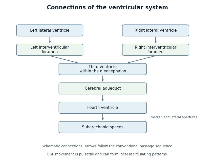
Anatomical connections of the ventricular system
. Original schematic showing paired lateral ventricles, the interventricular foramina, third ventricle, cerebral aqueduct, fourth ventricle and subarachnoid spaces. Arrows follow the conventional passage sequence; cerebrospinal fluid also undergoes pulsatile movement and local recirculation.[ref]Neuroscience, 2nd edition edited by D. Purves and others. The Ventricular System, Figure 1.17, anatomical connections
Details.
The third ventricle is the narrow midline cavity of the diencephalon, between the paired thalami and adjoining the hypothalamus. It contains cerebrospinal fluid and forms part of the ventricular system. The lateral ventricles communicate with it through the interventricular foramina; the cerebral aqueduct connects it to the fourth ventricle.[ref]
This cavity is more than a passive container. A 2016 study of mouse third ventricles found organized groups of cilia producing a network of cerebrospinal fluid flows, including a periodically changing pattern. Related complex flows were observed in rat and pig third ventricles.[ref]
Cilia-based flow network in the brain ventricles by Regina Faubel and others. Abstract; ciliary modules and time-dependent flow
An opening specifies a possible route between cavities; local cilia-driven transport specifies how material moves within a cavity. A single well-mixed compartment model can therefore conceal spatial differences in the delivery of signaling molecules.
The central canal of the spinal cord is the narrow cavity along the central spinal region, developmentally continuous with the ventricular system. In adults its patency varies. Anatomical continuity does not imply that it carries most cerebrospinal-fluid outflow from the fourth ventricle.
The cerebral aqueduct is the narrow midbrain passage connecting the third and fourth ventricles. Its small caliber makes the relation between geometry and fluid passage important. It is a conduit rather than the principal site of cerebrospinal-fluid production.
A choroid plexus is a vascular and epithelial structure projecting into a brain ventricle and contributing to cerebrospinal fluid production. Its epithelial barrier separates blood-associated and ventricular compartments. Secretion is a regulated transport process rather than simple unfiltered leakage from vessels.
The choroid plexus of the fourth ventricle is the vascular secretory structure associated with the tela choroidea of that ventricle. Its position near the roof distinguishes it from neural nuclei underlying the ventricular floor.
The cellular lining of the ventricular system and central canal of the spinal cord.
An ependymal cell is a cell of the ependyma, the lining of the brain ventricular system and central canal of the spinal cord. Many ventricular ependymal cells carry multiple motile cilia facing the cerebrospinal fluid. Their coordinated beating can move nearby fluid; the cells are distinct from the neurons whose signaling circuits surround the cavity.[ref]
Anatomy and Physiology 2e by J. Gordon Betts and others. Nervous tissue: ependymal cells and cerebrospinal-fluid circulation
In a 2016 study, organized cilia modules in the mouse third ventricle generated structured fluid-flow networks and a periodically switching flow pattern.[ref]
Cilia-based flow network in the brain ventricles by Regina Faubel and others. Abstract; collective ciliary transport
The result illustrates how an epithelial lining can actively organize its fluid environment rather than simply delimit it. A cilium on one cell acts locally; tissue-level organization determines the collective transport pattern.Tanycytes are specialized ependymal-related cells with long processes. Their geometry and functions should not be assigned indiscriminately to every ependymal cell.
Cerebrospinal fluid is the fluid occupying the brain ventricles and surrounding subarachnoid spaces. Production, movement and removal connect its composition and pressure to the function of several interfaces. It is not interchangeable with either blood or fluid inside neurons.
The fourth ventricle is the ventricular cavity between the dorsal pons and medulla oblongata anteriorly and the cerebellum posteriorly. It communicates with the cerebral aqueduct and with subarachnoid spaces through median and lateral apertures.
A lateral aperture of the fourth ventricle is an opening near the end of a lateral recess of the fourth ventricle. The paired apertures allow communication between ventricular and subarachnoid fluid spaces. They are distinct from the single median aperture of the fourth ventricle.
The paired lateral aperture of the fourth ventricle openings.
The median aperture of the fourth ventricle is the midline opening connecting the ventricular cavity with the cisterna magna region. It is a principal route into the subarachnoid space, together with the paired lateral apertures.
The tela choroidea is a thin vascular membrane associated with the roof of a ventricular cavity and its choroid plexus. In the fourth ventricle it contributes to the thin inferior roof. A vascular membrane and the neighboring neural sheet are different structural components.
The inferior medullary velum is a thin neural sheet associated with the inferior roof of the fourth ventricle, extending from the nodular region toward the floccular attachments. It is distinct from the adjacent vascular tela choroidea.
The superior medullary velum is a thin neural sheet spanning between the superior cerebellar peduncles and contributing to the upper roof of the fourth ventricle. Its ventricular surface and cerebellar attachment provide landmarks for neighboring pathways.
A lateral recess of the fourth ventricle is a lateral extension of the ventricular cavity toward a lateral aperture of the fourth ventricle. Its direction differs from the main cranial-to-caudal ventricular axis, making three-dimensional relations important in interpreting a section.
Plural name of the lateral recess of the fourth ventricle.
The fastigium is the apex of the tent-like roof of the fourth ventricle. It is a geometric landmark of the cavity, distinct from a neural nucleus or a fluid outlet. Its name helps orient descriptions of the superior and inferior roof.
The diamond-shaped floor of the fourth ventricle, formed by the dorsal pons and upper medulla oblongata.
The funiculus separans is a narrow ridge in the Caudal floor of the fourth ventricle, separating the vagal trigone from the area postrema. It is a surface landmark, not a complete boundary between all underlying neural pathways.
The area postrema is a specialized vascular region near the Caudal fourth ventricle, involved in detecting circulating signals and contributing to emetic responses. Its close relation to vagal sensory circuits connects blood-borne information with coordinated behavior.
The vagal trigone is a surface region of the floor of the fourth ventricle associated with the underlying dorsal motor nucleus of the vagus. Surface shape and the full extent of the nucleus are different anatomical descriptions.
The hypoglossal trigone is a triangular surface region near the Caudal midline of the floor of the fourth ventricle. It overlies the region of the hypoglossal nucleus, connecting an external landmark with a deeper motor structure.
The vestibular area is a lateral region of the floor of the fourth ventricle associated with vestibular nuclear structures. It helps orient the sensory side of the floor relative to more medial motor landmarks.
The superior fovea is a small depression associated with the sulcus limitans in the pontine part of the floor of the fourth ventricle. It is a regional surface landmark rather than an isolated functional center.
The facial colliculus is a dorsal pontine elevation in the floor of the fourth ventricle. It reflects the underlying abducens nucleus and looping fibers of the facial nerve. Its name therefore should not be read as indicating the location of the facial motor nucleus itself.
The striae medullares of the fourth ventricle are transversely running fiber markings across the ventricular floor, associated with auditory pathways. Their visibility and arrangement vary, so a surface specimen need not display one invariant pattern.
The sulcus limitans is a longitudinal groove associated with the developmental division between sensory and motor regions of the neural tube. In the floor of the fourth ventricle, it helps orient the medial eminence relative to lateral sensory regions.
The medial eminence is a longitudinal elevation on each side of the midline in the floor of the fourth ventricle. The sulcus limitans marks its lateral relation. Local elevations within it include the facial colliculus.
The obex is a landmark at the Caudal tip of the fourth ventricle, near its transition toward the central canal of the spinal cord. It locates the inferior end of the ventricular floor rather than serving as a separate large cerebrospinal-fluid outlet.
Articles by others on the same topic
There are currently no matching articles.