Topics (194k) Articles (196k) Users (296) Discussions (237) Comments (383) Files (715) New article

Fast neutron therapy is a type of radiation therapy that uses fast neutrons—high-energy particles that are not electrically charged—for the treatment of cancer. Unlike conventional radiation therapies that typically utilize X-rays or gamma rays (which are forms of electromagnetic radiation), fast neutron therapy employs neutrons to target and destroy cancerous cells. ### Key Features of Fast Neutron Therapy: 1. **Mechanism of Action**: Fast neutrons interact with atomic nuclei in a different manner than photons.
Electron therapy is a form of radiation therapy that uses electron beams to treat cancer. Unlike traditional X-ray radiation therapy, which uses high-energy photons, electron therapy specifically utilizes electrons, which have a lower penetration depth in tissues. This characteristic makes electron therapy particularly useful for treating superficial tumors, such as those found in skin cancers and certain types of breast cancer.
Electromagnetic radiation (EMR) refers to the waves of the electromagnetic field that propagate through space, carrying energy. This radiation encompasses a broad spectrum, ranging from low-frequency radio waves to high-frequency gamma rays. The electromagnetic spectrum includes: - **Radio waves**: Used for communication (radio, TV, cell phones). - **Microwaves**: Used in microwave ovens and various communication technologies. - **Infrared radiation**: Associated with heat and used in remote controls.
Electroencephalography functional magnetic resonance imaging (EEG-fMRI) is a neuroimaging technique that combines two distinct methodologies: electroencephalography (EEG) and functional magnetic resonance imaging (fMRI). Each technique has its strengths and limitations, and their combination aims to provide a more comprehensive understanding of brain activity. ### Electroencephalography (EEG): - **Nature of Measurement**: EEG measures the electrical activity of the brain through electrodes placed on the scalp.
Echogenicity refers to the ability of a tissue to reflect ultrasonic waves during an ultrasound examination. It is a key concept in diagnostic imaging that helps radiologists and clinicians differentiate between various types of tissues based on how they respond to ultrasound waves. Tissues with high echogenicity appear brighter on the ultrasound image because they reflect more sound waves, while tissues with low echogenicity appear darker, as they either absorb or transmit more sound waves.
EGS can refer to different things depending on the context. One common interpretation is "Educational Guidance Services," which focuses on providing support and resources for students in educational settings. In another context, EGS might stand for "Economic Growth Strategy" in relation to economic planning and development.
Dosimetry is the scientific measurement, calculation, and assessment of ionizing radiation doses absorbed by materials and biological tissues. It is primarily used in fields such as radiation therapy, radiology, nuclear medicine, and radiation protection. Dosimetry plays a crucial role in ensuring that individuals receive the appropriate amount of radiation for medical treatment while minimizing exposure to healthy tissues, as well as in monitoring radiation levels in occupational settings to protect workers from harmful exposure.
Dose Area Product (DAP) is a measure used in radiology to quantify the potential radiation exposure to patients during diagnostic imaging procedures, particularly in the context of X-ray and fluoroscopy examinations. It represents the product of the radiation dose (measured in Gray, Gy) received by the patient and the area of the irradiated field (measured in square centimeters, cm²). DAP is typically expressed in units of Gray-centimeters squared (Gy·cm²).
A Dose-Volume Histogram (DVH) is a graphical representation used primarily in radiotherapy and radiation treatment planning to assess and quantify the distribution of radiation dose within a given volume of tissue. It provides valuable information about how much of a specific volume of tissue receives a particular dose of radiation. ### Key Components of a DVH: 1. **X-Axis (Dose Axis)**: Represents the radiation dose delivered, usually measured in Gray (Gy).
Deuterium-depleted water (DDW) is water that has a lower concentration of deuterium, a stable isotope of hydrogen, compared to regular water (H2O). In regular water, most of the hydrogen atoms are protium (the most common isotope of hydrogen, with no neutrons), but a small percentage (approximately 0.0156%) are deuterium (D), which has one neutron in addition to the proton.
Deep Inspiration Breath-Hold (DIBH) is a technique primarily used in radiation therapy, especially in the treatment of cancers in the thoracic region, such as breast and lung cancer. The DIBH technique involves instructing patients to take a deep breath and hold it while the radiation is being delivered.
A DaT scan, or dopamine transporter scan, is a type of SPECT (single photon emission computed tomography) imaging used to assess the function of dopamine transporters in the brain. It is primarily utilized for the differential diagnosis of movement disorders, particularly to help differentiate between Parkinson’s disease (PD) and other conditions that can cause similar symptoms, such as essential tremor or other atypical parkinsonian syndromes.
Combined photothermal and photodynamic therapy (PTT and PDT) is a synergistic approach used primarily in cancer treatment that utilizes two different mechanisms of action to enhance the efficacy of tumor eradication. ### Photothermal Therapy (PTT) PTT involves the application of heat to cancer cells, typically using light-absorbing agents known as "photosensitizers" that are localized to the tumor.
Carbon-11-choline is a radiotracer used primarily in positron emission tomography (PET) imaging. It is a synthetic compound that incorporates the radioactive isotope Carbon-11 (C-11), which has a half-life of about 20.4 minutes. This rapid decay allows for imaging procedures to be conducted shortly after its administration. Carbon-11-choline is particularly useful in the detection and characterization of certain tumors, especially prostate cancer.
Brachytherapy is a form of radiation therapy used to treat various types of cancer. It involves placing a radioactive source directly inside or very close to the tumor, allowing for a higher dose of radiation to be delivered to the cancerous cells while minimizing exposure to surrounding healthy tissues. There are two main types of brachytherapy: 1. **Low-Dose Rate (LDR) Brachytherapy**: In this method, radioactive seeds are implanted in or near the tumor.
"Beam's eye view" is a term often used in relation to photography and cinematography to describe a perspective that mimics the viewpoint of a beam of light or a laser beam, typically emphasizing the direct line of sight from the beam's origin to its target. This concept can be applied in various contexts, such as highlighting how light interacts with objects in its path, creating dramatic visual effects or emphasizing perspectives in storytelling.
4DCT, or four-dimensional computed tomography, is an advanced imaging technique that captures both the anatomical structures of the body as well as the changes in those structures over time. The "4D" aspect refers to three-dimensional spatial dimensions (length, width, and height) plus one temporal dimension (time). In a typical 4DCT scan, a series of CT images are taken rapidly over a period, often while the patient is breathing.
Nuclear medicine is a specialized field of medical imaging and therapy that utilizes radioactive materials, or radiopharmaceuticals, for diagnosis and treatment of various health conditions. It involves the use of small amounts of radioactive substances to carry out imaging and therapeutic procedures. ### Diagnostic Uses In diagnostic applications, nuclear medicine techniques can visualize the function of organs and tissues.
Medical ultrasonography, commonly known as ultrasound, is a diagnostic imaging technique that uses high-frequency sound waves to create images of structures within the body. It is a non-invasive and safe procedure that is widely used in various medical fields to visualize organs, tissues, and blood flow. ### Key Features of Medical Ultrasonography: 1. **How it Works**: An ultrasound machine generates sound waves that are emitted through a transducer.
Medical physics journals are academic publications that focus on the study and application of physics principles in medicine, particularly in the fields of medical imaging, radiation therapy, and diagnostic procedures. These journals serve as platforms for researchers, clinicians, physicists, and engineers to publish their findings, reviews, and advancements in medical technology, techniques, and methodologies.
Pinned article: Introduction to the OurBigBook Project
Welcome to the OurBigBook Project! Our goal is to create the perfect publishing platform for STEM subjects, and get university-level students to write the best free STEM tutorials ever.
Everyone is welcome to create an account and play with the site: ourbigbook.com/go/register. We belive that students themselves can write amazing tutorials, but teachers are welcome too. You can write about anything you want, it doesn't have to be STEM or even educational. Silly test content is very welcome and you won't be penalized in any way. Just keep it legal!
Intro to OurBigBook
. Source. We have two killer features:
- topics: topics group articles by different users with the same title, e.g. here is the topic for the "Fundamental Theorem of Calculus" ourbigbook.com/go/topic/fundamental-theorem-of-calculusArticles of different users are sorted by upvote within each article page. This feature is a bit like:
- a Wikipedia where each user can have their own version of each article
- a Q&A website like Stack Overflow, where multiple people can give their views on a given topic, and the best ones are sorted by upvote. Except you don't need to wait for someone to ask first, and any topic goes, no matter how narrow or broad
This feature makes it possible for readers to find better explanations of any topic created by other writers. And it allows writers to create an explanation in a place that readers might actually find it.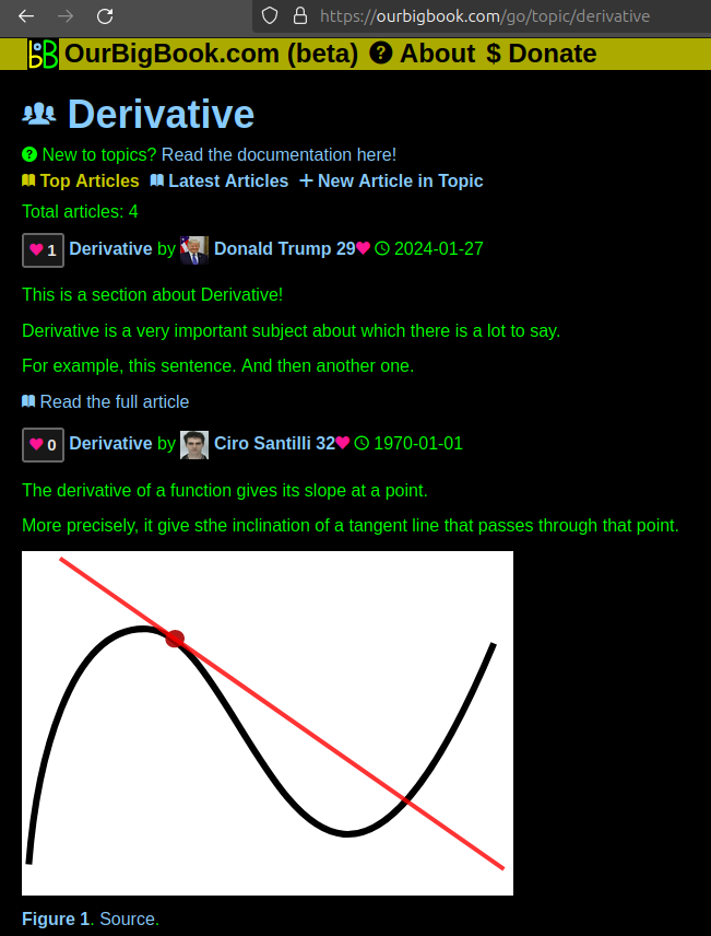
Figure 1. Screenshot of the "Derivative" topic page. View it live at: ourbigbook.com/go/topic/derivativeVideo 2. OurBigBook Web topics demo. Source. - local editing: you can store all your personal knowledge base content locally in a plaintext markup format that can be edited locally and published either:This way you can be sure that even if OurBigBook.com were to go down one day (which we have no plans to do as it is quite cheap to host!), your content will still be perfectly readable as a static site.
- to OurBigBook.com to get awesome multi-user features like topics and likes
- as HTML files to a static website, which you can host yourself for free on many external providers like GitHub Pages, and remain in full control

Figure 2. You can publish local OurBigBook lightweight markup files to either OurBigBook.com or as a static website.
Figure 3. Visual Studio Code extension installation.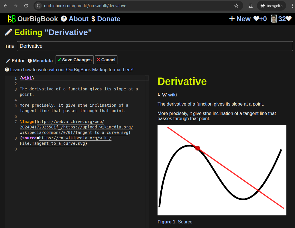
Figure 5. . You can also edit articles on the Web editor without installing anything locally. Video 3. Edit locally and publish demo. Source. This shows editing OurBigBook Markup and publishing it using the Visual Studio Code extension. 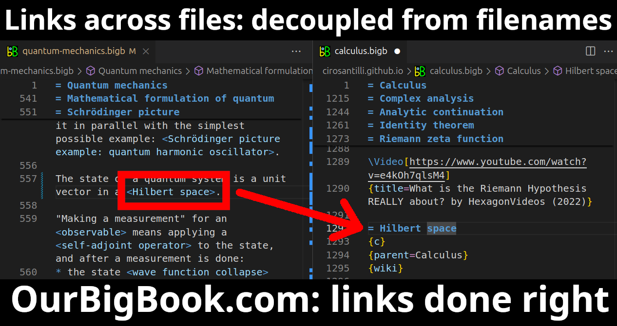
- Infinitely deep tables of contents:
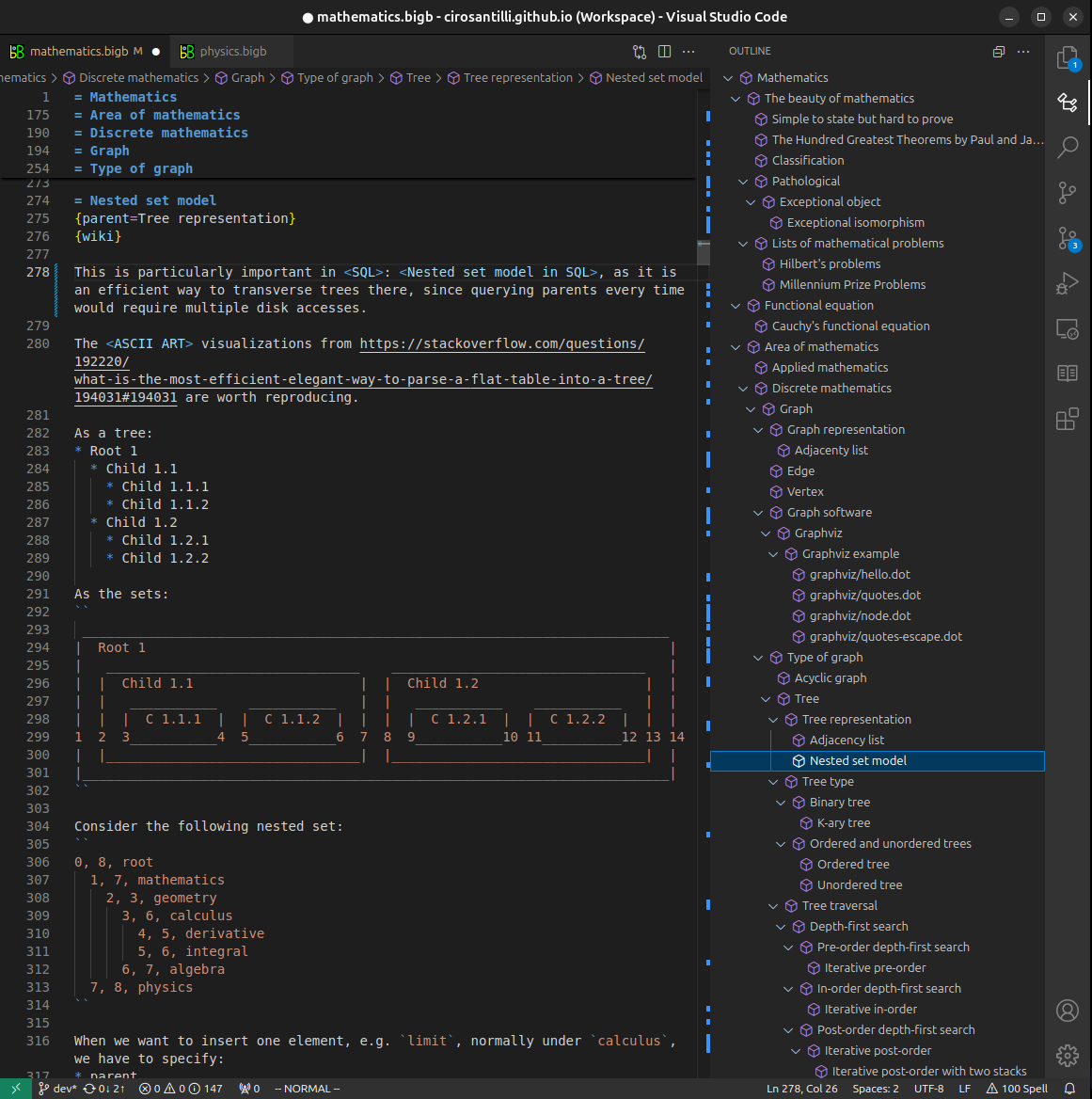
All our software is open source and hosted at: github.com/ourbigbook/ourbigbook
Further documentation can be found at: docs.ourbigbook.com
Feel free to reach our to us for any help or suggestions: docs.ourbigbook.com/#contact