mRNA display is an in vitro selection method in which a peptide or protein is covalently linked to the messenger RNA encoding it. Selecting the displayed molecule can therefore retain recoverable sequence information about that same molecule: a physical genotype-phenotype linkage.
In the original experiment, synthetic messenger RNA carried puromycin at its 3′ end. During translation, this peptide acceptor enabled fusion of the message to its encoded peptide. Richard W. Roberts and Jack W. Szostak tested the linkage by selecting an encoded myc epitope tag with an antibody.[ref]
RNA-peptide fusions for the in vitro selection of peptides and proteins by Richard W. Roberts and Jack W. Szostak. Abstract; Figures 1 and 4, fusion construction and immunoprecipitation
The experimental readout went beyond observing binding. Following immunoprecipitation, reverse transcription and polymerase chain reaction recovered the encoding information. A restriction enzyme distinguished the selected myc sequence from background pool sequences, allowing before-and-after enrichment to be measured.[ref]
RNA-peptide fusions for the in vitro selection of peptides and proteins by Richard W. Roberts and Jack W. Szostak. Selective Enrichment of an RNA-Peptide Fusion and Figure 5
This tests whether selection of a molecular property can recover the associated sequence; it does not establish that every possible sequence displays equally well.An original statistical sampling calculation distinguishes nominal input size from effective displayed-library coverage. Suppose independent input molecules contain a desired sequence class with probability , and each such molecule forms a usable fusion with probability . Before any binding selection,For , and stipulated , the expected usable desired count is only one and the chance of none is approximately 36.8%. Raising the hypothetical yield to 0.10 raises the expected count to ten and reduces that probability to approximately 0.00454%. These are constructed independent-sampling examples, not fusion yields claimed for the original experiment.
Sequence-dependent translation, fusion formation, recovery and amplification can each change representation. A large input sequence pool is therefore different from a large usable displayed pool. Repeating selection and amplification can enrich retained variants, while an independent functional or binding affinity measurement tests whether the recovered sequence has the intended property.
Effective displayed-library coverage describes which molecular sequence variants actually occur as usable displayed molecules, rather than merely being allowed by a library design or present in its input material. In mRNA display, successful translation, fusion formation and recovery connect input sequences to usable genotype-phenotype linkages. Duplicates of one sequence do not replace missing sequences.
An original occupancy model has equally likely sequence variants, independent input molecules and usable-display probability for each input. A particular variant is usable with probability per input. Thereforewhere is the number of distinct represented variants. The expected usable molecule count is , which is generally larger than the expected distinct count.
For , and stipulated , the expected usable count is 3000 but the expected distinct count is only about 950.2. An average 95% represented fraction does not imply 95% probability that every variant is represented. By the union bound,A sufficient condition for that probability to be at most is . With in this example, this requires at least 990,349 input molecules under the stated model.
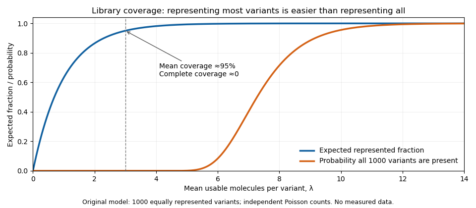
Expected represented fraction and probability of complete coverage answer different library-design questions
. Details. The figure uses a Poissonized displayed-library occupancy model: independently Poisson-distributed usable counts per variant with mean . The expected represented fraction is , while the probability of all variants being present is . That is an explicitly different count model, providing a convenient comparison rather than an exact fixed-input formula.
Real variants need not have equal abundance or display probability. In the original RNA-peptide fusion experiment, the selected and unselected encoding-sequence ratios were affected by preferential translation and reverse transcription.[ref]
RNA-peptide fusions for the in vitro selection of peptides and proteins by Richard W. Roberts and Jack W. Szostak. Figure 5 legend: input mixtures and biased unselected ratios
Coverage calculations therefore need assumptions about sequence representation and process losses; nominal library size alone does not validate them. Articles by others on the same topic


mRNA display is a technique used in molecular biology and biotechnology to select and analyze peptides or proteins based on the genetic information encoded in mRNA. The method combines aspects of mRNA and protein interactions to create a powerful platform for discovering new proteins, understanding protein functions, and developing therapeutic agents. ### Key Features of mRNA Display: 1. **Encoding Proteins**: In mRNA display, a library of mRNA molecules is linked to their corresponding peptides or proteins.