A protein is a biological molecule made of one or more polypeptide chains: ordered amino acid residues joined by peptide bonds. Biological translation assembles a chain in the order specified by messenger RNA codons. Protein folding then brings parts of that chain into spatial relationships that support its function. See Cooper, The Cell: protein composition and structure. These processes can overlap in a biological cell; the sequence-to-shape explanation does not require waiting until biological translation finishes before any folding begins. See Cooper, The Cell: protein folding and processing.
The useful connection is from information to physical work. A protein's protein primary structure records an order; its protein tertiary structure can bring distant amino acid residues together at an active site. An enzyme uses such an arrangement to recognize a substrate and accelerate a chemical reaction. Other proteins serve as receptors, membrane transport proteins or structural material. For example, actin assembles into filaments whose mechanical properties can be described by biopolymer mechanics. See Alberts et al., Molecular Biology of the Cell: protein function and binding.
Order matters beyond composition. Let A stand for alanine and M for methionine, and read from the N-terminus to the C-terminus. The illustrative strings AMAMAMAMAMAM and AAAAAAMMMMMM each contain exactly six A and six M amino acid residues. Each has peptide bonds when made into one unbranched polypeptide chain. In the first string all eleven neighboring pairs have different letters; in the second only the pair at positions 6 and 7 does. A composition histogram therefore loses information about which amino acid residues are neighbors. Their arrangements can affect protein folding and function; these short invented strings neither specify a known functional protein nor establish what shape either would adopt.
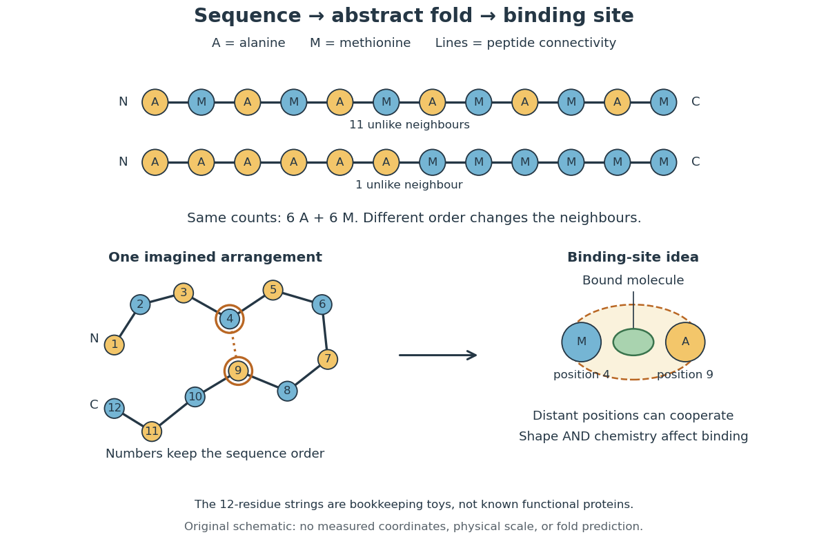
From amino acid order to an imagined fold and binding site
. Original schematic using two twelve-residue strings with equal composition. The imagined fold uses the first string: colors and numbers preserve its protein primary structure, while positions 4 and 9 approach one another. The binding-site cartoon explains spatial cooperation, not an actual enzyme or a measured protein structure. No actual coordinates, physical scale or exact protein folding predictor are implied. Details.Protein structure is a moving arrangement, not a permanently rigid outline. Substrate binding can change an enzyme's shape through induced fit, and a functional protein can include flexible regions. Sequence, surroundings and interacting molecules must therefore be considered together. See Alberts et al.: binding, conformational changes and protein function.
A protein obtained from cattle serum and widely used as a reference sample in biochemistry. It serves as a protein standard for assays such as the bicinchoninic acid assay, and as a model protein in dialysis experiments.
An antibody is a protein that recognizes particular molecular targets through binding. Antibodies participate in immunity; laboratory assays such as Western blots exploit their selective binding to detect chosen proteins.
An IgG gives a concrete architecture: two immunoglobulin heavy chains and two immunoglobulin light chains, joined by disulfide bonds. Each arm ends in a paratope assembled from an antibody variable region of each kind of chain. The two paratopes recognize the same epitope in this conventional IgG; the flexible hinge allows different arm separations. The antibody constant regions form the remaining portions. This is an illustrative IgG, rather than the architecture of every antibody format. [ref]
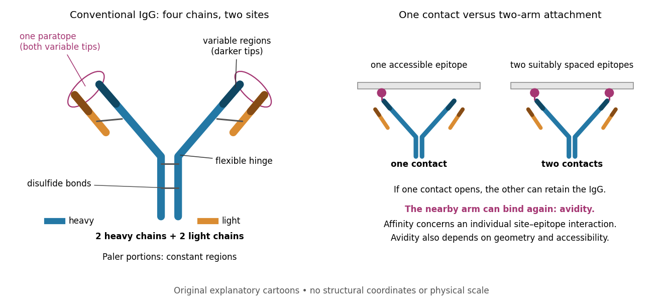
Three questions must be kept separate. Antibody specificity asks which antigens are recognized. Antibody affinity describes one paratope–epitope interaction. Avidity describes the combined attachment when several contacts can cooperate. A selective antibody can have modest antibody affinity, and a monoclonal antibody can still show cross-reactivity. For an assay, the practical question is whether the observed readout identifies the intended antigen under the tested conditions; Western blot antibody specificity controls make that question concrete.
In an indirect Western blot, the primary antibody binds an epitope on the proposed target protein. Its binding must be tested in the actual sample and assay conditions through Western blot antibody specificity controls.
A secondary antibody recognizes another antibody, rather than the proposed target protein directly. In Western blot antibody detection, its attached label makes retained primary antibodies visible. A secondary antibody must recognize the kind of primary antibody used.
An epitope is the molecular feature recognized by an antibody. Protein denaturation can expose an epitope or destroy the spatial arrangement required for its recognition; an antibody that works on a folded protein need not work in SDS-PAGE-based Western blot antibody detection.
Immunoglobulin G is an antibody class whose conventional four-chain architecture supplies the example above.
An immunoglobulin heavy chain extends through an arm and the stem of the illustrated IgG.
An immunoglobulin light chain accompanies an arm of the illustrated IgG, contributing to its paratope.
The paired antibody variable regions of an immunoglobulin heavy chain and an immunoglobulin light chain contribute to an antibody binding site. Variation refers to differences between antibody sequences, not to a tip continually changing identity while binding.
An antibody constant region is a chain portion outside the antibody variable region. Constant does not mean identical across every antibody class.
A Fab fragment contains one antibody binding site, allowing comparison with the two-site intact IgG in the antibody surface plasmon resonance experiment.
A paratope is the recognizing surface on an antibody; the epitope is the recognized feature on its antigen. Contacts depend on spatial arrangement and chemistry, including hydrogen bonds and Van der Waals forces. Thus a paratope is not simply a slot for a protein primary structure string. A folded protein can bring distant amino acid residues into one epitope. [ref]
In antibody binding, an antigen is material bearing a recognized epitope. An antigen need not be a protein; polysaccharides can also carry epitopes. One antigen may present several different epitopes, so recognizing one feature does not imply recognizing every part of the target. [ref]
The interaction of the antibody molecule with specific antigen. Immunobiology, 5th edition, §3-8: small recognized regions of protein and polysaccharide antigens
Antibody specificity describes preferential recognition of selected epitopes. It is established relative to tested alternatives and assay conditions, rather than inferred from a reagent's name. Western blot antibody specificity controls distinguish recognition of the proposed protein from retained label caused by other material.
Cross-reactivity occurs when an antibody also recognizes another antigen beyond the one against which it was raised. This may involve related features, but obvious overall similarity is not required. [ref]
Appendix I. Immunologists' Toolbox. Immunobiology, 5th edition, Appendix I, antiserum generation: cross-reactive antigens and absorption
For an imagined example, a paratope contacts a small surface patch shared by two otherwise different proteins. Both can bind even though their remaining surfaces differ. Increasing antibody concentration can reveal weak binding that was previously undetectable. Conversely, an extra Western blot band need not prove cross-reactivity: the intended protein may occur in multiple forms, or detection reagents may contribute background. The target-deleted and no-primary antibody comparisons under Western blot antibody specificity controls test different explanations.
A monoclonal antibody preparation derives from one selected antibody-producing biological cell and its descendants, or reproduces their specified antibody sequences. Monoclonal describes the origin and composition of the preparation; it does not guarantee absence of cross-reactivity or establish antibody affinity. [ref]
Glossary. Immunobiology, 5th edition, Glossary: Monoclonal antibodies
Avidity is the combined strength of multivalent attachment. An intact IgG can make two contacts when accessible epitopes have suitable spacing. [ref]
Molecular Biology of the Cell, 4th edition by Bruce Alberts and others. Chapter 24, B Cells and Antibodies, A Typical Antibody Has Two Identical Antigen-Binding Sites; The Strength of an Antibody-Antigen Interaction Depends on Both the Number and the Affinity of the Antigen-Binding Sites: avidity
If one contact breaks while the other remains, the detached arm stays nearby and can bind again before the whole antibody escapes. That geometric advantage does not require changing either paratope's intrinsic antibody affinity. Two arms binding two separate freely moving antigens do not automatically provide the same tethering advantage. Epitope spacing, accessibility and surface density matter; avidity is not obtained by adding two dissociation constants.Antibody affinity concerns one paratope binding one epitope. A Fab fragment supplies a single-site comparison. [ref]
Glossary. Immunobiology, 5th edition, Glossary: affinity, single-site binding
In the dilute chemical equilibrium model below, smaller dissociation constant means larger antibody equilibrium occupancy at the same free antigen concentration. State the antigen, temperature and buffer solution: a dissociation constant is not a context-free score. It does not alone specify how fast attachment or detachment occurs.Antibody equilibrium occupancy counts occupied antibody binding sites, rather than retained assay label. For an original 1:1 model, let denote an unoccupied site, free antigen bearing one accessible epitope, and their complex. At chemical equilibrium, writeSquare brackets mean concentrations. The dissociation constant here has concentration units. This is the same independent-site calculation as receptor binding occupancy, now applied to an antibody binding site. The last equality follows by substituting into the occupied fraction and cancelling .
Assume identical independent sites, reversible 1:1 binding, dilute chemical solution behavior, fixed temperature and buffer solution, and enough time for chemical equilibrium. Exclude two-arm bridging, cross-reactivity and inaccessible sites from this model. Crucially, is free antigen concentration; substituting the amount supplied per volume requires negligible depletion by binding. Independent IgG arms could obey this model site by site, but it does not describe avidity from bridging a surface.
Stipulate , with . Free antigen concentrations , and give , and . A second stipulated dissociation constant of instead gives at . Both curves approach one at large : a nearly saturated readout can conceal an antibody affinity difference. These are invented values, with no universal threshold for a useful antibody.
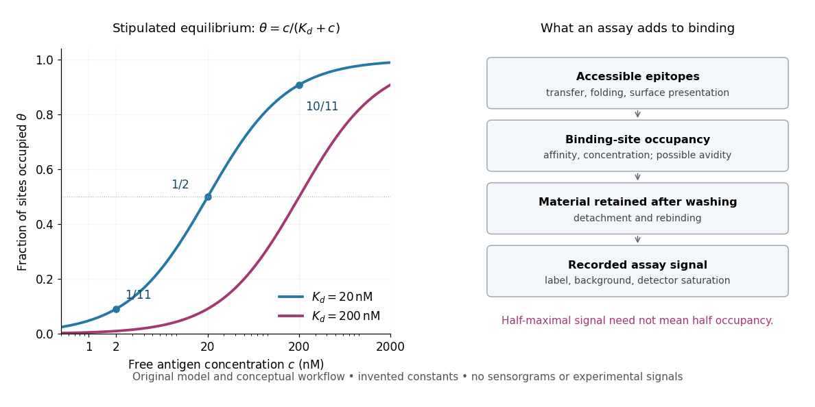
Antibody equilibrium occupancy and the additional steps in an assay readout
. Original analytic curves for stipulated dissociation constants of and ; marked examples belong to the first curve. The adjacent schematic separates epitope accessibility, binding, washing and detection. It contains no measured sensorgrams, fitted assay parameters or experimental signals. A logarithmic scale changes the display, not the independent-site assumptions. Details.A Western blot readout additionally depends on Western blot transfer, accessible epitopes, loss during washing and Western blot antibody detection. Western blot response saturation can flatten the final signal even when binding itself remains responsive. An ELISA also reports retained label through an enzyme readout, rather than automatically reporting . Thus half of an assay's maximum signal need not occur at . Varying free antigen through concentrations near the candidate dissociation constant, while checking depletion and the readout's response, is more informative than comparing a single saturated point.
A concrete comparison used a Biacore T100 with a CM5 chip bearing the target receptor; unused reactive surface groups were blocked. At in a pH 7.4 buffer solution, intact IgG and its Fab fragment were injected for at ; dissociation was monitored for . The optical response tracks changes in refractive index near the surface, rather than counting occupied paratopes directly. A reference surface and blank injections corrected background; association and dissociation responses were fitted with a 1:1 model. The reported intact-IgG detachment was slower than that of the Fab fragment. [ref]
Antibody-Unfolding and Metastable-State Binding in Force Spectroscopy and Recognition Imaging by Parminder Kaur and others. Kaur and others, Materials and Methods, Surface plasmon resonance; Table 1, whole IgG and Fab rows
This comparison supports format-dependent attachment, not an intrinsic single-site antibody affinity for intact IgG. The reference and blanks address background, not two-arm avidity, surface accessibility or delivery by diffusion. The paper's fitted 1:1 constants alone cannot distinguish those mechanisms. Its Fab fragment comparison therefore motivates specifying the binding format before interpreting a dissociation constant; a good fit does not establish a unique molecular model.
A Western blot detects selected proteins after their separation and transfer to a membrane, using antibodies. In the yeast cross-species complementation experiment, detection helps distinguish failed protein production from failure of an expressed protein to restore growth.
The useful idea is to preserve where proteins migrated in gel electrophoresis, then ask which positions retain an antibody. Western blot separation, Western blot transfer and Western blot antibody detection answer different questions. A band is evidence of retained label at a position; Western blot band interpretation requires Western blot controls before assigning identity or comparing abundance.
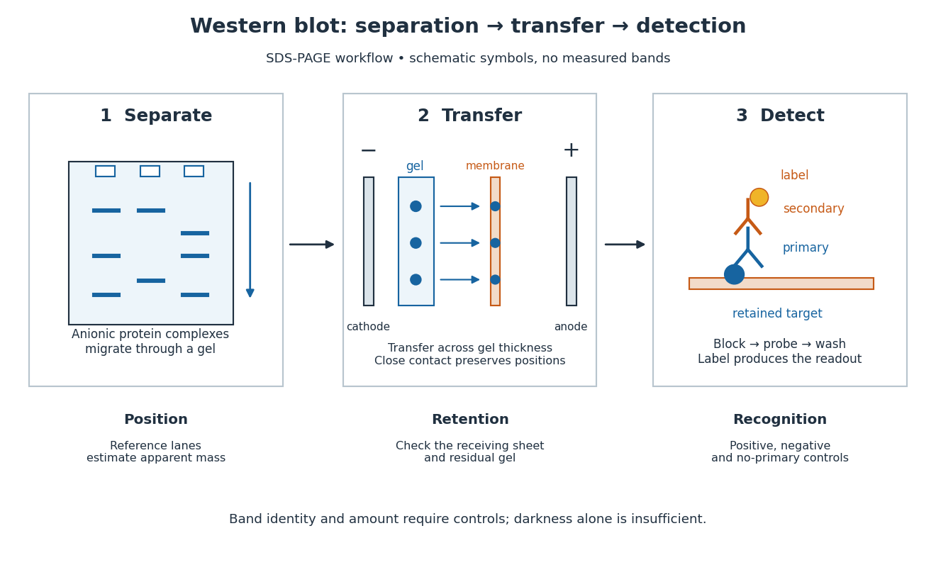
In the common SDS-PAGE version, protein denaturation and binding of an anionic surfactant reduce differences due to folded shape and intrinsic electric charge. An electric field drives the resulting protein complexes through the gel; smaller chains generally migrate farther. A lane of reference proteins supplies positions for estimating apparent mass. This estimate depends on the separation conditions: it is not a direct weighing of a molecule, and different proteins can migrate together.
Western blot transfer moves separated proteins out of the gel onto a Western blot membrane, using an electric field across the gel thickness. Maintaining close contact preserves the lane pattern. Contact gaps, incomplete removal from the gel and passage through the receiving sheet can change how much protein is retained.
The original 1979 experiment pressed a wetted, -pore nitrocellulose Western blot membrane against the gel between scouring pads and plastic pipette-tray grids, held by elastic bands. The urea-gel assembly faced the cathode in acidic solution, with applied for one hour; the SDS-PAGE variant reversed polarity and used a glycine/methanol buffer solution. Stained gel/blot pairs checked pattern preservation. Corresponding labelled ribosome protein spots from two gels were compared through radioactive decay readouts after combustion. The stained gel remaining after transfer supplied a removal check; a second receiving sheet revealed breakthrough on overloading. These tests supported recovery for the examined urea-gel proteins, within experimental variation, while the SDS-PAGE results showed unequal staining recovery. Preserved band positions therefore did not establish universal quantitative transfer. [ref]
Electrophoretic transfer of proteins from polyacrylamide gels to nitrocellulose sheets: procedure and some applications by Harry Towbin and others. Towbin, Staehelin and Gordon, pp. 4350–4352: Electrophoretic Blotting Procedures, Figure 1, Figures 2–3 and Table 1
A Western blot membrane is a thin protein-binding support that receives the separated pattern. It is a laboratory support, distinct from a cell membrane. Retaining proteins on its surface makes them accessible for Western blot antibody detection.
A nitrocellulose Western blot membrane is a Western blot membrane made from nitrated cellulose. Its retention capacity is finite; the original Western blot transfer experiment tested breakthrough with a second receiving sheet.
Blocking occupies unused binding sites on the Western blot membrane. The primary antibody is applied, unbound material is washed away, and a labelled secondary antibody detects retained primary antibody. A fluorescence label emits detectable light after excitation; an enzyme-linked label can instead generate a colored product or light. Washing reduces background but does not prove that remaining binding is selective.
In the original experiment, blocking used excess protein. An anti-ribosome-protein serum followed by labelled secondary antibody produced colored or fluorescence bands; preimmune serum supplied comparison strips. The color readout was an enzyme reaction product, rather than a measurement of target enzyme activity. [ref]
Electrophoretic transfer of proteins from polyacrylamide gels to nitrocellulose sheets: procedure and some applications by Harry Towbin and others. Towbin, Staehelin and Gordon, pp. 4351–4353: Immunological Detection of Proteins on Nitrocellulose and Figure 4, including preimmune-serum controls
Band position gives an apparent mass, not unique protein identity. Intensity depends on retention, epitope recognition and detection response; it does not establish folded state, enzyme activity or successful protein targeting. Missing bands can reflect missing targets or failed Western blot transfer or Western blot antibody detection. [ref]
Considerations when quantitating protein abundance by immunoblot by Alicia A. McDonough and others. McDonough and others, “Consider the Specificity of the Antibody for the Target” and “Consider the Linearity of the Detection System”
The yeast cross-species complementation experiment consequently combines two logically different readouts: Western blot evidence about production of an antibody-recognized protein, and growth evidence about functional complementation. A detectable protein need not perform the biological task required for growth.
Western blot loading controls ask how much sample entered each lane; Western blot transfer controls ask what reached the receiving sheet; Western blot antibody specificity controls ask what the antibodies recognized. Each closes a different gap in Western blot band interpretation.
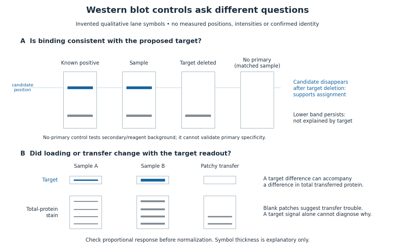
Conceptual lanes for Western blot controls
. Original schematic, with invented qualitative marks only. Candidate bands disappear in a target-deleted sample while a lower band persists; a separate panel shows how a loading or transfer difference can accompany a target-band difference. No lane, darkness, band position or relative amount represents a measurement, a specific real protein or confirmed identity. The no-primary comparison denotes a matched strip processed without primary antibody. Details.A Western blot loading control estimates lane-to-lane sample differences. A reference protein, such as actin, must have stable abundance and detection responsive to amount. Equal saturated bands fail to establish equal loading. [ref]
Considerations when quantitating protein abundance by immunoblot by Alicia A. McDonough and others. McDonough and others, “Consider the Loading Controls”, Figure 4: sample doubling changes claudin detection but not the actin signal
A Western blot transfer control tests retention after Western blot transfer. Total-protein staining of the Western blot membrane can reveal uneven lanes or blank patches; staining the gel after transfer tests what remained behind. Transferred reference bands check selected positions, not recovery of every target. A pretransfer loading image alone cannot establish the amount retained on the receiving sheet.
A known target-containing sample is a positive comparison for Western blot antibody detection. A matched sample with gene deletion supplies a negative comparison: disappearance of the proposed band supports the assignment, while a persisting band indicates binding unrelated to the deleted target. Deletion must actually remove the recognized protein; changes in other proteins can complicate the comparison. Omitting the primary antibody checks binding or background from the secondary antibody and detection reagents, but cannot exclude binding of the primary antibody to an unintended protein. Independent antibodies recognizing different epitopes add evidence. [ref]
Considerations when quantitating protein abundance by immunoblot by Alicia A. McDonough and others. McDonough and others, “Consider the Specificity of the Antibody for the Target”, Figure 2: positive tissue, knockout tissue and antibodies to different termini
Total-protein Western blot normalization compares a target-band signal with a signal representing many transferred proteins in that lane. It is distinct from measuring one reference protein, and from measuring the target itself. Its denominator is a loading proxy: normalization estimates target abundance relative to detected total protein, rather than target amount per biological cell. Different sample compositions, selective Western blot transfer losses or unequal stain responses can still matter.
For an original calculation, suppose background-corrected target signals , and total-protein signals , are in their respective proportional response ranges. ThenThe invented signals double together, so this calculation supplies no evidence for a higher target fraction. With but , the same calculation gives . These examples assume a common target response and a common total-protein response between lanes, negligible remaining background, and comparable target recovery; they do not equate total-protein staining with target abundance. Multiplying both signals in a lane by the same loading factor cancels in their ratio; a selective target loss does not cancel.
Western blot response saturation means that increasing target amount no longer increases the recorded signal proportionally. It can arise before imaging, through limited binding or reagent availability, as well as through detector limits. Shortening exposure can address a detector limit without repairing an already unresponsive assay. Test the target and normalization readouts with a dilution series under the actual assay conditions; merely seeing a gray band does not establish a proportional response.
In a 2013 methods experiment, a biological cell extract containing added purified yeast protein was separated on gels, transferred with a Trans-Blot Turbo to polymer membranes, and read with an enzyme-linked secondary antibody. A ChemiDoc MP imager and film recorded the same dilution-series blots. The target and an abundant reference protein had different usable response ranges; transferred total-protein imaging and the reference bands did not track loading equally well. This demonstrates the need for assay-specific response checks, rather than a universal conversion from darkness to amount. [ref]
An illustrative response model separates background , target amount , retained fraction , response scale and characteristic amount :This is an assumed model, not a fitted Western blot calibration. When , . When , approaches . For two otherwise identical lanes, doubling gives the background-corrected ratioAt the ratio is , despite a true twofold amount change. Thus Western blot normalization cannot recover a lost amount difference merely by dividing two saturated readouts.
Protein targeting directs a protein to its cellular destination. Synthesis in the cytosol does not imply that the protein works there.
Organelle protein import brings externally synthesized proteins into an organelle. Many plastid proteins made from nuclear genes carry targeting information recognized by machinery that transports them across the envelope membranes. This delivery enables nuclear genes to supply functions inside a plastid, but the presence of targeting information is not itself evidence that a gene was acquired through endosymbiotic gene transfer. [ref]
Origin and Evolution of Plastids and Photosynthesis in Eukaryotes by Geoffrey I. McFadden. “How to tame your symbiont”: targeting, membrane passage and host-derived proteins
A polypeptide is a chain of amino acid residues linked by peptide bonds. The term describes chain chemistry; calling a chain a polypeptide alone does not establish that it is folded or functional. The gene expression example N–methionine–alanine–C contains two amino acid residues and one peptide bond; the twelve-residue example under protein contains eleven peptide bonds. See IUPAC-IUB: amino-acid symbols and representation of residues in chains.
An amino acid residue is the portion of an amino acid incorporated into a polypeptide chain. For example, the A in the illustrative protein primary structure is an alanine residue, rather than a free alanine molecule floating beside the chain. Counting amino acid residues counts chain positions; repeated letters are separate positions. See IUPAC-IUB, section 3AA-16: symbols for amino-acid residues.
A tyrosine residue is an amino acid residue whose amino acid side chain can accept a phosphate group during receptor tyrosine kinase signaling. The conventional one-letter sequence symbol is .
A phosphotyrosine residue is a tyrosine residue modified by phosphorylation. Its location and surrounding protein primary structure help determine which SH2 domains can recognize it.
An amino acid side chain is the variable group attached to an amino acid alpha carbon, conventionally denoted . For alanine it is : a carbon bonded to three hydrogen chemical atoms. In a beta strand, successive amino acid side chains alternate between the two faces in the idealized arrangement. See Clark, Douglas and Choi: amino-acid R groups.
The amino acid alpha carbon is the backbone carbon carrying the amino acid side chain in the usual protein amino acid residue. Marking successive amino acid alpha carbons is a useful way to trace a polypeptide chain, but omits intervening backbone chemical atoms. See Clark, Douglas and Choi: the central alpha carbon.
A glutamic acid residue is an amino acid residue represented by Glu or E in polypeptide sequence notation. GAA and GAG specify glutamic acid residues in the standard genetic code; the ribosome example uses GAA as its third codon. See Elzanowski and Ostell, NCBI: standard genetic code, table 1. The name denotes an incorporated chain position, as distinguished from a free amino acid molecule.
A peptide bond is the covalent bond between the carboxyl carbon of one amino acid residue and the amino nitrogen of the next. The displayed group shows the usual linkage, with the central carbon–nitrogen connection being the peptide bond. In an intact unbranched chain of amino acid residues, adjacent positions supply peptide bonds. Cutting one internal peptide bond produces two fragments; merely changing the chain's shape need not cut it. See Clark, Douglas and Choi, Biology 2e: peptide bonds and protein structure.
The protein backbone is the repeating chain of chemical atoms that connects successive amino acid residues, including their peptide bonds. The attached groups that distinguish amino acids project from this shared framework. Protein folding changes the framework's spatial arrangement while ordinarily preserving its sequence connectivity. See Cooper: polypeptide backbone and attached groups.
Protein structure describes both the chain's chemical organization and its spatial arrangement. Four levels answer different questions: protein primary structure gives the order; protein secondary structure gives recurring local patterns; protein tertiary structure gives one chain's overall arrangement; protein quaternary structure gives the assembly of multiple chains. These are levels of description, not four mandatory stages that every protein must pass through. See Clark, Douglas and Choi: four levels of protein structure.
A protein domain is a distinguishable structural and functional region of a protein. A catalytic protein domain and a binding protein domain can carry out different tasks within one chain.
An SH2 domain is a protein domain that recognizes a phosphotyrosine residue together with surrounding sequence features. This selective binding recruits a protein to a modified receptor or docking protein; it does not itself imply catalysis.[ref]
Signaling through Enzyme-Linked Cell-Surface Receptors. “Phosphorylated Tyrosines Serve as Docking Sites For Proteins With SH2 Domains”, Figure 15-53
The protein primary structure specifies the ordered amino acid residues of each polypeptide chain, joined through peptide bonds. A chain is conventionally written from its N-terminus to its C-terminus. In the broader chemical sense, a complete protein primary structure also specifies covalent bond crosslinks between chain positions, including protein disulfide bonds; a bare sequence string leaves that information unstated. This describes chemical connectivity rather than the spatial arrangement of protein tertiary structure or protein quaternary structure. See IUPAC Gold Book: primary structure, sequence and chain crosslinking and IUPAC-IUB, 3AA-16: writing residue sequences from the amino end. Some conventions reserve protein primary structure for sequence alone and call the complete chemical connectivity the covalent structure; the distinction should be stated when comparing descriptions. See IUPAC-IUB polypeptide-conformation recommendations, section 6.1: the narrower sequence-only convention.
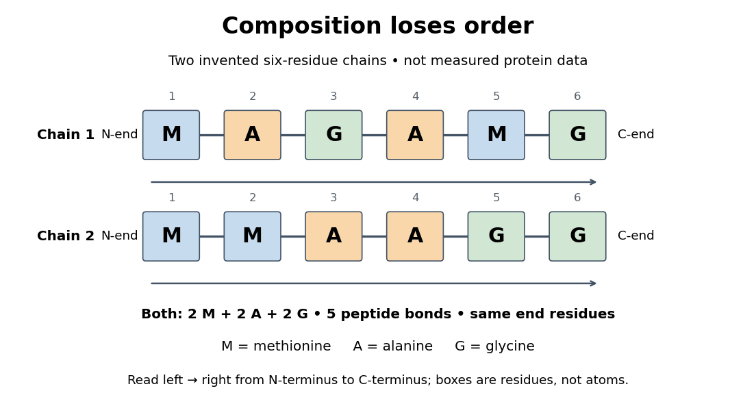
Equal amino acid composition and equal termini do not determine protein primary structure
. Original toy polypeptide chains MAGAMG and MMAAGG, read from the N-terminus to the C-terminus. Each contains two methionine, two alanine and two glycine amino acid residues, but their internal order differs. Letters denote chain positions rather than free amino acids; lines denote peptide bonds. Neither string is identified as a natural protein or a measured sequence. Details.In this toy example M denotes methionine, A alanine and G glycine. Both polypeptide chains have six amino acid residues, five peptide bonds, an M at the N-terminus and a G at the C-terminus. Their amino acid composition is identical, yet position 2 is A in MAGAMG and M in MMAAGG. There are distinct orders with these counts, a multinomial coefficient. Even fixing those two end residues leaves orders. The counts constrain the answer without determining it.
For multiple polypeptide chains, record each sequence separately and specify any protein disulfide bonds between cysteine positions. Mature insulin, for example, has an A chain of 21 amino acid residues and a B chain of 30 amino acid residues. Their combined 51 residues give peptide bonds, rather than the 50 of one unbroken 51-residue chain. See Cooper, The Cell: insulin's two chains and cysteine crosslinks, figure 2.16. A protein disulfide bond joins side groups; it does not concatenate the C-terminus of one chain with the N-terminus of another. The three insulin protein disulfide bonds connect A6 to A11 within the A chain, and A7 to B7 and A20 to B19 between chains. Positions are numbered from each chain's N-terminus. See Replacement of the interchain disulfide bridge-forming amino acids A7 and B7 by glutamate impairs the structure and activity of insulin: native disulfide positions.
Why keep this information? Protein primary structure lets us distinguish changes of chemical identity from changes of shape, and compare corresponding positions between proteins. Protein folding can bring distant amino acid residues together at a protein binding site while preserving their sequence. The attractive idea is that a readable linear record can constrain a complicated three-dimensional object; it is not, by itself, a guarantee of a unique fold in every environment. See protein folding and the ribonuclease A refolding experiment. Merely bending an intact chain preserves its protein primary structure; hydrolysis of a peptide bond, or changing which positions a protein disulfide bond connects, changes its chemical connectivity.
The amino acid composition lists amounts or proportions of the different amino acid residues in a protein or specified polypeptide chain, without giving their order. The toy MAGAMG and MMAAGG each have counts M:2, A:2, G:2, or a proportion of each. Knowing composition therefore does not supply protein primary structure. Sanger's account begins by distinguishing the already available composition measurements from the largely unknown residue order. See Sanger's authored 1985 account of the move from composition to terminal-residue analysis.
Glycine is an amino acid, represented by Gly or G for an incorporated amino acid residue. Its amino acid side chain is a hydrogen chemical atom. It was identified at one insulin N-terminus in Sanger's insulin sequencing experiment. See IUPAC Blue Book, P-103.1: amino-acid names, symbols and structures.
Phenylalanine is an amino acid, represented by Phe or F for an incorporated amino acid residue. It was identified at the other insulin N-terminus in Sanger's insulin sequencing experiment. See IUPAC Blue Book, P-103.1: amino-acid names and symbols.
Cysteine is an amino acid whose amino acid side chain contains sulfur, represented by Cys or C for an incorporated amino acid residue. Two cysteine positions can be joined through a protein disulfide bond. See IUPAC Blue Book, P-103.1: cysteine's structure and symbols and Cooper: cysteine crosslinks in proteins. The C in a sequence means cysteine, whereas the C-end label in the toy diagram means C-terminus.
The motivating question was experimental: how could one recover the order in a protein when breaking all its peptide bonds destroys that order? Frederick Sanger and colleagues used complementary tests on portions of insulin. The following is an explanation of their evidence, not a laboratory recipe.
First, mark ends before destroying the chains. Sanger used the colored reagent fluorodinitrobenzene, abbreviated FDNB, to label free amino groups. After acid hydrolysis, the attached label remained on the end-residue products. Separating these derivatives by their different movement through solvents and comparing them with reference derivatives made the labels readable. The N-terminus products identified glycine and phenylalanine. Sanger emphasized that finding a stable label and a way to separate its products were both essential. See Sanger, Citation Classic (1985), discussing The free amino groups of insulin (1945). FDNB also labels suitable free amino groups on amino acid side chains, so a labeled product is not automatically an N-terminus product. See Sanger, The Chemistry of Insulin, Nobel lecture (1958), pp. 546–547.
Next, separate chains and preserve local order. Oxidation with performic acid broke the protein disulfide bonds, allowing A and B chains to be isolated. Partial hydrolysis and enzyme treatments produced fragments; these were separated and their composition and local sequences characterized. Overlaps between different fragment sets linked smaller sequences into longer ones. Many more fragments than minimally necessary were checked for consistency. See Sanger's authored Nobel lecture, pp. 547–552: chain separation, fragment analysis and sequence reconstruction.
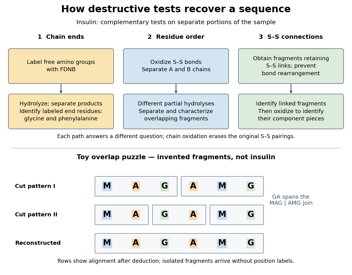
The toy fragments make the logic visible. Suppose one cut pattern yields MAG and AMG, and another yields MA, GA and MG. The first set alone permits MAGAMG or AMGMAG. The fragment GA in the second set crosses the MAG–AMG boundary in MAGAMG; it does not occur in AMGMAG. Among concatenations of the first set, therefore, the second set selects MAGAMG. This example assumes intact, correctly identified fragments from one homogeneous chain and known fragment membership in each cut pattern. Repeated stretches, missing fragments or sample mixtures can leave ambiguities; the picture illustrates inference from overlaps, not a universal guarantee of reconstruction.
Finally, determine the protein disulfide bonds separately. Oxidation used to separate whole chains erases their original connections. Sanger and colleagues instead studied fragments still joined by protein disulfide bonds, then oxidized the isolated joined fragments to identify their component pieces and locate the connected chain regions. An important trap was rearrangement of disulfide bonds during treatment: products can acquire connections absent from the starting protein. Their work therefore had to establish conditions that prevented rearrangement before interpreting a linked fragment as evidence. See Ryle and Sanger, A Disulphide Interchange Reaction, Nature 171, 1025–1026 (1953) and Sanger's Nobel lecture, pp. 553–554: preserving and identifying disulfide connections. The result was an experimentally constrained chemical structure rather than a composition list or a photograph of a fold.
Insulin is a protein hormone. Its mature form has two polypeptide chains; identifying their sequences and protein disulfide bonds provided the motivating example in Sanger's insulin sequencing experiment. See Cooper: insulin as a two-chain hormone, figure 2.16.
A meal illustrates the insulin side of blood glucose homeostasis: rising glucose stimulates beta cells in the pancreatic islets, and released insulin favors uptake and storage of fuel. Alpha cells produce glucagon, while delta cells produce somatostatin, which inhibits release of both insulin and glucagon. The pancreas therefore contains interacting sensor and secretory biological cells, rather than one undifferentiated source of hormones. [ref]
Insulin and glucagon are peptide hormones, built from amino acid residues joined by peptide bonds; glucose is a small carbohydrate molecule. A hormone changes signal transduction, not the identity of the fuel it regulates. Human mature insulin has 51 amino acid residues in two polypeptide chains. Beta cells process proinsulin to insulin and C-peptide, storing the products in secretory granules for exocytosis. Increased glucose metabolism raises the ATP-to-ADP ratio, tending to close ATP-sensitive potassium channels; depolarization and entry of calcium ions then trigger exocytosis. This is an important triggering pathway, with additional amplification and modulatory inputs.[ref]
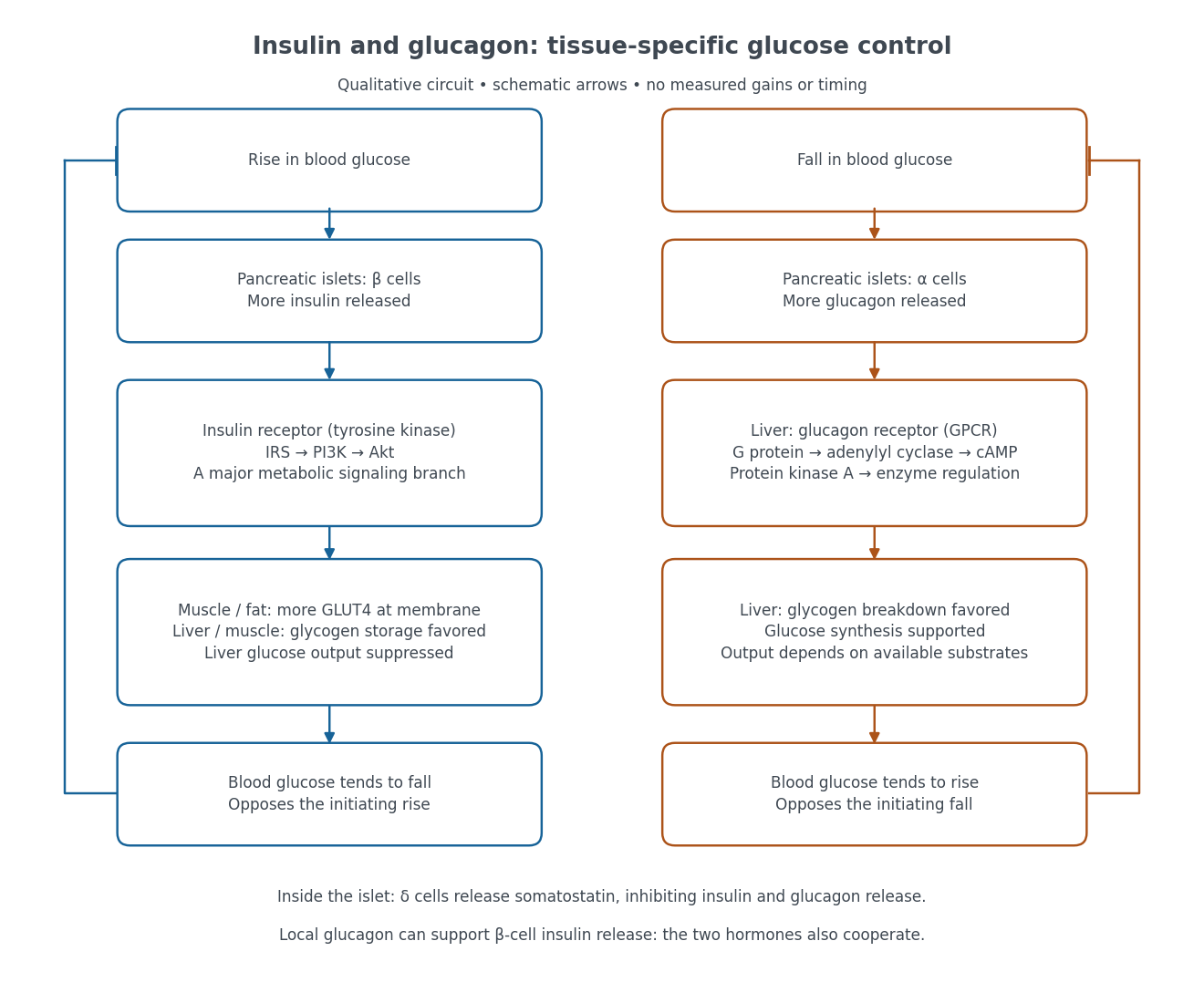
Binding to the insulin receptor, a receptor tyrosine kinase, starts phosphorylation of the receptor and insulin receptor substrates. Recruitment of phosphoinositide 3-kinase and activation of Akt transmit a major metabolic branch. In skeletal muscle and adipose tissue, that branch helps move GLUT4 to the cell membrane, increasing glucose uptake by facilitated diffusion. GLUT4 is a membrane transport protein, not an insulin receptor. Insulin also favors glycogenesis in skeletal muscle and the liver, and inhibits lipolysis in adipose tissue. In the liver, suppression of glucose output involves enzyme regulation, gene expression, and indirect changes in substrate supply. Its relative direct and indirect contributions are more complex than a single Akt arrow. [ref]
Insulin is not a universal permission slip for glucose entry: red blood cells and much of the nervous system use uptake mechanisms that are not acutely dependent on insulin. Nor does the liver use the same GLUT4 switch as skeletal muscle. This is why reduced insulin action can change blood glucose dramatically without stopping all glucose uptake. [ref]
Diabetes can disrupt production, response, and counterregulation. Type 1 diabetes usually involves destruction of beta cells and severe loss of endogenous insulin; type 2 diabetes combines insulin resistance with insufficient beta cell compensation. High circulating insulin can therefore coexist with impaired insulin action. Inappropriate glucagon release can add to excessive liver glucose output, and the response to low blood glucose can also be impaired: "more glucagon" is not a complete description of diabetes. These distinctions explain mechanisms, rather than prescribe treatment. [ref] [ref]
A single polypeptide precursor whose processing yields mature insulin and C-peptide; it is not the two-chain mature hormone. The biosynthetic distinction is described under insulin.
Glucose clamp technique: a method for quantifying insulin secretion and resistance. by R. A. DeFronzo and others separates two questions. In a hyperglycemic clamp, variable glucose infusion holds blood glucose above baseline while sampled insulin reports the response. The 1979 article's abstract reports an early release burst followed by a slower rise. In a euglycemic insulin clamp, infused insulin is held elevated and variable glucose infusion holds blood glucose at baseline. Frequent blood sampling, a glucose analyzer, and controlled infusion apparatus make the imposed concentration a maintained condition rather than an uncontrolled consequence. [ref][ref]
The maintained glucose level controls an important confounder: a change in blood glucose would itself change uptake and hormone release. At a steady state, conservation of mass gives uptake equal to infused supply plus endogenous production, after accounting for losses. Consequently, infusion alone estimates uptake only if endogenous production is negligible and the other balance assumptions hold. Labelled glucose measurements can estimate residual endogenous production; matching infused insulin across comparisons matters. Whole-body uptake cannot by itself identify the responding tissue, and plasma insulin reflects both release and removal. This interpretation uses the accessible 1979 abstract and the later authored methods chapter; the original article's full methods were not available for inspection.[ref]
Protein secondary structure consists of recurring local arrangements of the protein backbone, notably the alpha helix and beta sheet. Backbone hydrogen bonds help stabilize these patterns. A beta sheet can bring together stretches far apart in the protein primary structure; local refers to the pattern of backbone geometry, not necessarily neighboring sequence positions. See Alberts et al.: alpha helices, beta sheets and backbone interactions.
An alpha helix is a coiled protein secondary structure with a repeated pattern of backbone hydrogen bonds. In the standard pattern, the backbone group of amino acid residue bonds to that of residue : for example, positions 2 and 6. This is an additional spatial interaction, not a replacement peptide bond between those positions. Pauling, Corey and Branson proposed the helical arrangement in their 1951 paper by reasoning from the geometry of small molecules. See Pauling, Corey and Branson, The Structure of Proteins: Two Hydrogen-Bonded Helical Configurations of the Polypeptide Chain, PNAS 37, 205–211 (1951).
A beta sheet consists of extended stretches of protein backbone lying alongside one another, joined by backbone hydrogen bonds. The stretches can belong to different portions of the same polypeptide chain or to different chains.
Each extended stretch is a beta strand. Neighboring beta strands pack side by side, with repeated backbone hydrogen bonds between their backbone and groups: the oxygen is a hydrogen bond acceptor, and the backbone nitrogen with its attached hydrogen provides a hydrogen bond donor. These are interactions of the shared protein backbone, not merely contacts between amino acid side chains. Unlike an alpha helix, a beta sheet joins adjacent extended stretches rather than coiling one stretch around an axis. See Clark, Douglas and Choi, Biology 2e, 3.4: backbone groups in secondary structure.
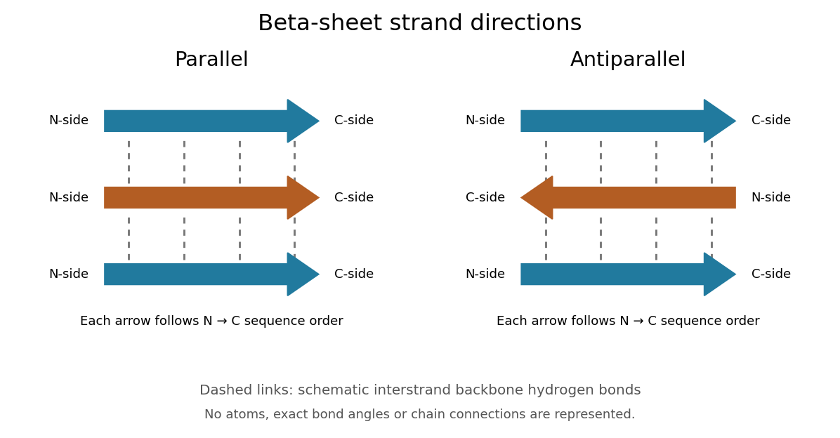
Read each beta strand in the sequence direction from the N-terminus toward the C-terminus, even when it is an internal part of a longer polypeptide chain. In a parallel beta sheet, neighboring beta strands run in the same N→C direction; in an antiparallel beta sheet, they run in opposite N→C directions. A three-strand example can therefore have arrows right–right–right or right–left–right. A beta sheet can also mix the two arrangements. Direction does not identify chain membership: a single polypeptide chain can fold back to supply several beta strands, and different polypeptide chains can contribute to one beta sheet. See Alberts et al., Molecular Biology of the Cell: beta-sheet strand arrangements.
The pleated appearance comes from the geometry of the protein backbone. Successive amino acid residues carry amino acid side chains on alternating faces of an ideal beta strand, conventionally drawn above and below the sheet. The attachment positions are the amino acid alpha carbons. Thus a six-position cartoon has groups 1, 3 and 5 on one face and 2, 4 and 6 on the other. This alternation is a spatial pattern, not a rule that alternating positions must contain identical amino acids. See Clark, Douglas and Choi: side chains above and below the pleats.
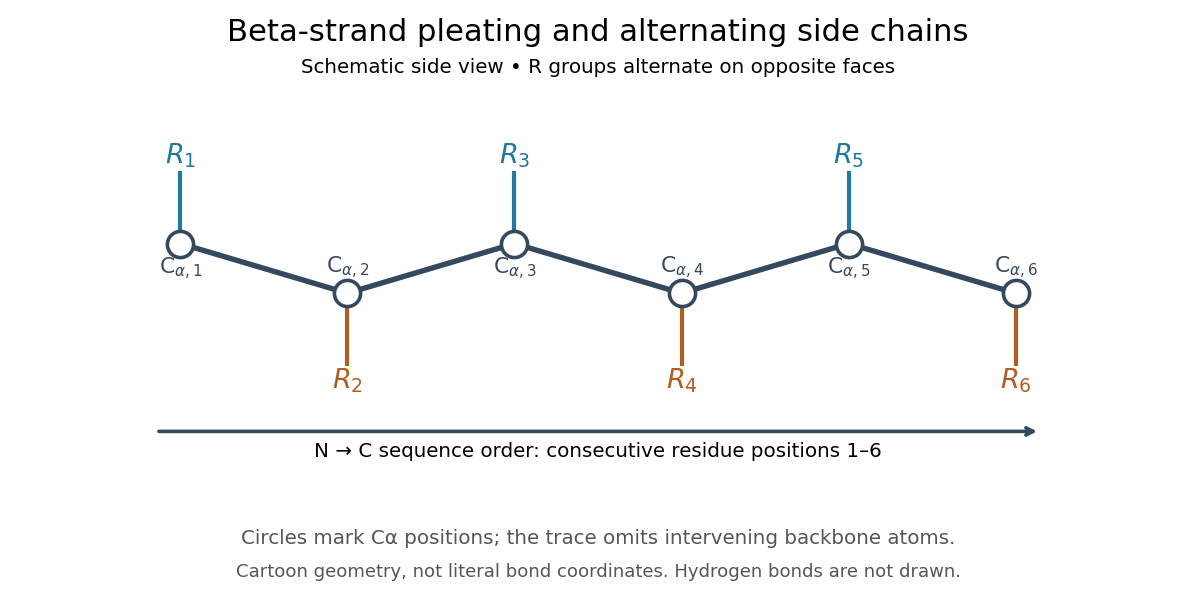
Real beta sheets commonly twist and curve; pleating does not make them perfectly flat plates. Which beta strands are neighbors, how connecting regions join them, and how their faces pack are features of the three-dimensional protein structure. An alternating pattern in the protein primary structure alone neither establishes a beta sheet nor specifies a unique fold independently of surroundings and interacting molecules. See Breda et al., Protein Structure, Modelling and Applications: extended strands, sheet twist and three-dimensional organization and protein folding.
Why this arrangement is useful: repeated backbone hydrogen bonds organize extended chain regions into a framework that can contribute to the interior of a folded protein. Amino acid side chains and connecting loops give that framework chemical variety and can shape a protein binding site for a ligand. Its attractive feature is the combination of a reusable backbone pattern with variable exposed groups: similar beta sheets can support different recognition surfaces. See Alberts et al.: beta-sheet cores and loops that form binding sites.
Historically, Linus Pauling and Robert B. Corey published The Pleated Sheet, a New Layer Configuration of Polypeptide Chains in PNAS 37, 251–256 (1951). They reasoned from peptide bond geometry and possible backbone hydrogen bonds, proposing an idealized layer rather than measuring every modern beta sheet. Their initial model should not be identified with all present-day beta sheet geometries; they published further pleated-sheet configurations later in 1951. See Pauling and Corey: original 1951 paper and scan, [ref]
https://williams.chemistry.gatech.edu/course_Information/6572/papers/Pauling_PNAS-1951_sheet.pdf. accessible scan of the original paper
, and Pauling and Corey, Configurations of Polypeptide Chains With Favored Orientations Around Single Bonds: Two New Pleated Sheets, PNAS 37, 729–740 (1951).A beta strand is an extended segment of protein backbone in the geometry characteristic of a beta sheet. Several beta strands can associate through backbone hydrogen bonds. A beta strand need not be an entire polypeptide chain.
A parallel beta sheet has neighboring beta strands running in the same direction along their amino acid residue sequences, from the N-terminus toward the C-terminus.
An antiparallel beta sheet has neighboring beta strands running in opposite N→C sequence directions. This specifies strand orientation rather than whether the beta strands belong to one polypeptide chain or several.
A backbone hydrogen bond is a hydrogen bond involving groups of the protein backbone, typically a backbone oxygen chemical atom and a hydrogen chemical atom already joined by a covalent bond to backbone nitrogen. It can connect positions that are not adjacent along the polypeptide chain. The peptide bonds still determine which amino acid residues are consecutive. See the original structural argument cited under alpha helix.
The protein tertiary structure is the overall spatial arrangement of one polypeptide chain. It includes how its protein secondary structures and intervening regions fit together. For example, the schematic under protein places positions 4 and 9 near each other despite their separation in protein primary structure. Such cooperation can help construct an active site; sequence distance and spatial separation answer different questions. Hydrophobicity, backbone hydrogen bonds and other interactions help stabilize folds, and some proteins additionally contain protein disulfide bonds. See Cooper: tertiary structure and interactions between chain regions.
A protein binding site is a region of a protein structure that interacts with a ligand. Its arrangement of amino acid residues helps determine recognition; connecting loops supported by a beta sheet can contribute to it. An enzyme's active site binds a substrate and accelerates a chemical reaction. See Alberts et al.: protein binding sites and active sites.
A protein disulfide bond is a disulfide bond linking two sulfur chemical atoms carried by suitable amino acid residues. It can connect distant portions of a polypeptide chain or different chains. See Cooper: sulfur-containing residues and disulfide crosslinks. The four internal protein disulfide bonds of ribonuclease A are central to the ribonuclease A refolding experiment; breaking these crosslinks need not break the peptide bonds along its protein backbone.
The protein quaternary structure is the arrangement of multiple polypeptide chains in a functional assembly. A single-chain protein has protein tertiary structure without requiring protein quaternary structure. For an exact bookkeeping example, two intact model chains of twelve amino acid residues each contain residues and peptide bonds. Assembly alone adds no peptide bond joining their ends. Stable functional assembly should also be distinguished from protein aggregation. See Alberts et al.: proteins as assemblies of polypeptide subunits.
Protein folding is the process by which a polypeptide chain acquires an organized spatial arrangement. The interactions of its amino acid residues with one another and the surroundings favor some arrangements over others. Protein folding can bring chemically useful groups together at an active site without changing their order along the protein backbone. Its importance is practical: knowing a sequence alone does not tell us whether a sample has the structure needed for function. See Cooper: acquiring functional protein structure.
The ribonuclease A refolding experiment showed that an isolated protein could recover its native properties without a pre-existing folded template under suitable laboratory conditions. Anfinsen interpreted this through a thermodynamic hypothesis: the native arrangement minimizes the whole system's Gibbs free energy in the appropriate environment. His Nobel lecture was delivered on 11 December 1972 and published in Science in 1973. The specified environment is part of the claim, not a disposable qualification. See Anfinsen, Principles that Govern the Folding of Protein Chains, Science 181, 223–230 (1973): Nobel lecture, especially p. 223.
This does not imply that every protein rapidly folds unaided. Molecular chaperones can assist protein folding and reduce protein aggregation; required partners, surroundings and time still matter. Goloubinoff and colleagues' 1989 reconstitution experiments demonstrated assistance from two molecular chaperones and ATP in recovering an active enzyme from unfolded chains. This is experimental evidence that assistance can matter even when the chains already have their specified sequences. See Goloubinoff, Christeller, Gatenby and Lorimer, 1989: chaperone-assisted reconstitution of an active enzyme.
A molecular chaperone is a protein that assists other proteins in reaching or maintaining useful arrangements, for example by limiting unwanted association during protein folding. Assistance does not rewrite the client's protein primary structure. In a crowded cytoplasm, reducing protein aggregation can make successful protein folding more reliable. See Cooper: molecular chaperones and folding assistance.
Protein denaturation is the loss of a protein's native spatial organization, often with loss of biological function. Changes in temperature, pH or surroundings can disrupt protein secondary structure, protein tertiary structure or protein quaternary structure. Protein denaturation ordinarily preserves the peptide bonds and protein primary structure; it is different from cutting the chain. Recovery by protein folding is possible in some cases, but removing the denaturing conditions does not guarantee that every protein recovers. See Clark, Douglas and Choi: denaturation, environmental conditions and loss of function.
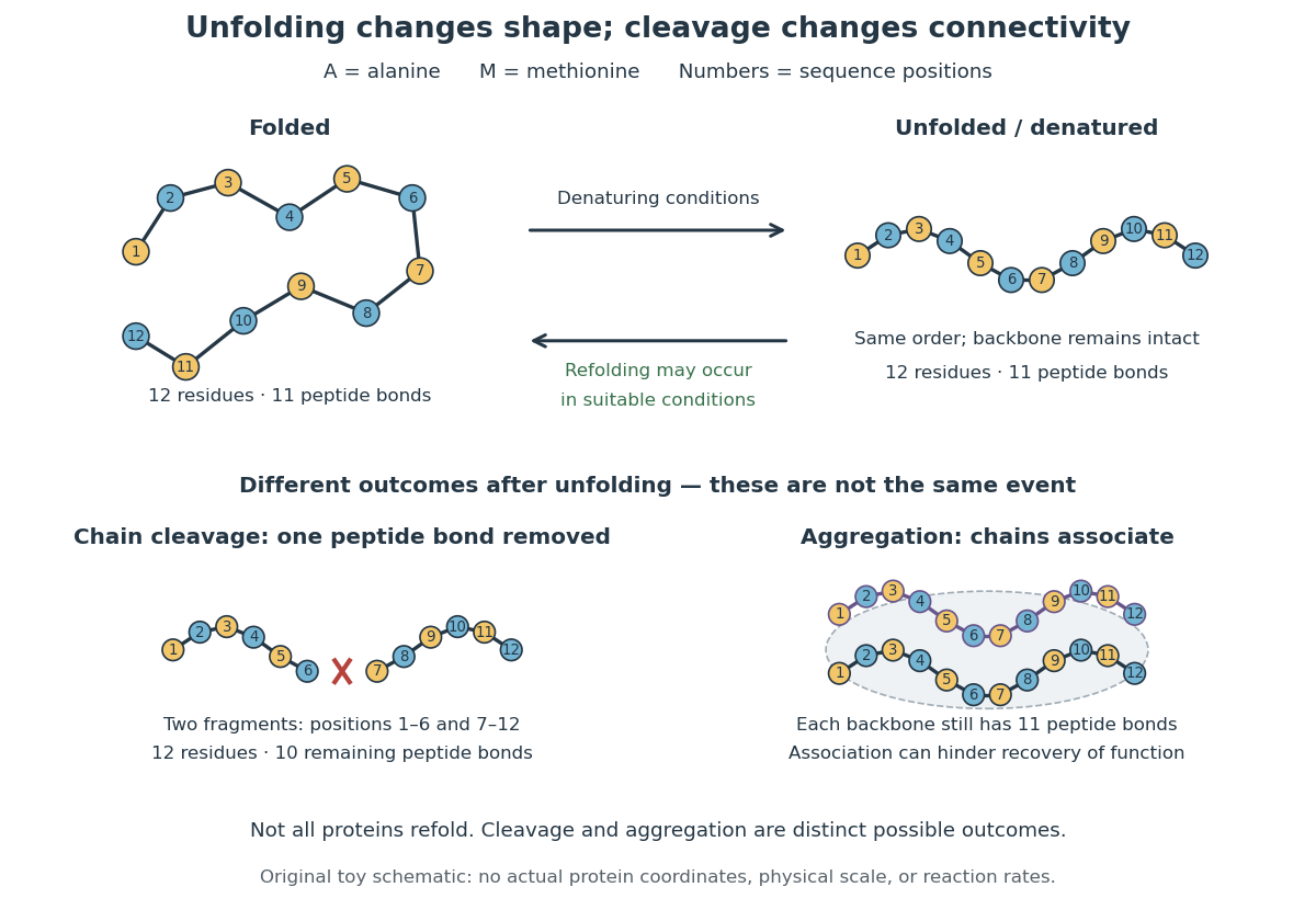
In the diagram, unfolding preserves all eleven peptide bonds. Cleaving the connection between positions 6 and 7 leaves two six-residue fragments: each contains five peptide bonds, so there are in total. Simply cooling or otherwise restoring suitable conditions cannot recover the original chain by protein folding alone; the missing covalent bond would have to be remade. Conversely, protein aggregation can impede recovery without breaking any peptide bond. These distinctions explain why loss of function does not by itself reveal whether a protein unfolded, associated with other chains, or suffered chemical damage.
Protein aggregation is the association of proteins into clusters, often involving exposed regions of partly folded or unfolded polypeptide chains. It can prevent recovery of normal function while leaving the protein primary structures intact. It is distinct from both unfolding one chain and the functional assembly described by protein quaternary structure. See Cooper: partly folded chains, aggregation and chaperone protection.
Ribonuclease A is an enzyme that catalyzes cleavage of RNA. The bovine pancreatic ribonuclease A studied by Anfinsen and colleagues has 124 amino acid residues and four internal protein disulfide bonds. It supplied a particularly informative test of the relationship between protein primary structure, protein folding and biological function: recovery could be measured through restored enzyme activity. See the primary sources under ribonuclease A refolding experiment.
Anfinsen, Haber, Sela and White's 1961 experiments began by treating bovine pancreatic ribonuclease A with mercaptoethanol in urea. Urea promotes unfolding; mercaptoethanol breaks protein disulfide bonds. The peptide bonds along the chain remain intact. After separating the chain from those reagents, the authors allowed its reactive sulfur groups to form protein disulfide bonds in the presence of molecular oxygen, at pH and temperature –. They examined protein concentrations of and . Measurements showed recovery of native properties under these conditions; the isolated major component had normal activity toward both RNA and a synthetic substrate. See Anfinsen, Haber, Sela and White, The Kinetics of Formation of Native Ribonuclease during Oxidation of the Reduced Polypeptide Chain, PNAS 47, 1309–1314 (1961), pp. 1309–1311.
The observation supports the sufficiency of that intact sequence to recover native protein structure in the tested surroundings without a folded template. It does not establish that every protein refolds, that the original living biological cell needed no assistance, or that activity always returns immediately. Protein folding, forming protein disulfide bonds and recovering an active site are related processes rather than interchangeable measurements.
Anfinsen's lecture also discusses forming protein disulfide bonds while the chain remains in concentrated urea. This gave mostly inactive material; after removing urea, a small amount of mercaptoethanol permitted rearrangement toward the native form. As an exact combinatorial check, eight labeled reactive sulfur groups have ways to be paired into four protein disulfide bonds: choose the partner of one group, then repeat with the unpaired groups. Only one pairing is the native one. Equal counting does not mean that all pairings are equally likely experimentally, nor that actual protein folding searches them uniformly. See Anfinsen's Nobel lecture, 1973 publication: scrambled ribonuclease and the 105 pairings, p. 223.
Lysozyme is a protein enzyme that attacks the peptidoglycan of bacterial cell walls. Its active site is a cleft in the folded protein structure, where a suitable substrate can interact with positioned chemical groups. This illustrates why possessing the right amino acid residues is insufficient if their spatial arrangement is lost. See Alberts et al.: lysozyme as a worked example of protein binding and enzyme function.
A concrete structural example is Cheetham, Artymiuk and Phillips's 1992 study of hen egg-white lysozyme bound to a short carbohydrate that inhibits its action. The bound carbohydrate occupies part of the active site; it is not a reacting substrate in this complex. The study describes specific interactions and changes of shape accompanying binding. Thus a real binding site has chemical interactions and flexibility as well as a geometric outline. See Cheetham, Artymiuk and Phillips, 1992: structure of a lysozyme complex with bound inhibitor and RCSB PDB entry 1HEW: the authors' structure and bound three-unit carbohydrate. The diagrams above illustrate the idea; their twelve-residue strings are not lysozyme sequences or coordinates.
The N-terminus is the amino end of a protein or amino acid chain. Sequence notation lists residues from the N-terminus toward the C-terminus, the same direction in which biological translation builds a chain. The two-residue example under gene expression places methionine at the N-terminal end before any subsequent processing. [ref]
https://openstax.org/books/biology-2e/pages/3-4-proteins. OpenStax Biology 2e, 3.4: amino acids and polypeptide direction
The C-terminus is the carboxyl end of a protein or amino acid chain. During biological translation, each new residue is added at this end. The example under gene expression gives N–methionine–alanine–C, placing alanine at the C-terminus; its stop codon supplies no further residue. [ref]
Actin is a protein whose monomers assemble into filaments. An idealized actin filament has a bending modulus and persistence length; inferring its effective elastic modulus from those measurements additionally requires a cross-sectional model.
A biochemical receptor is a protein that recognizes a signal and couples its detection to a biological effect. Its binding partner is a ligand; recognition depends on compatible shape and chemical interactions at a binding site within the protein structure. Specificity is selective recognition, not a guarantee that only one ligand can bind. Binding also need not activate the receptor: a competing ligand may occupy it while preventing activation by another ligand. See Alberts et al., Molecular Biology of the Cell, 4th edition: protein binding and specificity.
Compare a cell-surface receptor, accessible to an extracellular ligand, with an intracellular receptor, whose ligand must reach the interior. A cell-surface receptor can detect a hydrophilic ligand without transporting it through the cell membrane. In contrast, some membrane-permeable signals reach intracellular receptors that regulate gene expression. Location and downstream coupling are separate distinctions: an ionotropic receptor directly gates an ion channel, whereas a metabotropic receptor works through intracellular signal transduction. See Clark, Douglas and Choi, Biology 2e, 9.1: receptor locations and mechanisms.
Receptor binding occupancy quantifies recognition separately from response. A dissociation constant measures binding in a specified equilibrium model; it does not by itself predict the cellular effect. This separation is useful when comparing signals that bind equally well but produce different responses, or when interpreting a binding experiment alongside a functional measurement.
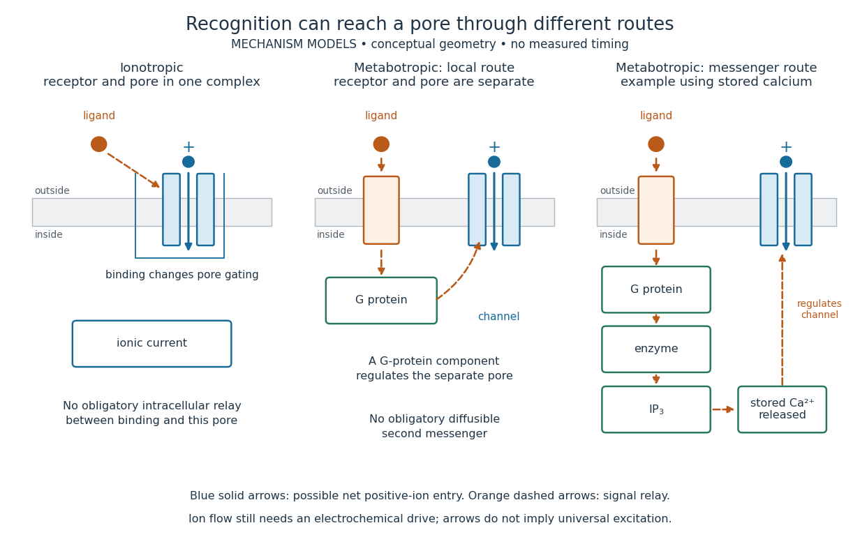
An ionotropic receptor is a receptor in which binding of a ligand directly gates an ion channel within the same protein complex. The binding site detects the chemical signal; the pore supplies a path across the cell membrane. At a chemical synapse, a neurotransmitter therefore changes synaptic conductance without an obligatory intervening G protein or second messenger. [ref]
A familiar example is the nicotinic acetylcholine receptor of skeletal muscle: acetylcholine binding opens a pore passing cations, including sodium and potassium. The muscarinic acetylcholine receptor instead uses intracellular signal transduction; the same neurotransmitter can engage different mechanisms. Direct gating does not mean that an ionotropic receptor pumps ions: electrochemical gradients drive passage through the open ion channel. Nor does “ionotropic” specify excitation. The reversal potential, initial membrane potential and other conductances determine whether the effect favors an action potential, causes hyperpolarization, or supplies shunting inhibition. [ref]
A concrete electrical approximation separates opening from drive. Let identical ion channels each have open-channel electrical conductance and opening probability . At fixed membrane potential , with common reversal potential , the expected value of inward-positive electric current isFor stipulated values , , , and , the mean open count is , the population electrical conductance is and . One open ion channel carries . These are illustrative values, not measurements. At , the net electric current vanishes despite open ion channels; neither binding nor opening alone determines the readout. The approximation omits changes in ion concentrations and voltage-dependent open-channel electrical conductance.
A motivating experiment made the elementary electrical events visible. As Bert Sakmann recounts, chronically denervated frog muscle fibers had acetylcholine-sensitive receptors spread beyond the original nerve contact. Mild enzyme treatment exposed the cell membrane; a polished glass pipette sealed over a small patch to reduce background noise. Two intracellular electrodes held the local membrane potential through voltage clamp, while the patch pipette recorded tiny electric currents. Applying suberyldicholine, a ligand that activates the acetylcholine receptor and prolongs openings, revealed abrupt, approximately rectangular current events. Their amplitudes agreed reasonably with estimates from whole-membrane current fluctuations. This established discrete opening and closing events; it did not make every ligand response instantaneous. Background noise and finite recording bandwidth concealed sufficiently small or brief events.[ref]
Recording an intact membrane patch alone does not exclude intracellular relays. Tank, Huganir, Greengard and Webb supplied a complementary isolation test in 1983: they purified nicotinic acetylcholine receptors from an electric ray's electric organ, incorporated them into artificial lipid membranes, and fused small vesicles into larger vesicles by freezing and thawing. Sealed glass-pipette recordings from isolated membrane patches resolved functional ion channels with properties resembling those in muscle. Removing the intact cytoplasm and its signaling machinery supports the sufficiency of the receptor complex for channel function; an artificial lipid environment still cannot reproduce every native regulatory interaction.[ref]
Direct gating supports millisecond communication at many chemical synapses. Response duration also depends on neurotransmitter clearance, binding and channel-state transitions. Continued ligand exposure can prolong activity. Classify an ionotropic receptor by its direct connection between recognition and pore gating, rather than a universal duration cutoff. [ref]
A metabotropic receptor couples ligand recognition to intracellular signal transduction, which can regulate a separate ion channel, alter enzyme activity or change gene expression. In the usual neurotransmitter context, metabotropic receptors are G protein-coupled receptors: receptor activation is relayed by a G protein. There need not be a change in membrane potential at all. [ref]
Two routes should be distinguished. An activated G protein can regulate an ion channel locally at the cell membrane, without an obligatory diffusible second messenger. Alternatively, it can regulate an enzyme that changes the availability of a second messenger, such as cAMP or inositol trisphosphate. Downstream proteins then change channel opening, phosphorylation or other cellular activities. The receptor, G protein, second messenger and final target are distinct components; an activated G protein is a relay protein, rather than itself a small-molecule second messenger.[ref]
General Principles of Cell Communication. Figure 15-15
Do not turn this useful chemical synapse classification into the claim that every receptor is a GPCR, or that every indirect response must pass through a G protein. For example, the insulin receptor is an enzyme-linked cell-surface receptor whose intracellular catalytic activity initiates protein phosphorylation. Intracellular receptors supply another category. “Metabotropic” commonly names the GPCR branch in neuroscience; the broader idea of biochemical coupling does not make all non-channel receptors structurally identical. [ref]
Separating recognition from execution allows signal amplification and branching. A persistently activated receptor can activate several relay proteins; a regulated enzyme can produce many second messenger molecules. As an explicitly invented count example, suppose one activated receptor activates four relay proteins during one interval, and each relay activates a distinct enzyme that produces fifteen second messenger molecules during the next interval. With sufficient substrates and no loss, this gives molecules. Those counts do not specify a physiological gain or response duration. A directly gated ion channel also passes many ions while open, so amplification is not an exclusive property of metabotropic receptors: always name the input and output being compared. [ref]
A particularly instructive electrophysiology experiment separated a chemical trigger from the ion channel carrying its eventual electric current. In 1987, Hirono, Ito and Sugiyama injected messenger RNA from rat brain into frog egg cells, then tested acquired responses to neurotensin and muscarinic acetylcholine stimulation. Under voltage clamp at , neurotensin elicited transient, oscillatory inward electric currents after a delay of several tens of seconds. This delay refers to that response and preparation. Reversal potential measurements implicated increased permeability to chloride anions, rather than a pore specific to the applied ligand. Both neurotensin and acetylcholine responses were inhibited by pertussis toxin, which interferes with susceptible G proteins, and by intracellular EGTA, which binds free calcium. Injecting inositol trisphosphate mimicked the responses; a large prior dose of inositol trisphosphate reduced subsequent responses to either extracellular signal. These interventions implicate a shared intracellular relay and calcium-dependent channel opening.[ref]
The informative comparison with the nicotinic acetylcholine receptor experiment is mechanistic: an extracellular ligand can produce an inward electric current either by directly gating the pore or by mobilizing an intracellular second messenger and stored calcium that activate another pore. Intracellular inositol trisphosphate bypasses reception, whereas EGTA tests the need for free calcium. However, neither inhibition nor mimicry alone identifies every molecular connection: a perturbation can affect several targets, and an injected mediator can activate a pathway without proving that it is the only natural route. The egg cell's endogenous machinery and the mixed messenger RNA preparation also limit extrapolation to an intact neuron. The experiment establishes functional coupling in its preparation, rather than the exact molecular composition or timing of every metabotropic receptor.
Even G protein-dependent channel regulation need not involve a lengthy second messenger cascade. Pfaffinger and colleagues reported in 1985 that acetylcholine-evoked potassium channel activation in embryonic chick atrial cells required intracellular GTP, a guanine-containing nucleotide, and was abolished by pertussis-toxin pretreatment. Changes in cyclic nucleotides, including cAMP, did not account for the channel response. The report discusses intrinsic activation delays of roughly –, illustrating relatively rapid metabotropic receptor action. These observations support a G protein coupling step without cyclic-nucleotide second messengers; they do not by themselves exclude every possible intermediary or prove the specific subunit-channel interaction.[ref]
A small timing model makes the extra steps visible without inventing an experimental trace. Let jump from zero to one at and remain on. For a direct-gating model output , one relay variable , and its downstream output , take zero initial conditions andAll outputs are normalized dimensionless variables; they are neither recorded electric currents nor fitted concentrations. With illustrative time constants , and , their exponential function solutions for areInitially , while because its relay must first build up. Both outputs eventually approach one. Equal normalized final values do not imply equal biological amplitudes or absence of signal amplification; normalization deliberately removes that question.
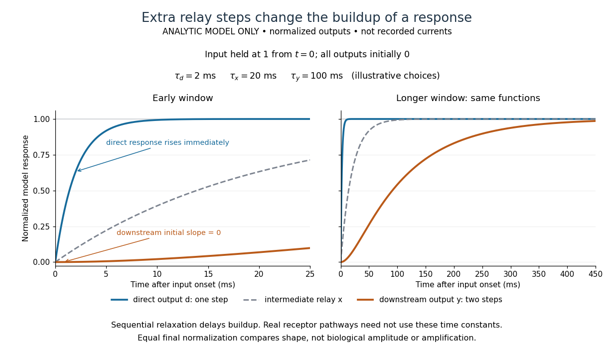
The model's useful feature is the delayed buildup created by sequential relaxation, not its arbitrary numbers. Actual kinetics also depend on ligand delivery, relay deactivation, neurotransmitter clearance and the final target. A local G protein route can act promptly, while an ionotropic receptor can remain active during continued exposure or feed a longer-lasting intracellular response. The beauty of metabotropic receptor coupling is its flexibility: the same recognition event can adjust electrical responsiveness and other cellular tasks through different targets. To classify an observed response, combine timing with molecular identity and controlled perturbations.
A G protein-coupled receptor is a cell-surface receptor that relays activation through a G protein on the intracellular side of the cell membrane. The activated G protein can regulate an enzyme or ion channel, so a GPCR supplies a concrete metabotropic receptor example: ligand recognition and the eventual response are connected through distinct components. Different G proteins couple to different targets. See Alberts et al., Molecular Biology of the Cell, 4th edition: signaling through G-protein-linked cell-surface receptors.
A cell-surface receptor detects an extracellular signal at the cell membrane. Its changed state affects the intracellular side, coupling reception to signal transduction or directly to an ion channel's opening.
A receptor tyrosine kinase combines extracellular ligand recognition with an intracellular protein kinase protein domain. A membrane-spanning segment connects these parts across the cell membrane: the signal changes the intracellular machinery without requiring the ligand to enter the cytosol. The protein kinase transfers a phosphate group from ATP to selected tyrosine residues on proteins, including the receptor itself.[ref]
Signaling through Enzyme-Linked Cell-Surface Receptors. “Activated Receptor Tyrosine Kinases Phosphorylate Themselves”: extracellular binding, intracellular catalytic domains and ATP-dependent phosphate transfer
This architecture makes a reception event useful in two ways: it can change catalytic activity and assemble a local group of signaling proteins. Trans-autophosphorylation can create phosphotyrosine residues recognized by SH2 domains. Recruitment positions a downstream enzyme or relay protein near its partners. Neither every modified site nor every recruited protein has the same effect; association is a specific molecular connection, rather than a universal command to activate everything nearby.[ref]
Signaling through Enzyme-Linked Cell-Surface Receptors. “Phosphorylated Tyrosines Serve as Docking Sites For Proteins With SH2 Domains”, Figure 15-53: signaling enzymes, adaptors and negative regulators
Compare the epidermal growth factor receptor with the insulin receptor. Their common topology does not impose one activation geometry or make assembly equivalent to activity. The receptor phosphorylation assay consequently needs more than evidence that two protein chains associate.
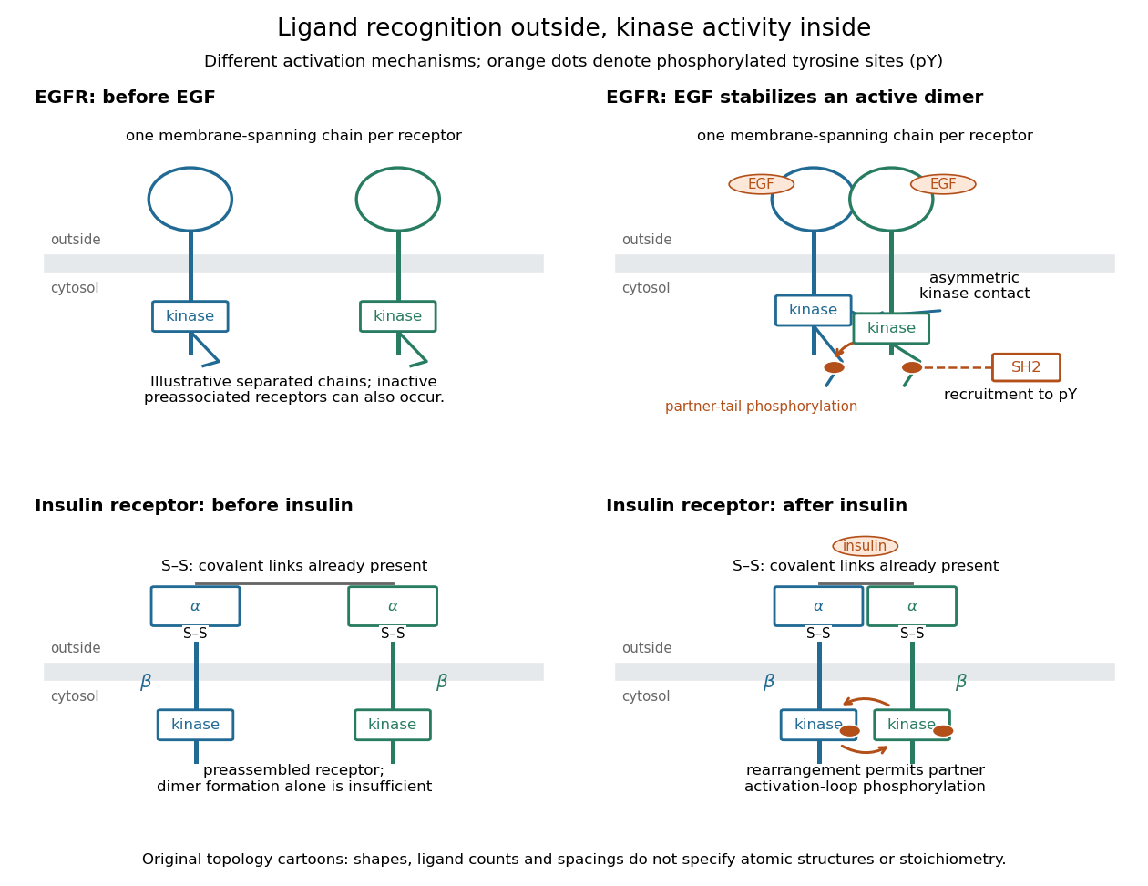
Receptor tyrosine kinase architecture and distinct activation mechanisms
. Original topology cartoons contrasting EGFR with the preassembled insulin receptor. Orange dots mark phosphotyrosine residues; S–S marks protein disulfide bonds. The EGFR panel separates asymmetric catalytic contact from partner-tail phosphorylation. Shapes, distances and drawn ligand counts are explanatory, rather than atomic structures or binding stoichiometry. Details.In an illustrative EGFR activation route, EGF stabilizes an associated pair of receptor chains. Asymmetric contact between their protein kinase protein domains activates one catalytic partner without requiring its activation-loop phosphorylation. Subsequent phosphorylation of cytoplasmic tails creates recruitment sites. Preassociated inactive EGFR populations can also occur: the cartoon's separated starting chains are not a universal initial condition.[ref]
Cell signaling by receptor-tyrosine kinases by Mark A Lemmon and Joseph Schlessinger. Lemmon and Schlessinger, “Mechanisms of Receptor Activation” and “Allosteric activation of TKDs”, Figure 2F
The mature insulin receptor already contains two extracellular chains and two membrane-spanning chains, joined through protein disulfide bonds, before insulin binds. Insulin induces rearrangement of this assembly. Partner-mediated phosphorylation of the inhibitory activation loop permits increased protein kinase activity. Ligand-induced creation of a previously absent dimer is therefore an unsuitable universal explanation.[ref]
Cell signaling by receptor-tyrosine kinases by Mark A Lemmon and Joseph Schlessinger. “Mechanisms of Receptor Activation” and “TKD autoinhibition by the activation loop: insulin and FGF receptors”, Figure 2E
An insulin receptor substrate is a docking protein phosphorylated by the activated insulin receptor. Its phosphotyrosine residues recruit downstream signaling proteins, including SH2 domain-containing components of phosphoinositide 3-kinase. This connects reception to the pathway described under insulin, rather than making the insulin receptor substrate another extracellular ligand.[ref]
Cell signaling by receptor-tyrosine kinases by Mark A Lemmon and Joseph Schlessinger. “Linking RTK activation to cell signaling”: IRS docking proteins and Figure 3A
A receptor phosphorylation assay asks whether, and how much, a specified receptor is modified by phosphorylation under defined conditions. It is distinct from a binding assay for receptor binding occupancy, a test of protein kinase activity on a supplied substrate, and a test of downstream signal transduction. Those measurements answer related but different questions.
For a Western blot implementation, compare matched samples with ligand omitted or added, detect a specified phosphorylated epitope, and separately detect total receptor abundance. Preserve the sampled state by stopping further enzyme reactions during sample preparation; otherwise protein kinase or protein phosphatase activity after sampling can change the result. A general anti-phosphotyrosine residue antibody recognizes many modified proteins; a site-selective antibody gives a narrower readout, but still needs Western blot antibody specificity controls.
Reason through the controls before interpreting the difference. Loss of the proposed signal after receptor-specific gene deletion supports protein identity. Treatment with a protein phosphatase, alongside preservation of the total-receptor signal, tests phosphate-dependent recognition. Western blot loading controls and Western blot transfer controls address sample recovery; a usable detection range addresses Western blot response saturation. Lower signal after perturbing protein kinase activity supports dependence on that perturbation, but alternative targets and altered receptor abundance can complicate the inference. These are proposed assay comparisons, not outcomes attributed to the historical membrane experiment.
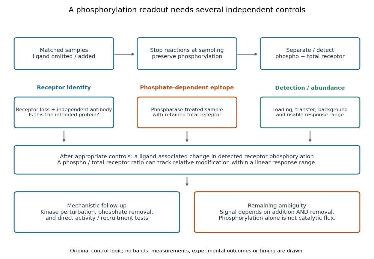
An original idealized measurement model explains the normalization. Let be the retained number of receptor molecules, the fraction carrying the selected modified epitope, and fixed detection gains. After subtracting their respective backgrounds, suppose the phosphorylated and total-receptor signals lie in a linear response range:Dividing by total receptor cancels under these assumptions. It differs from total-protein Western blot normalization, whose denominator includes many other proteins. For invented values , samples with and give and , but both give . Twice the phospho signal here means twice the retained receptor amount, with no change in fractional modification. Unequal recovery, changed epitope accessibility, nonzero uncorrected background or Western blot response saturation invalidates the cancellation. Unknown relative gains prevent reading an absolute modified fraction directly from the ratio.
A motivating experiment used isolated membranes from A-431 biological cells, which supplied abundant EGFR. Separate membrane aliquots were incubated at with ATP carrying radioactive phosphorus in its terminal phosphate group, plus magnesium or manganese ions. Radioactivity in material recovered by chemical acid-induced precipitation supplied the readout. Adding EGF beforehand enhanced incorporation relative to the unstimulated preparation; this placed a ligand-dependent biochemical response in a cell-free membrane system.[ref]
Epidermal Growth Factor by Stanley Cohen. Cohen, Epidermal Growth Factor, printed p. 339: A-431 membrane aliquots, terminally labeled ATP, divalent ions and trichloroacetic acid-insoluble incorporation
The follow-up comparisons separated increased addition from suppressed removal: phosphate removal was rapid but unaffected by EGF, and anti-EGF antibody reversed activation by removing the ligand. These observations support reversible regulation of membrane-associated protein kinase activity.[ref]
Epidermal Growth Factor by Stanley Cohen. printed p. 341, numbered observations 1–4: rapid activation, dephosphorylation, lack of soluble kinase release and anti-EGF reversal
Total incorporation alone cannot identify which protein or amino acid residue received the label, prove intrinsic receptor catalysis, or predict an intact biological cell's response.The same account records an instructive identification error: receptor phosphorylation was initially assigned to the wrong amino acid residue because released modified amino acids migrated together in the electrophoretic electric field. Reanalysis identified phosphotyrosine residues. Retained radioactivity is evidence about the assay's separation and detection, not unique chemical identity.[ref]
Epidermal Growth Factor by Stanley Cohen. printed p. 341: co-migration in the electrophoretic system and subsequent identification of tyrosine phosphorylation
Protein kinase activity adds modification, while protein tyrosine phosphatase activity can remove it. An original one-site model shows why a receptor phosphorylation assay cannot determine either process from a single modified fraction. Let be the fraction of a fixed receptor population carrying one selected phosphotyrosine residue. Stipulate constant effective first-order rate constants , with ATP availability absorbed into , and no receptor synthesis, degradation or trafficking:Here is the steady state fraction and the time constant. The ordinary differential equation has the exponential function solutionThe balance equation and a single relaxation step give this result; it is an illustrative model, rather than a fit or a claim about every RTK.
With stipulated rate constants , and . Doubling both rate constants leaves but changes to . The balanced phosphate turnover per site,doubles from to . Identical steady state modification therefore permits different catalytic fluxes. Removing the addition term instead gives ; this illustrates why a perturbation that stops protein kinase activity can expose removal dynamics, subject to the model's unchanged-removal assumption. A maintained steady state with ongoing ATP consumption is not chemical equilibrium.
Articles were limited to the first 100 out of 104 total.
Articles by others on the same topic



Protein is a macromolecule that is essential for the structure, function, and regulation of the body's tissues and organs. It is made up of long chains of amino acids, which are organic compounds composed of carbon, hydrogen, oxygen, nitrogen, and sometimes sulfur. There are 20 different amino acids that combine in various sequences to form proteins, each of which has a specific function in the body.