An objective aperture selects outgoing Electron directions in the objective lens's back focal plane. Passing the central transmitted beam gives bright-field transmission electron microscopy; selecting a separated diffracted beam gives dark-field transmission electron microscopy.[ref]
In a paraxial approximation, an outgoing angle maps approximately to position in this focal plane, where is the objective focal length. A centred opening of radius therefore admits . Selecting a direction selects a component of the outgoing wavefunction; the subsequent image still assigns intensity to specimen positions. The opening is not a hole cut into a real-space specimen image. Its angular selection must be distinguished from selecting a specimen region for electron diffraction.
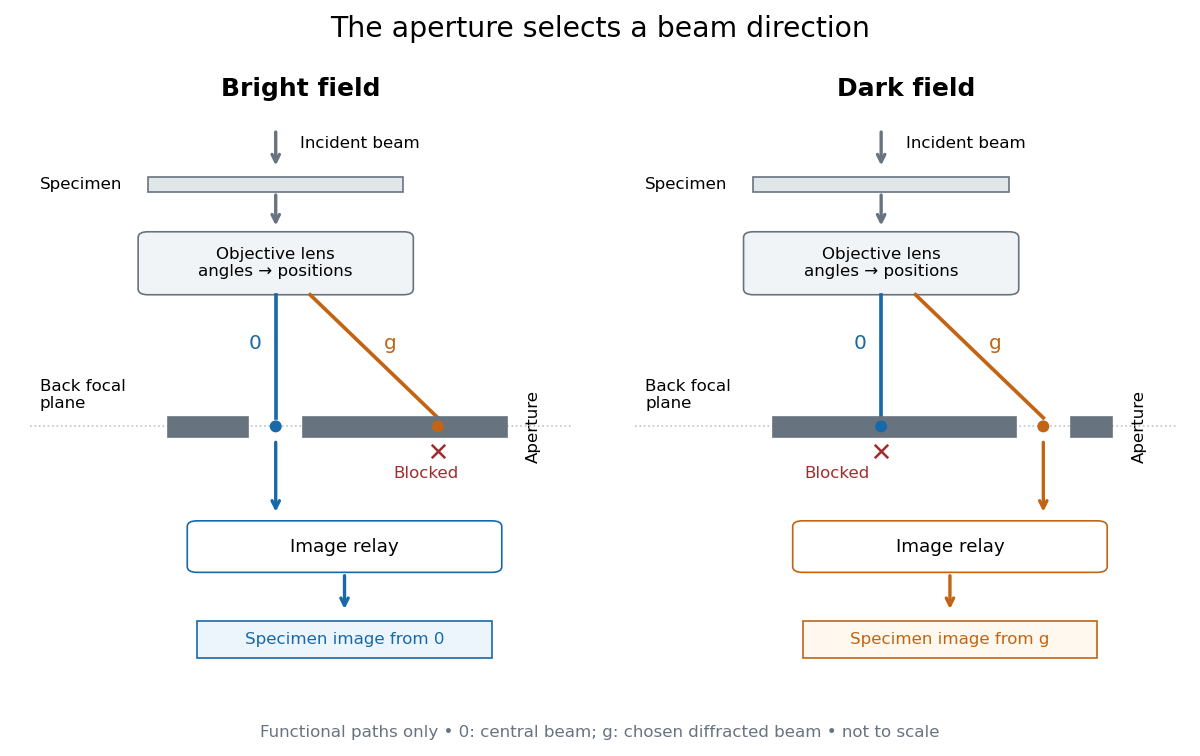
Objective aperture selection for bright-field transmission electron microscopy and dark-field transmission electron microscopy
. Original functional diagram. Lower relay boxes denote image formation, not literal ray paths. Scales and beam angles are illustrative. Source. Details.
Bright-field transmission electron microscopy selects the central transmitted beam; deflection into excluded electron diffraction beams can make a region dark.[ref]
Bright-field image by JEOL. Definition: transmitted beam and dark diffracting regions
Dark-field transmission electron microscopy selects a diffracted beam while excluding the central beam. Regions contributing to that selected electron diffraction beam can appear bright.[ref]
Dark-field image by JEOL. Definition: selected diffracted beam and bright contributing regions
Articles by others on the same topic
There are currently no matching articles.