Mouse-human heterokaryon membrane-mixing experiment
ID: mouse-human-heterokaryon-membrane-mixing-experiment

In 1970 Larry Frye and Michael Edidin fused mouse C11D and human VA-2 cell lines with Sendai virus, forming heterokaryons. Suspensions were maintained in a shaking water bath at . Removed aliquots were stained by indirect immunofluorescence: antibodies against parental surface antigens, then differently fluorescent secondary antibodies. A Leitz Ortholux fluorescence microscope used a mercury lamp and color-selecting filters. Green mouse and red human surface patterns became intermixed.[ref]
The rapid intermixing of cell surface antigens after formation of mouse-human heterokaryons by Larry Frye and Michael Edidin. Frye and Edidin, The rapid intermixing of cell surface antigens after formation of mouse-human heterokaryons, pp. 319–325: Summary, Cell fusion, Fluorescent staining, Fluorescence microscopy, Table 1, time-course results
Same-species fusions tested antibody specificity; stable hybrid lines supplied a two-color control. Inhibitors of biological translation did not prevent mixing; lowering temperature inhibited it. These observations supported lateral redistribution over rapid synthesis. The authors could not conclusively exclude passage through cytoplasm and return. The readout compared populations, not trajectories of a continuously observed individual biological cell, and did not establish unrestricted motion of every membrane protein.[ref]
The rapid intermixing of cell surface antigens after formation of mouse-human heterokaryons by Larry Frye and Michael Edidin. pp. 324–331: specificity controls, inhibitor studies, temperature dependence and Discussion of mechanisms iii and iv
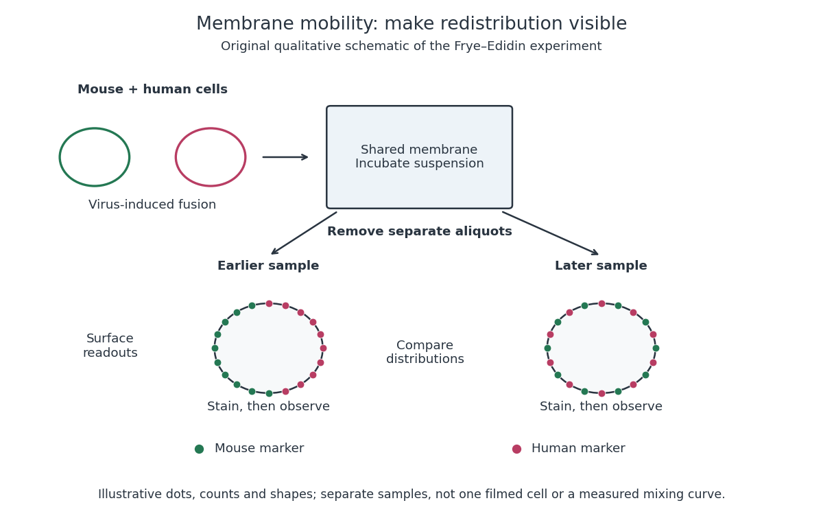
Separate time-point samples reveal changing surface distributions
. Original qualitative schematic of the mouse-human heterokaryon membrane-mixing experiment. Dot counts and shapes are illustrative; separate samples show distributions rather than individual trajectories or measured mixing curves. Details.
The decisive design idea is to begin with spatially distinguishable material. A uniformly labeled surface can look unchanged while its components move. Bringing together differently identifiable parental surfaces creates an initial pattern whose loss reveals redistribution; antibody specificity makes that loss interpretable. Membrane topology asks which side a part faces, whereas this experiment asks where it can move along that side.
New to topics? Read the docs here!