A biological cell is a membrane-bounded basic unit of living organization. In a cell-level experiment it can be an experimental unit, provided that treatment assignment and relevant interactions are handled at that level.
The shared architecture of cellular life combines a selective cell membrane, internal cytoplasm, inherited information in DNA, and ribosomes that assemble proteins. Metabolism supplies transformations of matter and energy needed for maintenance and growth. These components form a coupled system: a boundary retains useful molecules, enzymes organize chemical reactions, and inherited information supports rebuilding the machinery. See Clark, Douglas and Choi, Biology 2e: prokaryotic cells.
A worked structural comparison: in a typical prokaryote, DNA occupies a region without a surrounding cell nucleus; in a typical nucleated eukaryote, much of the DNA lies inside a membrane-bounded cell nucleus. Both still have a cell membrane, cytoplasm and ribosomes. The extra boundary compartmentalizes processes; it does not create a different basic principle of living organization. See Clark, Douglas and Choi, Biology 2e: eukaryotic cells.
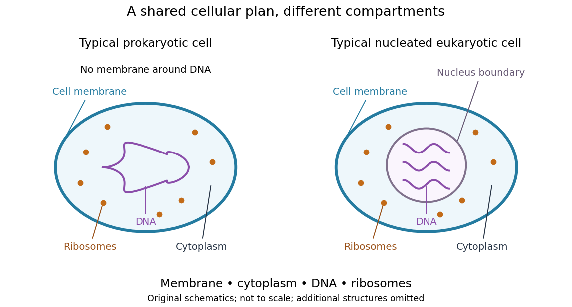
Shared cellular architecture with different DNA compartmentalization
. Original simplified schematics of a prokaryote and a nucleated eukaryote. Colors identify corresponding components; sizes and component counts are illustrative, and additional structures are omitted. Details.
Shared architecture describes cellular life and its developmental machinery, rather than requiring every specialized biological cell to retain every component. A mature mammalian red blood cell, for example, lacks a cell nucleus, although it develops from nucleated precursors. See Clark, Douglas and Choi, Biology 2e: components of blood. This distinction makes cellular unity compatible with striking specialization.
A phagocyte is a biological cell that takes up particles, such as bacteria or cell debris, by phagocytosis: the cell membrane encloses the material in an intracellular phagosome. Macrophages and neutrophils are important examples. The term describes an uptake capability, rather than a single developmental lineage. Contact with a particle, internalization and destruction of the ingested material are distinct events.
A useful experimental distinction is between phagocytosis and production of reactive oxygen compounds. S. D. Wright and S. C. Silverstein measured hydrogen peroxide released by human granulocytes, monocytes and macrophages spreading on surfaces coated with defined recognition molecules. Surfaces carrying IgG stimulated peroxide release, whereas surfaces carrying C3b or iC3b did not appreciably do so. Even when fibronectin supported a strong complement-receptor-associated phagocytic response in cultured monocytes, measurable peroxide release did not follow.[ref]
Receptors for C3b and C3bi promote phagocytosis but not the release of toxic oxygen from human phagocytes by S. D. Wright and S. C. Silverstein. Abstract and Figure1: ligand-coated surfaces, cultured monocytes and separate uptake/oxidant endpoints.
For example, counting ten particle-containing phagosomes would establish uptake in the observed phagocyte; it would not count ten killed bacteria. A killing assay needs a further endpoint, such as recovery of viable organisms. Likewise, a low extracellular peroxide measurement alone does not establish absence of phagocytosis. Choosing an endpoint that matches the biological question is essential when comparing phagocytes.
A biological membrane is a thin, organized boundary built from lipids and associated proteins that separates cellular compartments while controlling exchange and communication. The cell membrane bounds the whole biological cell; internal biological membranes surround structures such as the endoplasmic reticulum and mitochondrion. A biological membrane is therefore more general than the outer boundary of a cell.
The basic lipid bilayer puts water-compatible groups toward the surrounding water and hydrophobic portions toward its interior. Its barrier properties make transport selective: crossing a biological membrane through the lipid interior differs from moving through an ion channel or a transporter protein. Such selectivity allows different chemical conditions on the two sides, including an electrochemical gradient that can store usable energy. Membrane proteins also provide receptors, enzymes and attachment sites.
A biological membrane is organized but can remain fluid. In the mouse-human heterokaryon membrane-mixing experiment, Larry Frye and Michael Edidin fused cells whose surface antigens could be distinguished by different fluorescent antibody labels. Initially separate patterns became intermixed, providing evidence for lateral mobility of membrane proteins. This helped motivate the fluid mosaic model: many components can move within a shared membrane rather than occupying a completely rigid shell.[ref]
The rapid intermixing of cell surface antigens after formation of mouse-human heterokaryons by Larry Frye and Michael Edidin. Journal of Cell Science 7 (1970), 319–335: distinguishable parental surface antigens redistribute after cell fusion.
The experiment tests lateral redistribution, not unrestricted movement across the lipid bilayer or unlimited motion of every membrane protein. The distinction is useful: a boundary can preserve different environments across its thickness while permitting substantial rearrangement within its plane.
An eyespot is a subcellular light-sensing apparatus that helps a motile organism determine the direction of illumination. In Chlamydomonas reinhardtii, it combines carotenoid-rich granules in the chloroplast with channelrhodopsin proteins in the adjacent cell membrane. Its optical arrangement gives the light-sensitive machinery a directional response; it does not form a visual image.[ref]
Eyespot-dependent determination of the phototactic sign in Chlamydomonas reinhardtii by Noriko Ueki and others. Introduction and Fig.1, eyespot granules and membrane photoreceptors
During swimming, flagella propel the rotating biological cell, so the eyespot samples changing orientations to the light. Differences in light response can then guide phototaxis. Experiments with mutants lacking the visible pigment layers found that they could still show phototaxis, but with the direction reversed under the tested conditions. Optical observations showed that the cell body could concentrate light onto the rear of the receptors; pigment-layer shielding helps determine the direction of the response.[ref]
Eyespot-dependent determination of the phototactic sign in Chlamydomonas reinhardtii by Noriko Ueki and others. Abstract, mutant comparisons and Fig.5, cellular lens effect
This distinguishes detecting light from correctly interpreting its direction.Cell survival means that a biological cell remains alive over a specified interval and under specified conditions. It differs from cell proliferation: a neuron can survive for years without dividing, while an increasing cell population can conceal simultaneous cell division and cell death. A cell viability assay measures a chosen property, such as retention of an intact cell membrane or continuing metabolism, rather than every possible future function. [ref]
Molecular Biology of the Cell, 4th edition by Bruce Alberts and others. Chapter 17, Programmed Cell Death: survival signals and regulation of apoptosis
The balance is actively regulated. Survival signals can restrain apoptosis; loss of a required signal can permit a death programme. In a cell-free experiment, removing cytochrome c suppressed apoptotic activity and adding it back restored that activity in the presence of dATP. This linked a normally compartmentalized mitochondrial protein to activation of an apoptosis pathway, rather than identifying survival merely with a high metabolic readout. [ref]
Induction of apoptotic program in cell-free extracts: requirement for dATP and cytochrome c by X. Liu and others. 1996, abstract: immunodepletion, add-back and cytosolic release
A cell population is a defined collection of biological cells, for example the cells in a culture, tissue compartment or measured cell type. Its definition specifies which cells belong, rather than assuming that every included cell is identical. Cells can differ in cell cycle stage, differentiation, molecular abundance or future fate.
For a counted compartment, a binary cell division adds one cell to the total, while cell death removes one; migration and differentiation can also change membership. A net change in cell count does not reveal how many divisions and deaths occurred. A hundred divisions accompanied by ninety deaths gives the same net increase as ten divisions without death.
Cell lineage measurements resolve some of this ambiguity. In the primary reconstruction of Caenorhabditis elegans embryogenesis, investigators followed living cells with differential interference contrast microscopy and recorded division histories and cell fates.[ref]
The embryonic cell lineage of the nematode Caenorhabditis elegans by John Sulston and others. Introduction, lineage reconstruction and Figure 3: live Nomarski observations and the embryonic pedigree
Flow cytometry and single-cell RNA sequencing instead characterize sampled cells by measured properties. Their observed fractions need not represent the entire starting cell population if survival, dissociation or detection differs between cell types.A germ cell belongs to the cell lineage that produces gametes and transmits a genome to the next generation. The term includes immature precursors, so a germ cell need not already be a sperm or an ovum, or already be haploid. In a mammal, proliferating precursors precede meiosis and subsequent gamete differentiation. The distinction from a somatic cell concerns developmental fate and ancestry, not simply where a cell is located. [ref]
Molecular Biology of the Cell, 4th edition by Bruce Alberts and others. Chapter 20, Germ Cells and Fertilization
Germ cell specification can depend on signals from neighbouring tissues. In a mouse experiment, embryos lacking functional BMP4 lacked primordial germ cells; the study used mutants and chimaeras to identify an essential contribution from extraembryonic tissue. This makes the inherited lineage an outcome of developmental signalling, rather than a compartment that must always be isolated at the first cleavage. [ref]
A vesicle is a small compartment enclosed by a lipid bilayer. Its vesicle lumen can carry soluble cargo, while its lipid bilayer carries lipids and membrane proteins. In a transport pathway, budding separates a carrier from one compartment and membrane fusion joins it to another. Orientation matters: fusion of a secretory vesicle with the cell membrane connects its vesicle lumen to the extracellular space while incorporating its membrane proteins into the surface. [ref]
Molecular Biology of the Cell, 4th edition by Bruce Alberts and others. Chapter 13, Intracellular Vesicular Traffic: topology of membrane transport
A vesicle is therefore more than a freely diffusing package. Recognition, movement and membrane fusion require distinct interactions. In a reconstitution experiment, complementary SNARE proteins placed in separate liposomes brought opposing lipid bilayers together and promoted fusion; the experiment isolated one part of transport from the rest of a living cell. [ref]
SNAREpins: minimal machinery for membrane fusion by T. Weber and others. 1998, abstract: separate v-SNARE and t-SNARE vesicles
The vesicle lumen is the enclosed interior of a vesicle, separated from the cytosol by its lipid bilayer. It can contain dissolved cargo, ions and portions of membrane proteins facing inward. In a synaptic vesicle, for example, the neurotransmitter is stored in the vesicle lumen before exocytosis.
Membrane topology makes this interior useful to track. When a vesicle buds from the endoplasmic reticulum, its vesicle lumen derives from the interior of the endoplasmic reticulum; fusion with the Golgi apparatus delivers the cargo into the next lumen without exposing it to the cytosol. When a secretory vesicle undergoes exocytosis, the vesicle lumen opens to the extracellular space. Thus an enclosed compartment can transport material through a biological cell while keeping it on the same side of a cell membrane throughout.[ref]
Molecular Biology of the Cell, 4th edition by Bruce Alberts and others. Chapter 13, The Molecular Mechanisms of Membrane Transport and the Maintenance of Compartmental Diversity: conservation of membrane orientation during budding and fusion
A melanosome is a specialized, membrane-bounded organelle in which melanin is synthesized and stored. Melanocytes produce melanosomes; pigment can subsequently be transferred to neighbouring keratinocytes. The melanosome is the compartment, whereas melanin is its pigment cargo. Maturation involves changes in internal organization as well as accumulation of pigment. [ref]
Molecular Biology of the Cell, 4th edition by Bruce Alberts and others. Chapter 22, skin: melanocytes and pigment distribution
An experiment connected that organization to a particular protein. Expression of Pmel17 in nonpigmented cells produced internal striations resembling those of immature melanosomes, within multivesicular compartments. The result supported a structural role for Pmel17 in constructing a scaffold before extensive pigment deposition. It did not show that expression of one protein produces every function of a mature melanosome. [ref]
Pmel17 initiates premelanosome morphogenesis within multivesicular bodies by Joanne F. Berson and others. 2001, abstract and Pmel17 expression experiments
A premelanosome is an immature melanosome whose internal architecture is developing before the compartment becomes heavily filled with melanin. In a characteristic maturation sequence, an initially vesicular compartment develops internal striations and then accumulates pigment on the resulting scaffold. “Immature” therefore describes a stage of an organelle's development, rather than a different kind of pigment.
Pmel17 helps construct that scaffold. Joanne F. Berson and colleagues localized processed Pmel17 to internal vesicles and striations of premelanosomes. Expressing the protein in nonpigmented cells also generated striation-like structures in multivesicular bodies.[ref]
Pmel17 initiates premelanosome morphogenesis within multivesicular bodies by Joanne F. Berson and others. 2001, abstract and comparison of pigmented with transfected nonpigmented cells
Building the scaffold can thus be distinguished experimentally from synthesizing the pigment that later occupies it.An animal cell is a eukaryotic biological cell belonging to an animal. Its cell membrane surrounds cytoplasm containing specialized compartments, usually including a cell nucleus and mitochondria. Unlike a typical plant cell, it has no rigid cell wall or chloroplasts. This permits flexible shapes: a macrophage can extend its surface around material during phagocytosis, whereas a red blood cell is specialized for transport. Specialized exceptions matter: mature mammalian red blood cells lose their cell nucleus. [ref]
Molecular Biology of the Cell, 4th edition by Bruce Alberts and others. Chapter 1, Cells and Genomes: eukaryotic cellular organization
A plant cell is a eukaryotic biological cell belonging to a plant. A cell wall outside the cell membrane provides mechanical support, while a large vacuole often occupies much of the interior. Photosynthetic plant cells contain chloroplasts; root cells and other nonphotosynthetic plant cells need not. Water entry can generate turgor pressure against the cell wall, explaining why cellular water balance contributes to the firmness of a leaf. [ref]
Historical name for the living peripheral protoplasmic layer in a vacuolated plant cell, not a synonym of its rigid cell wall.
A giant cell is a conspicuously large biological cell, often containing several cell nuclei. It can arise by fusion of separate biological cells or by repeated nuclear division without completed cytokinesis. These routes differ experimentally: more DNA or more nuclei does not establish that cell fusion occurred. Macrophage fusion produces some inflammatory giant cells, whereas normal specialized large cells also occur. The term describes morphology rather than one developmental lineage. [ref]
Molecular Biology of the Cell, 4th edition by Bruce Alberts and others. Chapter 18, Cytokinesis: nuclear division and cytoplasmic separation
A specialized contact connecting neighbouring biological cells or connecting a cell to its surroundings.
Different junctions perform different tasks: an adherens junction mechanically couples cells, a tight junction regulates passage between them, and a gap junction permits selected intercellular exchange. The shared category therefore specifies an organized contact rather than one universal permeability or mechanical function. [ref]
A tight junction is a belt-like cell junction near the apical boundary of many epithelial cells. Interacting strands of membrane proteins regulate passage between neighbouring cells, while helping keep apical-basal polarity in membrane components in different domains. A tight junction can be selectively permeable to ions and small solutes; tight does not mean universally impermeable. Its transport function must be measured rather than inferred solely from an apparently continuous contact. [ref]
Molecular Biology of the Cell, 4th edition by Bruce Alberts and others. Chapter 19, Tight Junctions Form a Selective Permeability Barrier Across Epithelial Cell Sheets
To test strand assembly, investigators expressed claudin-1 or claudin-2 in fibroblasts lacking tight junctions. Freeze-fracture electron microscopy revealed networks of strands at the new contacts, and coexpressed occludin was recruited into the claudin-based structures. This gain-of-function experiment identified major structural components, rather than merely finding proteins near an existing junction. [ref]
A cell-adhesion junction mechanically coupling adjacent biological cells through their cytoskeletons.
Links from the adhesion machinery to intermediate filaments distribute mechanical load across a tissue. This is useful in tissues exposed to stretching, such as skin: adhesion at one cell boundary is supported by a larger connected intracellular network. [ref]
Molecular Biology of the Cell, 4th edition by Bruce Alberts and others. Chapters 12, 18 and 19, compartments, division and cell junctions
A melanocyte is a pigment-producing biological cell that synthesizes melanin in specialized compartments called melanosomes. In skin, melanosomes are transferred to neighbouring keratinocytes, so the site of pigment production differs from much of its final distribution. Melanin helps attenuate ultraviolet radiation; differences in pigmentation depend on pigment production and handling, rather than simply the number of melanocytes. [ref]
A pericyte is a vessel-associated biological cell located along the outside of small blood vessels, closely associated with endothelial cells. Pericytes help regulate vessel maturation and barrier properties. In mice with reduced pericyte coverage, vascular permeability and transport across the blood-brain barrier change; this connects a surrounding support cell to the function of the endothelial cell barrier. Coverage, anatomical location and functional evidence should be considered together when identifying pericytes. [ref]
Pericytes regulate the blood–brain barrier by A. Armulik and others. Armulik and others, 2010, abstract and barrier-permeability experiments
An appendage used for movement or other functions. Bacterial flagella are driven by a bacterial flagellar motor; their construction differs from eukaryotic motile cilia.
A bacterial filament rotates as part of a motor-driven propeller, whereas a typical eukaryotic flagellum bends through microtubule-associated motor activity. The shared functional name therefore does not imply identical architecture or a single evolutionary origin for every appendage called a flagellum. [ref]
Molecular Biology of the Cell, 4th edition by Bruce Alberts and others. Chapter 16, Cilia and Flagella
A bacterial flagellum is a rotary swimming appendage comprising a filament, a flexible hook and an envelope-associated bacterial flagellar motor. Rotation of the helical filament converts motor torque into propulsion through a liquid. It differs structurally from a eukaryotic cilium, which bends through the action of dynein on microtubules. In the classic tethered-cell experiment, attachment through a flagellar filament lets the cell body rotate instead, making otherwise hard-to-see motor rotation visible by microscopy. [ref]
Flagellar rotation and the mechanism of bacterial motility by M. Silverman and M. Simon. Silverman and Simon, 1974, Flagellar rotation and the mechanism of bacterial motility
A cellular projection whose structure and functions differ from the bacterial flagellar apparatus. Motile cilia can move fluid; a transport estimate must compare advection with diffusion.
A eukaryotic cilium contains an organized microtubule-based core. Motile forms use dynein-dependent sliding and bending, while many nonmotile primary cilia participate in sensing. These functions distinguish cilia from the rotary filament of a bacterial flagellum. [ref]
Molecular Biology of the Cell, 4th edition by Bruce Alberts and others. Chapter 16, Cilia and Flagella
An axoneme is the organized microtubule-based core of a eukaryotic cilium or flagellum. It supplies a structural framework and, in motile forms, carries motor and linking components that turn relative microtubule sliding into movement. The axoneme is an internal assembly, not the entire membrane-covered projection.[ref]
Molecular Biology of the Cell, 4th edition by Bruce Alberts and others. Chapter 16, Cilia and Flagella: microtubule organization and sliding motors.
The familiar 9+2 arrangement contains nine outer doublets surrounding two central singlets. It describes spatial organization, not eleven identical independent tubules: each outer doublet has two associated tubules. Other axonemal arrangements occur, so the term does not require every cilium to have a central pair or to be motile.
An important mechanical test used isolated sea-urchin sperm axonemes. After brief treatment with trypsin, the preparations initially retained the characteristic organization, but addition of ATP caused active separation into individual tubules and groups. K. E. Summers and I. R. Gibbons interpreted these observations as evidence for sliding between the structural elements.[ref]
Adenosine triphosphate-induced sliding of tubules in trypsin-treated flagella of sea-urchin sperm by K. E. Summers and I. R. Gibbons. 1971 abstract: preserved starting architecture and ATP-induced active disintegration after digestion.
The experiment separates two roles that can otherwise be conflated. Motors such as dynein generate relative motion, while structural constraints normally keep sliding components in an organized assembly. Weakening the constraints reveals sliding as separation; preserving them permits bending. The disintegration experiment does not imply that a normally beating motile cilium repeatedly falls apart and rebuilds itself.
A motile cilium is a eukaryotic cilium that actively bends or rotates to move the cell or the surrounding fluid. Its internal axoneme contains organized microtubules and motor machinery. In familiar bending cilia, dynein uses energy from ATP to drive relative sliding of neighboring microtubule doublets; constraints on that sliding convert motor activity into a change of shape.[ref]
Molecular Biology of the Cell, 4th edition by Bruce Alberts and others. Chapter 16, Cilia and Flagella: axonemal motors, sliding and bending.
Many motile cilia have a 9+2 arrangement: nine outer doublets around two central singlets. The nine counts doublets, rather than nine individual outer tubules. Motility should not be defined solely by this pattern, since some motile cilia have other arrangements. Conversely, a ciliary-looking projection is not necessarily motile.
A classic experiment separated the force-producing mechanism from the intact structure's constraints. K. E. Summers and I. R. Gibbons isolated sea-urchin sperm axonemes and briefly treated them with trypsin. Adding ATP then caused active separation into tubules and groups of tubules. The experiment supported sliding between structural elements as a source of motion, rather than requiring each tubule to shorten like a contracting thread.[ref]
Adenosine triphosphate-induced sliding of tubules in trypsin-treated flagella of sea-urchin sperm by K. E. Summers and I. R. Gibbons. Abstract: retained initial 9+2 architecture and ATP-induced disintegration after limited digestion.
The long beating projection of a eukaryotic sperm cell and short motile cilia share this general mechanical framework. A bacterial flagellum has a different construction and motor. Comparing their functions alone would hide the difference between bending a microtubule assembly and rotating a bacterial filament.
The account that organisms are organized from biological cells and that cellular continuity is fundamental to their growth and reproduction. Its nineteenth-century formulation developed alongside changing accounts of cell division.
Its familiar modern formulation combines three claims: organisms consist of one or more cells, the cell is a basic unit of living organization, and new cells arise through pre-existing cellular lineages. The last claim concerns continuity of present-day life and does not by itself explain the earliest origin of cells. [ref]
Cell viability is the capacity of a biological cell to remain alive under specified conditions. Measurements use operational indicators such as intact cell membranes, metabolic activity or the ability to produce descendants. These indicators answer different questions: a membrane-intact biological cell can be alive yet unable to complete cell division. Consequently, a viability percentage needs an assay definition and a denominator, and should be distinguished from cell proliferation. [ref]
Animal Cell Culture Guide by ATCC. Cell viability: dye exclusion, recovery and culture assessment
The MTT assay measures formation of colored formazan when metabolically active biological cells reduce the tetrazolium salt MTT. The initially yellow reagent produces purple crystals; those crystals are solubilized before optical measurement with a multiwell spectrophotometer or microplate reader. The Roche instructions describe this chemical conversion and readout explicitly.[ref]
Cell Proliferation Kit I MTT instructions by Roche. §3.1, printed p. 9 and Figure 4; printed p. 10, solubilization and optical measurement
The measured endpoint is a metabolic color response. Under a calibrated linear model it can track viable-cell number, but per-cell activity also matters. Tim R. Mosmann's original 1983 paper noted that the signal depends on cellular activation as well as the presence of living cells.[ref]
Rapid colorimetric assay for cellular growth and survival: application to proliferation and cytotoxicity assays by T. Mosmann. Abstract, activation dependence and multiwell readout
For an original normalized comparison, suppose treated wells give absorbance , matched untreated controls and the reagent blank . The blank-corrected signal ratio isThis means of the control's measured MTT signal. It does not establish that of cells died. In , fewer viable cells and lower response per cell can produce the same result.
A calibration series with independently known cell numbers tests the useful response range. Cell-free wells containing the tested material help identify direct color, light scattering or reagent interactions; a vehicle control separates treatment-solvent effects. Complete and comparable solubilization matters because the readout concerns the colored solution rather than an arbitrary suspension of crystals.
For a cytotoxicity study, complementing this assay with a cell count or membrane-integrity endpoint can distinguish some ambiguities. Incubation history, cell state and observation time belong in the interpretation. MTT color formation, cell proliferation and cell death are related experimental questions, not interchangeable definitions.
An immune-cell lineage associated with antibody responses. Lineage-specific proliferation and clonal expansion should be distinguished from changes in a mixed sample.
A B cell displays an antigen-binding receptor and can differentiate into an antibody-secreting plasma cell or a memory biological population. Activation selects and expands particular binding specificities, allowing a rare lineage to contribute substantially to an immune response. [ref]
A plasma cell is an antibody-secreting differentiated B cell. Extensive rough endoplasmic reticulum and a developed Golgi apparatus support the production and secretion of large amounts of antibody. Recognition of a particular antigen is inherited from the activated B cell lineage; cell differentiation changes the secretory programme rather than inventing specificity from scratch. Long-lived plasma cells can maintain antibody production after the initiating response has subsided. [ref]
Cell death is irreversible loss of the integrated functions that sustain a biological cell. It can follow catastrophic injury or regulated processes such as apoptosis. Apoptosis typically packages cellular material for removal by other cells; uncontrolled membrane rupture can release intracellular contents. A transient fall in ATP or a single altered marker does not by itself establish irreversible cell death. In development, selective cell death helps shape tissues, illustrating that death of individual cells can serve the organism. [ref]
Molecular Biology of the Cell, 4th edition by Bruce Alberts and others. Chapter 17, Programmed Cell Death (Apoptosis)
Cell division partitions a parent biological cell into daughter biological cells. In a typical proliferating eukaryote, DNA replication precedes mitosis, which allocates chromosomes, and cytokinesis separates the cytoplasm. These events can be uncoupled: nuclear division without cytokinesis produces a cell with multiple nuclei. In many bacteria, binary fission instead coordinates chromosome segregation with formation of a new division boundary. [ref]
Molecular Biology of the Cell, 4th edition by Bruce Alberts and others. Chapter 18, The Mechanics of Cell Division: mitosis and cytokinesis
Asymmetric cell division produces daughter cells with different developmental fates. Unequal size can accompany it, but the defining distinction is the daughters' outcomes. An asymmetry may arise from unequal inheritance of intracellular components or from different access to a stem-cell niche after division. For example, one daughter of a germline stem cell can retain its niche contact while the other begins cell differentiation. This couples cell polarity, spindle apparatus orientation and local signalling. [ref]
Molecular Biology of the Cell, 4th edition by Bruce Alberts and others. Chapter 22, Stem Cells: asymmetric division and self-renewal
In the male Drosophila germline, a localized Bazooka patch provides a centrosome docking platform. Reducing Bazooka disrupted centrosome orientation; ectopic patches could associate with misplaced centrosomes. Measurements of both interphase centrosomes and mitotic spindle apparatus helped separate orientation defects from failure of the checkpoint that normally delays division. [ref]
The polarity protein Baz forms a platform for the centrosome orientation during asymmetric stem cell division in the Drosophila male germline by Mayu Inaba and others. 2015, Figure 3 and results: Baz knockdown, overexpression and orientation checkpoint
Components assemble around a selected site, coordinate membrane constriction and build the new cell-envelope boundary. FtsZ helps organize that machinery in many bacteria. A division defect can therefore arise from faulty placement, assembly or envelope synthesis. [ref]
Molecular Biology of the Cell, 4th edition by Bruce Alberts and others. Chapters 12, 18 and 19, compartments, division and cell junctions
A somatic cell belongs to the body of a multicellular organism rather than its germline. Keratinocytes and cardiomyocytes are examples. The distinction concerns contribution to reproduction rather than a universal difference in chromosome number: many somatic cells are diploid, but specialized exceptions occur. A mutation in a somatic cell can spread through its descendants within the body without necessarily being inherited by offspring. [ref]
Molecular Biology of the Cell, 4th edition by Bruce Alberts and others. Chapter 20, Germ Cells and Fertilization: germ line and soma
A stress response is a coordinated change in cellular activity caused by conditions that threaten normal function, such as excessive temperature, nutrient shortage or damaged proteins. It can alter gene expression, protein degradation and metabolism. For example, increased production of molecular chaperones helps handle unfolded proteins. The response is protective only within limits: prolonged or severe stress can cause cell death. Measuring response markers therefore establishes detection of stress, not necessarily successful recovery. [ref]
Molecular Biology of the Cell, 4th edition by Bruce Alberts and others. Chapter 6, From RNA to Protein: protein folding and quality control
An innate cytotoxic lymphocyte whose activation integrates inhibitory and activating signals. Reduced class-I display can remove inhibitory signalling, but is not by itself a universal guarantee of killing.
Vesicle trafficking moves membrane and cargo between cellular compartments through transport carriers. Cargo selection and budding are followed by targeting, vesicle docking and membrane fusion. A carrier from the endoplasmic reticulum can deliver newly synthesized proteins to the Golgi apparatus, while other carriers retrieve resident components. Directional transport maintains distinct compartments despite continual membrane exchange; it depends on selective recognition rather than random mixing. [ref]
Molecular Biology of the Cell, 4th edition by Bruce Alberts and others. Chapter 13, Intracellular Vesicular Traffic
Preferential delivery, recycling or removal of membrane components in different cellular domains. Domain-specific traffic converts positional information into unequal membrane growth and composition.
A cardiac muscle cell. Embryonic cell differentiation and contractile activity are compatible with continued proliferation; adult cell-cycle re-entry does not necessarily entail completed division.
Enlargement of individual cardiac muscle cells, distinct from an increase in cardiomyocyte number through completed division.
Neighbour-dependent selection among cell biological populations in which relatively less-fit cells are lost or displaced. Homogeneous toxicity or slower autonomous growth alone does not establish competitive elimination.
A proliferative descendant biological population that expands cell output before cell differentiation. The number of divisions and capacity for reversal depend on tissue and context.
A cellular phenotype attributable to the affected cell's own genotype or state under the tested conditions. Mosaic experiments distinguish this from effects transmitted through neighbouring cells or the environment.
A bacteriocyte is a specialized host cell containing bacterial symbionts. The intracellular environment and transmission route shape selection on both partners.
A reticulate body is the growth-associated form in the chlamydial developmental cycle. genome replication alone does not establish successful production of infectious elementary bodies.
An elementary body is the infectious form in the chlamydial developmental cycle. Environmental persistence during transmission is distinct from productive extracellular replication.
A chlamydial inclusion is the membrane-bound intracellular compartment supporting chlamydial development. Host trafficking interactions and nutrient access are regulated there.
A lipid droplet is a cellular lipid-storage structure. Delivery of an engineered fatty acid into storage can be limited independently of the pathway that synthesizes it.
A membrane contact site closely apposes distinct membranes without fusing them. Demonstrating apposition establishes architecture, while transfer or signaling requires separate functional evidence.
An endoplasmic reticulum-mitochondrial contact site brings ER and mitochondrial membranes close while preserving their separate compartments. Specific contacts can contribute to calcium handling, lipid exchange and organelle organization.
A Chinese hamster ovary cell is a mammalian cell type widely used in recombinant-protein production. Product glycosylation, assembly and quality depend on the engineered line and culture process.
HAT selection uses hypoxanthine, aminopterin and thymidine to distinguish cells able to maintain nucleotide supply through salvage metabolism under de novo synthesis blockade. In a conventional hybridoma scheme it selects suitable fusion products, not antigen specificity.
A myeloma is a malignancy of the plasma-cell lineage. Selected cultured myeloma lines provide an immortal fusion partner in hybridoma production; their metabolic selection properties depend on the particular line.
A B lymphocyte belongs to the antibody-producing lineage of the adaptive immune system. Recovery of an antigen-binding clone does not alone establish a useful therapeutic antibody or an immortal production line.
A lymphocyte recovered from within a tumour specimen. Presence in a specimen does not alone establish effective recognition or killing of malignant cells.
A stromal cell of the pancreas that can adopt activated states and contribute to extracellular matrix and metabolic interactions in pancreatic disease.
A lymphocyte using a T-cell receptor for antigen recognition. Distinct biological populations have cytotoxic, helper, regulatory and memory functions.
A coreceptor usually associated with recognition of peptide–MHC-class-II complexes. CD4 T-cell biological populations coordinate and regulate immune responses in addition to other context-dependent activities.
A T-cell costimulatory receptor. Its interaction with B7 ligands on activated antigen-presenting cells contributes to naive-cell priming.
A coreceptor associated with recognition of peptide–MHC-class-I complexes by cytotoxic T cells. It contributes to binding and signalling without replacing T-cell-receptor specificity.
An antigen-presenting immune cell that can help initiate T-cell responses. Antigen uptake alone does not establish effective activating presentation.
A myeloid biological population defined partly by its ability to suppress immune responses, particularly in disease-associated settings such as cancer.
A T-cell biological population with immune-suppressive functions that can restrain both pathological inflammation and antitumour responses.
A cell lining a blood or lymphatic vessel. Endothelial state influences barrier function, perfusion and interactions with circulating cells.
A macrophage within or associated with a tumour microenvironment. Its contribution to invasion, vascular remodelling or immunity depends on the particular functional state.
An innate immune cell that can phagocytose material and regulate tissue and immune responses. Its behaviour varies with local signals and cellular history.
An epithelioid macrophage is an activated macrophage with an enlarged, epithelial-like appearance. Such macrophages can form tightly organized groups in a granuloma, a tissue response that contains persistent material or infection. The resemblance concerns cell shape and arrangement: it does not make the cell an epithelial cell. Some epithelioid macrophages participate in the formation of multinucleated giant cells. [ref]
Immunobiology: The Immune System in Health and Disease, 5th edition by Charles A. Janeway, Jr. and others. Chapter 8, T Cell-Mediated Immunity: macrophage activation and granuloma formation
A fibroblast in a tumour-associated state. Some functions support tumour progression while other fibroblast biological populations or functions can restrain it; the label alone does not establish the causal effect.
A connective-tissue cell with roles in extracellular matrix production and tissue organisation. Fibroblast biological populations and activation states are heterogeneous.
An increase in cellular biomass or size. It differs from cell proliferation, which increases the number of cells.
A cell participating in an organised epithelial layer, with context-dependent polarity, adhesion and barrier or transport functions.
The cultured epithelial model can form polarized layers or epithelial cysts, making it useful for testing vesicle trafficking and apical-basal organization. Cell-line origin, culture geometry and the particular clone matter when comparing results across experiments. [ref]
Molecular Biology of the Cell, 4th edition by Bruce Alberts and others. Chapters 12, 18 and 19, compartments, division and cell junctions
A keratinocyte is the principal epithelial cell of the epidermis and produces keratin intermediate filaments. Dividing basal keratinocytes supply descendants that differentiate, move toward the surface and form the protective barrier. Terminal differentiation changes both protein expression and cellular architecture; the outermost mature cells lose their nuclei. This continual renewal separates a proliferative compartment from a mechanically protective surface. [ref]
Molecular Biology of the Cell, 4th edition by Bruce Alberts and others. Chapter 22, Epidermis and Its Renewal by Stem Cells
A keratinocyte in the basement-membrane-associated compartment of a stratified epithelium. Basal position does not establish that every such cell is actively dividing.
A specialized secretory cell normally found at the base of mammalian small-intestinal crypts. Its local antimicrobial and niche-support roles do not imply that all intestinal stem-cell systems require a Paneth-cell arrangement.
A specialized epithelial sensory cell biological population with characteristic apical morphology and context-dependent signalling roles.
A specialized intestinal epithelial cell that senses local conditions and releases signalling products, including hormones.
A specialized mucus-secreting epithelial cell, present in the intestinal lining and other mucosal tissues.
An absorptive epithelial cell of the intestinal lining. Enterocyte-lineage progenitors and terminally differentiated villus enterocytes need not have the same regenerative potential.
Partition of one cell into descendant cells. Copying and allocating inherited material must be coordinated with physical separation, but nuclear division and cytokinesis are distinct events.
A biological cell division that produces two daughter cells. It is an asexual reproduction mechanism in many unicellular organisms and is distinct from the exchange or recombination of genetic material during sexual processes.
The endomembrane system comprises functionally connected internal membranes such as the endoplasmic reticulum, Golgi apparatus, endosomes and lysosomes, together with their transport carriers. Connections through carrier traffic differ from direct continuity of the membrane sheets.
Ordinary vesicle budding and fusion preserve which side of a membrane a protein domain faces: a cytoplasmic domain remains cytoplasmic, and a luminal domain remains luminal. Invagination can enclose previously external space without transporting its contents through a bilayer. The endoplasmic reticulum lumen is continuous with the space between the two nuclear envelope membranes; it is not the chromosome-containing nucleoplasm. Any proposed evolutionary remodelling must account for these distinct spaces and the membrane connections between them.
A leukocyte, or white blood cell, is a biological cell involved in immune defence. The category includes lymphocytes and cells such as macrophages and neutrophils, whose recognition and effector mechanisms differ. Many leukocytes circulate in blood but act within tissues, so a blood count samples only part of their distribution. Unlike the name might suggest, the category describes cellular identity rather than requiring a visibly white individual cell. [ref]
Immunobiology: The Immune System in Health and Disease, 5th edition by Charles A. Janeway, Jr. and others. Chapter 1, The Components of the Immune System
A monocyte is a circulating leukocyte of the mononuclear phagocyte system. Monocytes participate in recognition, uptake and signaling during immune responses and can contribute cells that differentiate into tissue macrophages. The circulating monocyte and the tissue macrophage are related cell categories, not interchangeable names for every cell in either category.[ref]
https://meshb.nlm.nih.gov/record/ui?ui=D009000. NLM Monocytes scope note and monocyte/macrophage hierarchy
A cell name need not imply a uniform population. In 1989, B. Passlick and colleagues used two-color immunofluorescence and flow cytometry to identify human blood cells coexpressing CD14 and CD16. The cells were assigned to the monocyte lineage using morphology, other markers and functional properties. Their abundance was about 13 percent of the CD14-defined monocytes in the reported samples, not 13 percent of all blood cells.[ref]
For an illustrative count, a sample with 1,000 monocytes, of which 130 meet a specified subset definition, has a subset fraction . If the same tube contains 10,000 leukocytes, those 130 cells are only 1.3 percent of leukocytes. Changing the denominator changes the biological statement.
The originating comparison also found differences in adherence and uptake of antibody-coated red blood cells. A monocyte marker panel therefore identifies populations for comparison; it does not guarantee that all identified cells have the same phagocytosis rate or response to stimulation.[ref]
A granulocyte is a leukocyte characterized by abundant cytoplasmic granules. The major circulating categories include neutrophils, eosinophils and basophils. The term groups cells by characteristic morphology and related biology; it does not mean that every granule has the same contents or that every granulocyte performs the same immune function.[ref]
https://www.ncbi.nlm.nih.gov/mesh?term=Granulocytes. NLM Granulocytes scope note and child categories
For a simple classification example, imagine a differential count of 100 leukocytes containing 60 neutrophils,3 eosinophils and 1 basophil. The total is 64 granulocytes, not 60, even though neutrophils are the largest component in this invented sample. The example separates a broad category from its most numerous member; it is not a reference interval for a patient.
Morphology also should not be substituted for a functional measurement. S. D. Wright and S. C. Silverstein measured hydrogen peroxide release when human granulocytes and other phagocytes spread on surfaces bearing different recognition molecules. Surfaces coated with IgG produced a strong oxidant response, whereas C3b- or iC3b-coated surfaces did not produce appreciable peroxide release under the tested conditions.[ref]
Thus the presence of granules or a capacity for phagocytosis does not by itself establish which signaling pathway was activated. Cell identity, receptor stimulation, uptake and extracellular release are different experimental variables.
A lymphocyte is an immune-cell class that includes B cells, T cells and natural killer cells. B cells and T cells generate antigen-specific biological populations central to adaptive immunity; natural killer cells use a different balance of activating and inhibitory receptors. After activation, selected lymphocytes can proliferate and differentiate, allowing a rare precursor biological population to generate a substantial response. The broad class therefore includes cells with distinct recognition and effector functions. [ref]
Immunobiology: The Immune System in Health and Disease, 5th edition by Charles A. Janeway, Jr. and others. Chapter 1, The Components of the Immune System
Cell adhesion is attachment of a biological cell to another cell or to its surroundings through molecular interactions. Adhesion strength and turnover help determine how cells transmit forces and rearrange.
A cell-cell junction coupling adhesion to the cytoskeleton. It can transmit mechanical load while being remodelled during epithelial movements.
A focal adhesion is a multiprotein connection between integrins and the actin cytoskeleton. It can transmit traction and change its assembly in response to loading.
A class of biological cells distinguished by characteristic structure, function or molecular state. Comparing mixed biological tissues can confound changes within a cell type with changes in the abundance of that class.
subcellular localization is the distribution of a component among cellular regions or compartments. Changing localization can change a protein's partners even when its total abundance is constant.
Early cleavage can increase cell number while dividing a largely fixed initial egg volume into smaller units. The developmental contribution of an isolated blastomere depends on species, stage and interactions with its neighbours, rather than following from its size alone. [ref]
Molecular Biology of the Cell, 4th edition by Bruce Alberts and others. Chapter 21, Early Animal Development
Isolated blastomeres are reapposed in chosen combinations to test induction. Reciprocal wild-type and mutant combinations can separate a signalling-cell defect from a responding-cell defect.
An early posterior Caenorhabditis elegans blastomere that contacts EMS and provides inductive information required for normal endoderm specification.
The sister of the E blastomere in Caenorhabditis elegans. Its descendants include muscle and part of the pharynx; it is not the normal intestinal founder.
The founder of the Caenorhabditis elegans intestinal lineage. Following its descendants distinguishes failure of endoderm specification from a failure of terminal differentiation.
The early Caenorhabditis elegans blastomere that divides into MS and E. Its daughters normally produce predominantly mesodermal derivatives and the intestine, respectively.
Articles were limited to the first 100 out of 601 total.
Articles by others on the same topic
There are currently no matching articles.