A membrane protein is a protein associated with a biological membrane. Its association, orientation and mobility are separate properties: being embedded does not imply spanning, and spanning does not imply moving freely. Membrane proteins supply routes for membrane transport and recognition for signal transduction.[ref]
Cell Membranes. Cooper, The Cell: A Molecular Approach, 2nd edition, Cell Membranes, opening paragraph and Membrane Proteins
An ion channel supplies a pathway for ions; a carrier protein changes which side can reach a bound solute; a cell-surface receptor connects an external ligand to intracellular signal transduction. These examples distinguish passage from communication: a signal can cross the boundary as a change of protein activity without the ligand itself crossing.[ref]
Membrane Proteins. Alberts and others, Molecular Biology of the Cell, 4th edition, Membrane Proteins Can Be Associated with the Lipid Bilayer in Various Ways: association reflects function
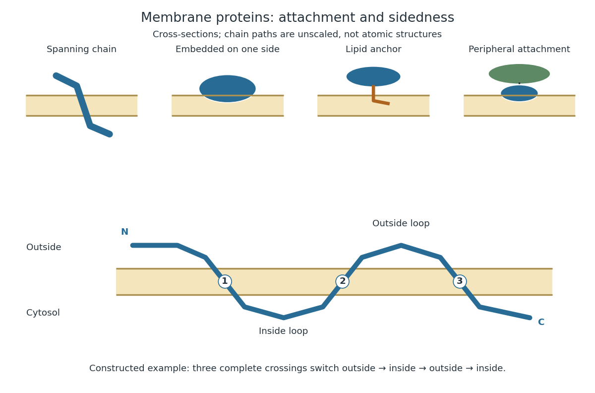
Membrane association and the orientation of a spanning chain
. Original schematic cross-sections of a lipid bilayer, followed by a constructed three-crossing polypeptide chain. Colored paths represent placement, not atomic protein structures. The N-terminus starts outside; each complete crossing switches sides, leaving the C-terminus inside. Drawn dimensions are unscaled. Details.
The same boundary can therefore retain substances and communicate selectively with its surroundings. Membrane topology tells us which parts can meet a given ligand; lateral mobility of membrane proteins tells us whether two parts can approach each other along the boundary. A diagram of a cell membrane becomes more useful when it answers both questions.
A light-driven proton pump with a seven-helix membrane-spanning structure and a retinal chromophore. Its mechanical unfolding involves membrane anchoring as well as protein and lipid interactions.
An iron-export protein. IRE-containing transcripts can be regulated at biological translation, while protein-level and systemic regulation provide additional controls.
A receptor supporting uptake of transferrin-bound iron. RNA regulation involving its 3′ region can increase transcript stability during iron shortage.
An experimental environment designed to shield hydrophobic protein surfaces while retaining relevant membrane properties. Detergent micelles, bicelles and lipid bilayer nanodiscs have different limitations.
A cargo receptor links a transported component to sorting machinery. An ER export receptor can bind a soluble lumenal cargo while exposing a cytoplasmic recognition site to the coat.
An integrin is a transmembrane adhesion receptor linking extracellular ligands to intracellular signalling and cytoskeleton interactions. Integrin-dependent contacts transmit force in many forms of cell migration; their necessity depends on the physical environment.
hydrophobic mismatch is an energetic incompatibility between a membrane protein's hydrophobic span and the hydrophobic thickness of its surrounding lipid bilayer. Local deformation, altered conformation or changed association can partly accommodate it.
An integral membrane protein is firmly incorporated into a membrane, commonly through contact with the hydrophobic interior of the lipid bilayer. Extraction behavior is an operational distinction: it resists treatments that remove many peripheral membrane proteins. Transmembrane proteins are one class; integral does not mean spanning.[ref]
Membrane Proteins. Molecular Biology of the Cell, 4th edition by Bruce Alberts and others; Membrane Proteins Can Be Associated with the Lipid Bilayer in Various Ways, Figure 10-17 and extraction-based distinction
A transmembrane protein has at least one polypeptide chain crossing the complete lipid bilayer. One crossing is single-pass; several crossings in one chain are multipass. Hydrophobic regions face the membrane interior, while hydrophilic regions can face water.[ref]
Cell Membranes. The Cell: A Molecular Approach, 2nd edition by Geoffrey M. Cooper; Cell Membranes, Membrane Proteins: spanning regions, single and multiple crossings
A tail-anchored protein has a membrane-spanning segment near its C-terminus; many reach the endoplasmic reticulum after biological translation.
A peripheral membrane protein associates with a membrane face through noncovalent interactions, without spanning its hydrophobic interior. Association with another membrane protein can suffice; peripheral describes attachment, not an unimportant function.[ref]
Membrane Proteins. Molecular Biology of the Cell, 4th edition by Bruce Alberts and others; Membrane Proteins Can Be Associated with the Lipid Bilayer in Various Ways: noncovalent attachment and peripheral proteins
A lipid-anchored protein attaches through a covalently linked lipid. The anchor inserts into the lipid bilayer; the polypeptide chain need not cross it. This is a description of attachment rather than a synonym for transmembrane protein.[ref]
Cell Membranes. The Cell: A Molecular Approach, 2nd edition by Geoffrey M. Cooper; Cell Membranes, Membrane Proteins: covalently attached lipid anchors on either membrane face
Membrane topology specifies the sidedness of a membrane-associated polypeptide chain: where its N-terminus, C-terminus and intervening protein domains lie relative to the lipid bilayer. Side-specific placement is essential to interpreting a cell-surface receptor.[ref]
Membrane Proteins. Molecular Biology of the Cell, 4th edition by Bruce Alberts and others; Membrane Proteins Can Be Associated with the Lipid Bilayer in Various Ways: aqueous domains on either side and receptor orientation
For an original worked example, stipulate a continuous polypeptide chain with its N-terminus outside a biological cell, exactly three complete crossings of the cell membrane, and no additional excursions into the lipid bilayer. Its segments lie, in order, outside, inside, outside, inside. A protein domain between the first and second crossings faces the cytosol; one between the second and third faces the exterior; the C-terminus faces the cytosol. Counting crossings determines these placements without specifying an atomic protein structure.
The proof is a side-switching argument. Assign exterior the sign and interior . Each complete crossing multiplies the sign by , so after crossings,An odd number puts the termini on opposite sides, an even number on the same side. This assumes complete crossings of one boundary; a chain that enters and returns to the same side without crossing must not be counted as a complete crossing. Nor does the rule determine which side the N-terminus starts on.
As a constructed accessibility test, put an epitope on the outside segment between crossings two and three, and another on the inside segment between crossings one and two. Stipulate an intact cell membrane that excludes the test antibody and accessible epitopes that bind it. Applying the antibody outside can label only the first epitope. If both become accessible after opening the boundary, the difference tests sidedness under those assumptions. Failure to label an epitope alone is insufficient: it could also be inaccessible because of protein folding or poor antibody affinity. This is a proposed reasoning exercise, separate from the historical mixing experiment below.
Lateral mobility of membrane proteins is movement along a membrane surface. It differs from membrane transport through the boundary and from reversing membrane topology. Attachment, crowding and barriers can restrict diffusion; the fluid mosaic model does not require every membrane protein to wander freely.[ref]
Structure of the Plasma Membrane. Cooper, The Cell: A Molecular Approach, 2nd edition, Structure of the Plasma Membrane, Mobility of Membrane Proteins: lateral movement and restrictions
An original local model treats a small, approximately flat patch as an unbounded plane with uniform diffusion coefficient , no drift and no trapping. Represent the two displacement coordinates by independent Brownian motions, each with zero expected value and variance . Since , addition of the two coordinate expectations givesThis is a root-mean-square displacement, not distance traveled along a jagged trajectory or the displacement of every protein. For a stipulated and , and . Reaching a stipulated root-mean-square scale gives . These model values are not measured diffusion coefficients or mixing times from the historical experiment.
Multiplying the target displacement scale by two multiplies the model time by four. This quadratic scaling is a useful way to see why local encounters and redistribution over an entire biological cell need not happen on the same timescale. A curved, bounded cell membrane, restricted paths and different mobile populations require more than this flat-patch model; disappearance of an initial pattern alone does not determine .
In 1970 Larry Frye and Michael Edidin fused mouse C11D and human VA-2 cell lines with Sendai virus, forming heterokaryons. Suspensions were maintained in a shaking water bath at . Removed aliquots were stained by indirect immunofluorescence: antibodies against parental surface antigens, then differently fluorescent secondary antibodies. A Leitz Ortholux fluorescence microscope used a mercury lamp and color-selecting filters. Green mouse and red human surface patterns became intermixed.[ref]
The rapid intermixing of cell surface antigens after formation of mouse-human heterokaryons by Larry Frye and Michael Edidin. Frye and Edidin, The rapid intermixing of cell surface antigens after formation of mouse-human heterokaryons, pp. 319–325: Summary, Cell fusion, Fluorescent staining, Fluorescence microscopy, Table 1, time-course results
Same-species fusions tested antibody specificity; stable hybrid lines supplied a two-color control. Inhibitors of biological translation did not prevent mixing; lowering temperature inhibited it. These observations supported lateral redistribution over rapid synthesis. The authors could not conclusively exclude passage through cytoplasm and return. The readout compared populations, not trajectories of a continuously observed individual biological cell, and did not establish unrestricted motion of every membrane protein.[ref]
The rapid intermixing of cell surface antigens after formation of mouse-human heterokaryons by Larry Frye and Michael Edidin. pp. 324–331: specificity controls, inhibitor studies, temperature dependence and Discussion of mechanisms iii and iv
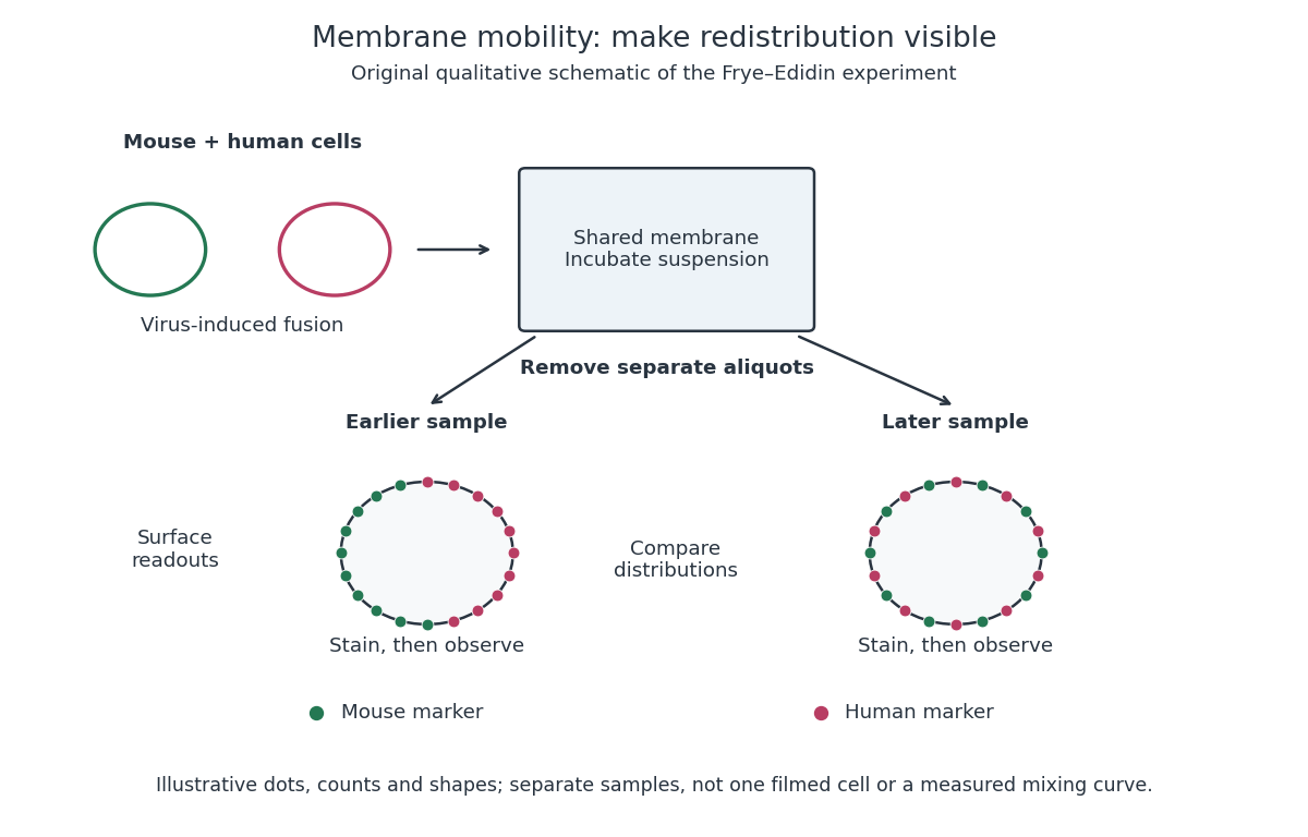
Separate time-point samples reveal changing surface distributions
. Original qualitative schematic of the mouse-human heterokaryon membrane-mixing experiment. Dot counts and shapes are illustrative; separate samples show distributions rather than individual trajectories or measured mixing curves. Details.
The decisive design idea is to begin with spatially distinguishable material. A uniformly labeled surface can look unchanged while its components move. Bringing together differently identifiable parental surfaces creates an initial pattern whose loss reveals redistribution; antibody specificity makes that loss interpretable. Membrane topology asks which side a part faces, whereas this experiment asks where it can move along that side.
A nucleotide-sugar transporter supplies an activated donor across a membrane to a compartment containing glycosyltransferases. Whole-cell donor abundance does not establish donor availability at a lumen-facing enzyme.
A scramblase facilitates relatively nonselective bidirectional movement of lipids between membrane leaflets, tending to dissipate asymmetry when active. Its action differs from directional transport used to maintain asymmetry.
A flippase catalyzes directional movement of selected lipids between membrane leaflets. Such activity can establish or maintain lipid asymmetry, subject to specificity and energy coupling.
An ATP-binding cassette transporter couples nucleotide-binding-domain activity to transmembrane transport. For an exporter, productive substrate movement must be measured separately from substrate binding and ATP hydrolysis.
AcrB is a bacterial proton-coupled multidrug transporter that functions as part of the AcrAB–TolC export assembly. Its asymmetric trimer and multiple drug-binding regions are structural models for studying broad specificity.[ref]
Crystal structures of a multidrug transporter reveal a functionally rotating mechanism by Satoshi Murakami and others. Figs. 1–5, drug-bound asymmetric AcrB trimer
A multidrug efflux pump exports multiple chemical substrates using a coupled source of free energy. Broad recognition can arise from overlapping binding sites and conformational flexibility, but binding alone does not establish productive transport.
A membrane carrier protein binds a solute and changes its exposed binding site from one side of a cell membrane to the other. Depending on its mechanism and coupling, it can mediate passive transport or active transport.
Articles by others on the same topic
There are currently no matching articles.