Cell counting estimates the number of biological cells in a preparation. A count per calibrated volume gives number density; multiplying by the whole preparation's volume estimates its total count. A trypan blue exclusion test adds an operational distinction between dye-excluding and dye-stained biological cells.
An original constructed hemocytometer count uses four such squares after mixing the biological cell preparation with the same volume of stain. The dye-excluding counts are ; the dye-stained counts are . The dilution factor is . With the sampled volume per square and the arithmetic mean count per square,Hence the undiluted preparation has dye-excluding number density and total number density . Its dye-excluding fraction is . If the well-mixed preparation contains , it contains an estimated dye-excluding biological cells. The chamber sample itself contains only counted dye-excluding biological cells.
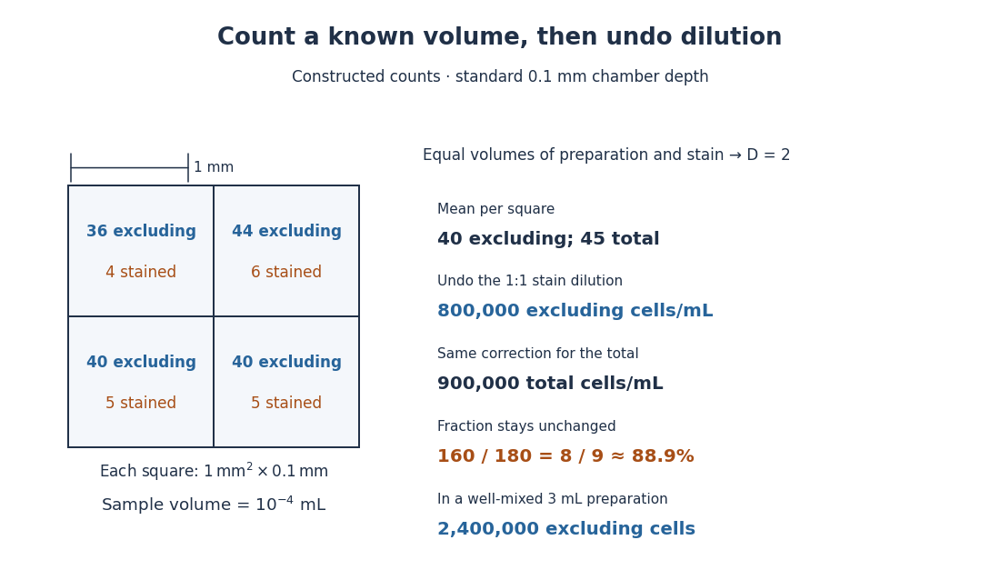

The relation follows from counting material in a known volume, then undoing dilution. It assumes a representative mixed sample and correctly identified single biological cells. Clumps, inconsistent decisions at grid edges, wrong chamber depth and nonrepresentative sampling introduce measurement error. A fixed convention such as including top and left edges while excluding bottom and right edges assigns shared-edge centers to just one square. Adding ten times as much suspension to a correctly filled chamber does not make each square's calibrated volume ten times larger.
The trypan blue exclusion test reads cell membrane integrity through trypan blue exclusion. Prolonged dye exposure can itself increase staining.[ref]
An unstained biological cell is dye-excluding at the time of this assay. That observation alone does not demonstrate future division, sustained metabolism or long-term renewal of a cell line. Conversely, a smaller unstained count can reflect both staining and loss of biological cells from the recovered preparation. Report the denominator and recovery procedure alongside the fraction.
Articles by others on the same topic
There are currently no matching articles.