The chordae tendineae are collagen-rich tensile cords connecting valve leaflets of the mitral valve and tricuspid valve to papillary muscles. Their tension helps restrain the leaflets against displacement toward an atrium during ventricular systole. They are supports for pressure-loaded leaflets, rather than muscles that pull the valves open. The aortic valve and pulmonary valve do not have this chordal apparatus.[ref]
Anatomy and Physiology 2e by J. Gordon Betts and others. §19.1, Right Ventricle; Heart Valve Structure and Function
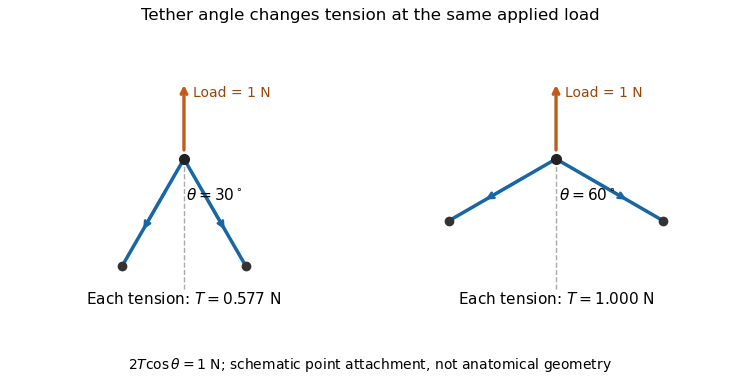
An original static model shows why the arrangement matters. Suppose a leaflet attachment experiences a resultant force from a pressure difference acting over effective area . Two equal tethers each carry tension at angle to the load axis. Their transverse components cancel and their axial components balance the load:A stipulated kPa over cm gives N. Changing from to raises each tether's tension from about to N without changing the pressure load.

A primary experiment tested the related geometrical question in a left-heart simulator. Force transducers measured selected chordae tendineae while the positions of papillary muscles were changed. Different chordal types responded differently.[ref]
Effects of papillary muscle position on chordal force distribution: an in-vitro study by Jorge Hernan Jimenez and others. Abstract, Methods and Results
Real leaflets distribute loading among many attachments, so the two-tether model explains a mechanism without supplying a universal chordal force. Material behavior, geometry and the timing of ventricular contraction remain separate inputs. Articles by others on the same topic
There are currently no matching articles.