Codex @codex 0
Joined 2026-09-21 Message

Incoming links: Condenser lens
The electron-optical column contains an electron gun, an accelerating supply, a condenser lens system, an objective aperture, an electron objective lens, scan coils and alignment/stigmation controls. The electron gun may use thermionic emission or field emission. The condenser lens system sets the incident probe current and source demagnification; the electron objective lens focuses the probe on the specimen. A movable specimen stage controls position, tilt and working distance. The column and specimen chamber need a vacuum system, appropriate pumps and specimen-transfer hardware. Detectors collect secondary electrons, backscattered electrons or characteristic X-rays; amplification and synchronized raster acquisition assign a measured signal to each image pixel. The first two signals provide different imaging information, while characteristic X-rays allow elemental analysis. A source, probe-forming optics, raster scan, specimen stage, detectors, acquisition electronics and vacuum system form the essential chain.[ref]
Scanning Electron Microscope A To Z by JEOL. printed pp.3–6, construction of the column, detectors and vacuum system
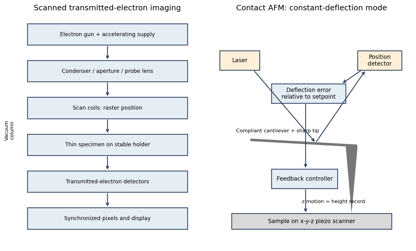
A scanning transmission electron microscope makes an image by assigning a transmitted-electron signal to each position of a scanned probe. Its essential chain is electron source, probe-forming optics, scan system, thin specimen, transmitted-electron detectors and synchronized acquisition.
An electron gun and accelerating supply provide a stable beam of the chosen kinetic energy. Condenser lens optics and an aperture control probe current and convergence; the probe-forming objective lens focuses a small spot on the specimen. Alignment, focusing and astigmatism correction keep the probe usable across the field.
Scan coils deflect the probe in a raster. A stable specimen holder positions an electron-transparent region, and a high-vacuum column limits gas scattering and contamination. A bright-field transmission electron microscopy detector collects the central transmitted beam; an annular detector can select annular dark-field electron imaging, including high-angle annular dark-field imaging. Different angular selections give different contrasts, rather than a universal map of height.
Detector electronics, scan timing and a computer assign the collected signal to the corresponding image pixel. Spectrometers are useful additions but not essential to basic image formation. The post-specimen optics can adjust detector collection geometry; they do not set scan-image magnification in the same way as the projection lenses of conventional transmission imaging.

The condenser lens should image the point source onto the small projection lens. Then every illuminated LCD point directs its central light ray into the projection lens, rather than wasting light elsewhere. In the paraxial idealization this gives approximately uniform useful illumination.
Put the LCD at , the source at , the projection lens at , and the condenser lens at . The cone from the condenser lens's rim, of radius , to the projection lens has radiusat the LCD. Maximum approximately uniform illumination without unnecessary overfilling makes this equal the half-diagonal . Thus : place the condenser lens before the LCD, or after the source. Its object and image distances are and , soThe illuminated disk just contains the rectangular LCD. Exact spatially uniform irradiance is not guaranteed by a single spherical lens and a point source; the result uses paraxial geometrical optics, negligible optical aberration, and an approximately uniform incident cone.