
Chordae tendineae 2026-11-05
The chordae tendineae are collagen-rich tensile cords connecting valve leaflets of the mitral valve and tricuspid valve to papillary muscles. Their tension helps restrain the leaflets against displacement toward an atrium during ventricular systole. They are supports for pressure-loaded leaflets, rather than muscles that pull the valves open. The aortic valve and pulmonary valve do not have this chordal apparatus.[ref]
Anatomy and Physiology 2e by J. Gordon Betts and others. §19.1, Right Ventricle; Heart Valve Structure and Function
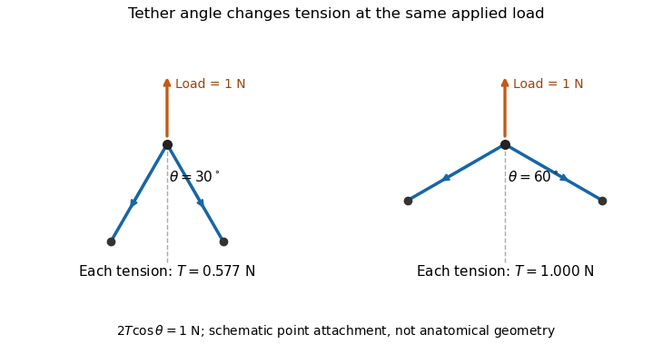
An original static model shows why the arrangement matters. Suppose a leaflet attachment experiences a resultant force from a pressure difference acting over effective area . Two equal tethers each carry tension at angle to the load axis. Their transverse components cancel and their axial components balance the load:A stipulated kPa over cm gives N. Changing from to raises each tether's tension from about to N without changing the pressure load.

A primary experiment tested the related geometrical question in a left-heart simulator. Force transducers measured selected chordae tendineae while the positions of papillary muscles were changed. Different chordal types responded differently.[ref]
Effects of papillary muscle position on chordal force distribution: an in-vitro study by Jorge Hernan Jimenez and others. Abstract, Methods and Results
Real leaflets distribute loading among many attachments, so the two-tether model explains a mechanism without supplying a universal chordal force. Material behavior, geometry and the timing of ventricular contraction remain separate inputs. Jorge Hernan Jimenez 2026-11-05
Jorge Hernan Jimenez is a biomedical-engineering researcher whose primary publications investigate the mechanics of the mitral valve and its supporting chordae tendineae. His 2005 first-author study lists the Wallace H. Coulter Department of Biomedical Engineering at Georgia Tech and Emory as the affiliation for that work.[ref]
Effects of papillary muscle position on chordal force distribution: an in-vitro study by Jorge Hernan Jimenez and others. Primary byline and affiliation
The experiment connected valve geometry to measured forces. Jimenez and colleagues placed six human and five porcine mitral valves in a physiological left-heart simulator, recorded cardiac output and transmitral pressure, and fitted force transducers to six chordae. They tested eight papillary-muscle positions constructed from vector displacements from the normal position. The tension response depended on chordal type: marginal chords were comparatively insensitive, while other classes showed larger changes.[ref]
The scientific point is that moving a support does not change every tether's load in the same way. In an idealized mechanical representation, a chordal force is , with tension and direction . Moving its attachment can change both. A change in one measured tension therefore does not, by itself, determine the complete resultant load or leaflet motion; several forces and the valve geometry must be considered together.
The simulator allowed a controlled geometric perturbation while observing forces during the cardiac cycle. It supports an in-vitro mechanical relationship under the stated conditions, rather than a universal prediction of clinical outcome from one displacement. The verified full byline is retained without attaching an unverified career history or namesake portrait.
Papillary muscle 2026-11-05
A papillary muscle is a muscular projection inside a ventricle that anchors chordae tendineae attached to the mitral valve or tricuspid valve. During ventricular systole, its contraction helps maintain the supporting geometry and tension that restrain the valve leaflets. Valve opening is driven by the appropriate pressure difference; a papillary muscle does not act as a cord-operated opener.[ref]
Anatomy and Physiology 2e by J. Gordon Betts and others. §19.1, Right Ventricle and Heart Valve Structure and Function
The anchor's position matters as well as its ability to contract. In a primary simulator study, investigators examined six human and five porcine specimens of the mitral valve, measuring six selected chordae tendineae with force transducers. Eight tested anchor configurations used 5 mm displacement components relative to a reference position. The resulting tension changes depended on which chord was measured.[ref] Moving an anchor therefore need not distribute extra load equally through the apparatus.
An original mechanical approximation illustrates the distinction. If a taut cord has reference length , cross-sectional area and effective Young's modulus , a small extra extension givesThis is a local Hooke's law approximation, not a claim that biological cords remain linear at every strain. Anchor displacement changes the cord's extension and direction, while active muscle contraction changes the moving support itself. In the equal-tether example under chordae tendineae, changing direction also changes the tension needed to balance a fixed leaflet load. The apparatus is consequently a coupled geometrical and muscular system; naming its parts alone does not predict its forces.
Valve leaflet 2026-11-05
A valve leaflet is a mobile tissue flap forming part of a heart valve. Its movement and contact with neighboring leaflets help permit forward blood flow while limiting backward flow. The supporting cardiac valve annulus, the pressure difference and the leaflet's geometry all contribute to its behavior. The meeting of leaflet surfaces that closes a valve is called coaptation.[ref]
The mitral valve and tricuspid valve have leaflets supported by chordae tendineae and papillary muscles. The aortic valve and pulmonary valve instead have pocket-like cusps without that chordal apparatus. A leaflet is therefore a functional part of a particular valve structure, not an isolated rigid door.
An original force estimate illustrates the loading scale. A uniform pressure difference over an effective area gives a resultant . For stipulated kPa and cm, N. Doubling the area at the same pressure doubles this resultant, but it does not specify how the load is divided among attachments or distributed as tissue stress.
A primary left-heart simulator experiment measured chordae tendineae forces while moving papillary muscles and found responses that depended on the chordal type.[ref]
Effects of papillary muscle position on chordal force distribution: an in-vitro study by Jorge Hernan Jimenez and others. Abstract, Methods and Results
This gives a concrete reason to examine leaflet geometry and supporting connections together. Complete coaptation is a contact-and-motion condition; an accurate measurement of one chordal force does not by itself show that the entire valve seals.