
Golgi tracer interpretation 2026-10-18
The pancreatic secretory-protein pulse-chase experiment of James D. Jamieson and George E. Palade used guinea pig pancreas slices and a short pulse of tritium-labeled leucine followed by a chase. Thin sections on coated grids received Ilford L-4 emulsion; developed grains were read against structures by electron microscopy. Their Table II localized label to peripheral Golgi apparatus vesicles and condensing vacuoles, without significant labeling of the piled flattened Golgi cisternae.[ref]
Intracellular Transport of Secretory Proteins in the Pancreatic Exocrine Cell. II. Transport to Condensing Vacuoles and Zymogen Granules by James D. Jamieson and George E. Palade. p. 598 Methods; p. 609 Table II and its footnote
An important comparison checked the isolated granule fraction by autoradiography and electron microscopy: early labeling came substantially from contaminating condensing vacuoles, rather than mature secretory granules. Thus a fraction's name did not uniquely identify the labeled structure. This supported transport into the Golgi apparatus's condensing region and onward storage, while fixed samples and finite grain localization limited interpretation. It did not establish a resolved cis-to-trans itinerary across every Golgi cisterna. The preparation's labeling also did not mark secretory cargo exclusively.[ref]
Intracellular Transport of Secretory Proteins in the Pancreatic Exocrine Cell. II. Transport to Condensing Vacuoles and Zymogen Granules by James D. Jamieson and George E. Palade. p. 609 Figure 14; p. 613, Transport of Labeled Proteins to Condensing Vacuoles, Zymogen Granules, and the Acinar Lumen: comparison of fraction labeling with microscopy; p. 611, General Premises
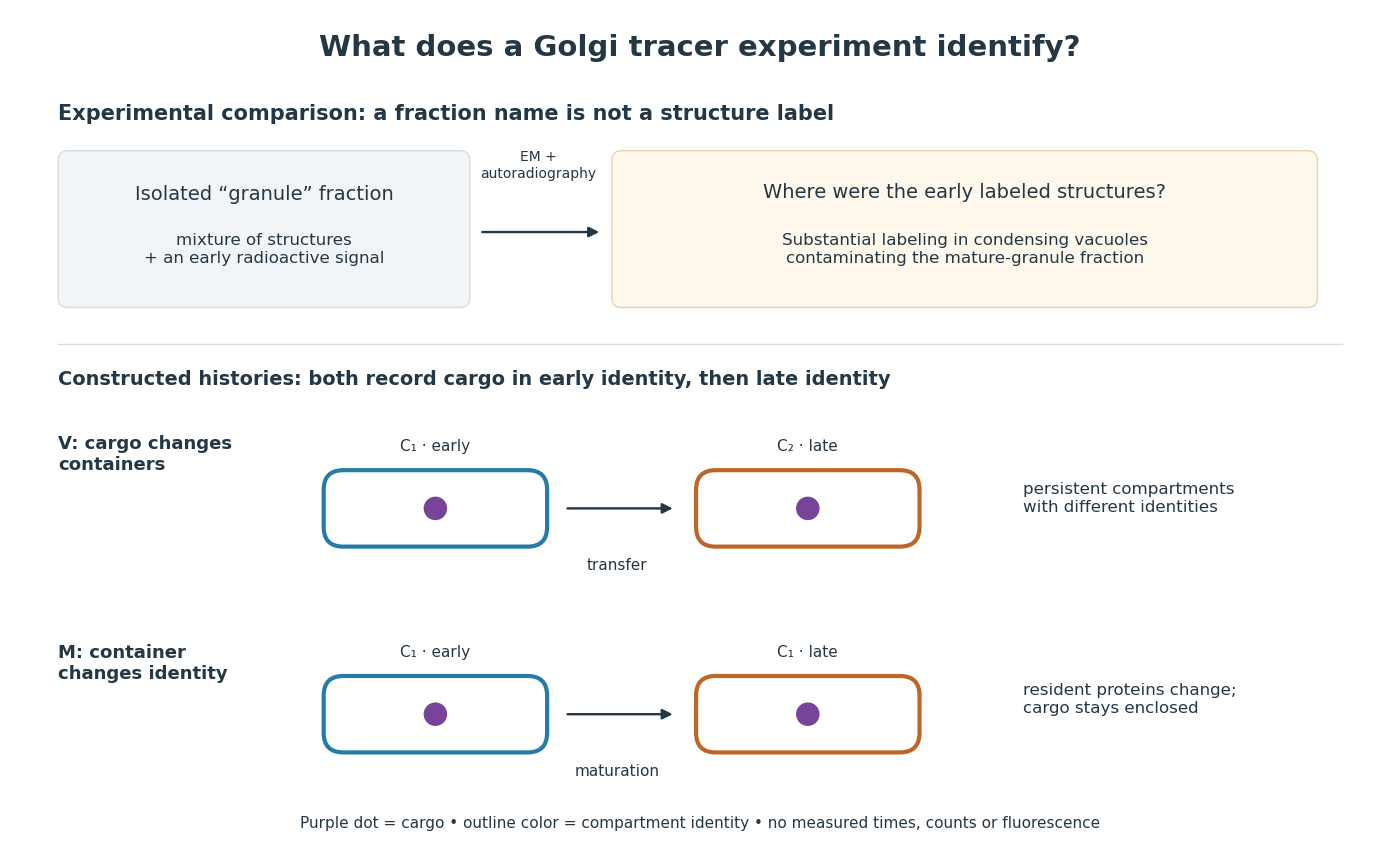

For a minimal worked inference, record only two outcomes for one labeled cargo cohort: early identity first, late identity later. Model V transfers cargo from container to container ; model M keeps it in while changes identity. Both produce the same recorded outcomes, so those outcomes alone cannot distinguish V from M. The proof is constructive: exhibit both histories and discard container identity from each record. More informative tracking would retain both cargo and resident-compartment markers, while resolving whether the same compartment persists. This is why pulse-chase analysis and compartment tracking answer complementary questions about the Golgi apparatus.
In 1967, James D. Jamieson and George E. Palade incubated guinea pig pancreas slices, following a three-minute pulse of tritium-labeled leucine with excess unlabeled leucine. This pulse-chase analysis marked newly made proteins while reducing subsequent incorporation. Fixed, Epon-embedded thin sections were mounted on coated grids, covered with Ilford L-4 photographic emulsion, developed and stained for electron microscopy. Autoradiography located photographic grains relative to pancreatic acinar cell structures.[ref]
Intracellular Transport of Secretory Proteins in the Pancreatic Exocrine Cell. II. Transport to Condensing Vacuoles and Zymogen Granules by James D. Jamieson and George E. Palade. Jamieson and Palade, Intracellular Transport of Secretory Proteins in the Pancreatic Exocrine Cell. II. Transport to Condensing Vacuoles and Zymogen Granules (1967), p. 598 Methods; Figure 10: three-minute pulse
Pulse-end samples provided a temporal reference: label lay mainly over rough endoplasmic reticulum; later samples shifted toward peripheral Golgi apparatus elements, condensing vacuoles and secretory granules. As a contamination check, isolated granule pellets were also examined by autoradiography: early pellet labeling chiefly marked condensing vacuoles. Grids from the compared pellet samples were coated together to control photographic processing. This supported transport of a previously labeled cohort rather than separate synthesis at each location. Fixed samples did not track individual proteins; localization was finite, and labeling was not absolutely exclusive to secretory cargo. The result concerns this preparation, not every protein in every biological cell.[ref]
Intracellular Transport of Secretory Proteins in the Pancreatic Exocrine Cell. II. Transport to Condensing Vacuoles and Zymogen Granules by James D. Jamieson and George E. Palade. pp. 603–606 and Figures 10–13: changing localization; pp. 610–611 and Figures 14–15, Table III: pellet check and matched emulsion processing; p. 611 General Premises: nonexportable-protein labeling