View the backbone along N–C to display in a Newman projection. The front amide nitrogen carries the preceding carbonyl and H, while the back C carries the next carbonyl, H and side chain. Rotation changes the contacts among these groups. A corresponding view along C–C displays . The peptide C–N bond itself is approximately planar and is not the freely rotating bond in either definition.
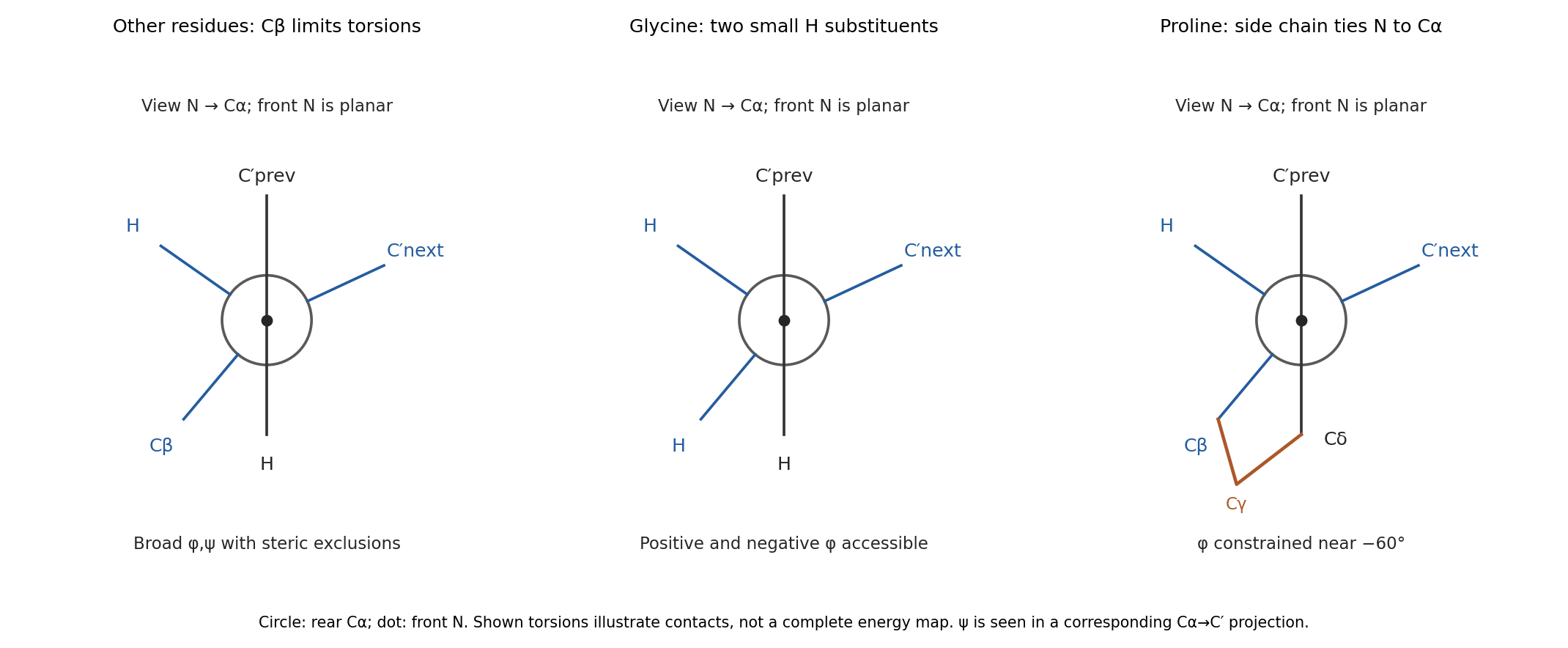

glycine replaces the side chain by a second hydrogen. Removing C removes many contacts that exclude backbone arrangements for other residues, so its allowed region is much broader and includes substantial positive- space. Its alpha carbon is also achiral, so the steric map is approximately mirror-symmetric between positive and negative torsions in the simple isolated-residue model.
proline closes a five-membered ring from C through three methylenes back to nitrogen. In the N–C projection, the nitrogen's side-chain arm and C's C arm are tied together, preventing unrestricted relative rotation. Its is consequently confined near the usual negative- region rather than exploring the broad range of an acyclic amino acid. proline also lacks a backbone N–H donor when incorporated into an ordinary internal peptide. Ring closure restricts strongly, but does not freeze every backbone or ring degree of freedom. These differences are naturally represented by distinct Ramachandran plots; the original torsion/steric analysis established the basis for such comparisons.[ref]
Stereochemistry of polypeptide chain configurations by G. N. Ramachandran and others. 1963, pp. 95–99, backbone torsions and steric restrictions
Articles by others on the same topic
There are currently no matching articles.