An enolate is an ambident nucleophile: the negative charge is concentrated toward oxygen, while its occupied conjugated orbital also has substantial reactive amplitude at the alpha carbon. An alkyl iodide presents an accessible, polarizable carbon electrophile. carbon attack through an SN2 reaction is generally kinetically favorable, while the more strongly charged oxygen is often coordinated to the counterion or solvated. The carbonyl reforms as the new C–C bond is made.carbon alkylation restores the carbonyl and makes the new C–C bond. oxygen alkylation instead gives an enol ether. The two outcomes are not resonance contributors of one product, and their ratio is not determined just by where the largest formal negative charge is drawn. electrophile, counterion, solvent and aggregation can change selectivity; hard silicon electrophiles and suitable acylating reagents often favor oxygen trapping. Product stability alone would not explain an irreversible kinetic choice.
Lithium diisopropylamide removes an alpha proton from the ethyl side of the ketone. The C–H electrons form the C=C part of a lithium enolate, while the C=O pi electrons move to oxygen. The phenyl side has no competing alpha C–H site. Diisopropylamine is the conjugate-acid byproduct.The carbon end of this enolate attacks the primary carbon bearing iodine in . The C–I bond breaks to iodide in an SN2 reaction, and carbonyl reformation givesThus a three-carbon methoxypropyl chain is attached at the ketone alpha carbon. The remote methyl ether survives. The hindered amide base is chosen to generate the enolate rather than to add to the carbonyl. No absolute stereochemical preference is specified for the new stereocenter under these achiral conditions.
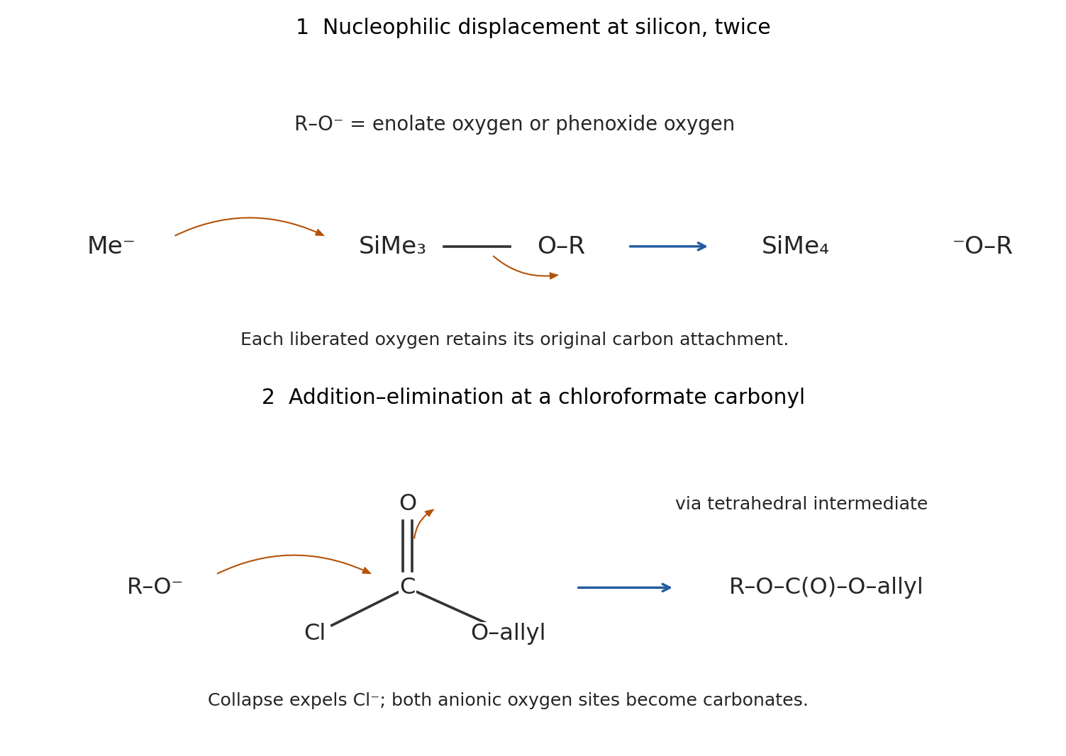
The initial nucleophile attacks silicon, not the nitrile or an alkene carbon. Each trimethylsilyl group has an electrophilic Si center. A methyl group from methyllithium attacks Si, passing through a pentacoordinate silicon arrangement; Si–O cleavage releases and the lithium salt of the oxygen-containing fragment. Cleaving the silyl enol ether generates a lithium enolate; cleaving the aryl silyl ether generates a phenoxide. Neither operation needs a new deprotonation at a carbon stereocenter.Here represents either the phenoxide or the oxygen-centered enolate contributor. Excess reagent permits both cleavages. The pre-existing methyl/nitrile stereocenter retains its configuration: it has no hydrogen to remove, and neither bond attached to that center is broken.
After HMPA is added, each anionic oxygen attacks the carbonyl carbon of allyl chloroformate. The C=O pi electrons move to the carbonyl oxygen, giving a tetrahedral intermediate; collapse expels chloride. The enolate becomes an allyl enol carbonate, and the phenoxide becomes an aryl allyl carbonate, each containing . oxygen acylation accounts for preservation of the enol double bond and for the two newly installed carbonate groups. carbon attack would give a different carbon skeleton. The two oxygens are trapped because they were selectively unmasked by desilylation, rather than because every ether oxygen in the molecule is equally reactive.
HMPA coordinates Li through its phosphoryl oxygen and makes the anionic oxygen sites more available for acylation. Strong cation solvation changes contact ion pairing, chelation and aggregate structure. It can therefore accelerate trapping of both the enolate and phenoxide by the chloroformate, helping realize the observed O-acylation pathway.
It is too strong to assert that HMPA necessarily produces completely free, monomeric anions. In a primary study of a lithium enolate in THF, low-temperature multinuclear NMR and reaction kinetics identified an HMPA-coordinated dimer as the reactive form, even with excess HMPA.[ref]
Effects of HMPA on the Structure and Reactivity of the Lithium Enolate of Cyclopentanone in THF: The Dimer is Responsible for Alkylation and Proton Exchange Reactions by Masaaki Suzuki and others. 2004, pp. 259–268, HMPA titration, aggregate assignment and alkylation kinetics
The useful conclusion here is altered lithium coordination and enhanced oxygen accessibility; the exact aggregate of this bifunctional substrate is not supplied.The carbamate carbonyl oxygen attacks electrophilic sulfur of triflic anhydride, with cleavage of the anhydride S–O bond to release triflate. Donation of the nitrogen lone pair toward the carbonyl carbon then gives the indicated imidoyl-type cation: the former carbonyl oxygen now bears Tf, and the N–H nitrogen participates in a C=N bond. This converts a relatively unreactive carbamate carbonyl into a strongly electrophilic carbon bearing an excellent leaving group.
The aromatic pi bond para to ethoxy attacks that carbon. The C=N pi electrons move to nitrogen as the new aryl–carbon bond forms. Loss of the aromatic proton restores aromaticity; nitrogen donation and departure of the O-triflylated group restore the imine bond. proton transfers during the sequence and carbonate workup remove the remaining N–H proton. The surviving five-membered ring is an oxazoline: the product is 2-(4-ethoxyphenyl)-2-Oxazoline. An equivalent arrow order can eliminate the triflyloxy leaving group before arene capture; these are limiting descriptions of the activated electrophile, not a need to postulate an unactivated carbamate undergoing ordinary Friedel–Crafts acylation.
Ethoxy donates a lone pair into the ring, stabilizing the ortho and para Wheland intermediates. The para pathway avoids the crowding of an incoming heterocycle beside ethoxy, giving the observed position. Mechanistic experiments on this transformation include in situ carbon NMR of the activated reagent, competition experiments and isotope-effect measurements supporting electrophilic aromatic substitution; they do not directly image every intermediate.[ref]
Simple and Efficient Aromatic C–H Oxazolination by Qiu Shi and others. 2023, Figure 7 and supporting information, product 3a and electrophilic-substitution evidence
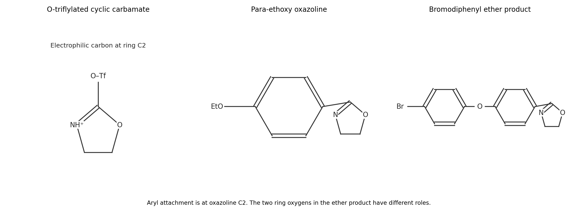
Oxazolination occurs para to oxygen on the ring that does not bear bromine. The product is 2-[4-(4-bromophenoxy)phenyl]-2-Oxazoline, shown in the final panel above. The brominated ring retains its para bromine; the other ring receives the new oxazolinyl group at its open para position.
The diaryl ether oxygen activates both rings toward electrophilic aromatic substitution. On the brominated ring, its least crowded para position is blocked, and bromine also decreases ring reactivity inductively. The unbrominated ring has an accessible para position, favored over its ortho sites by steric effects. This is substitution of an aromatic hydrogen, not displacement of the aryl bromide.
The formula provides a useful consistency check: loses one aromatic H and acquires , giving . It cannot by itself distinguish positional isomers; the directing and steric arguments select the connectivity.
In the strongly acidic nitrating mixture, the dimethylamino nitrogen is predominantly protonated. The substituent is therefore during aromatic substitution, rather than the neutral electron-donating group. Its lone pair is unavailable for conjugation. Ortho or para attack would generate a sigma-complex contributor with positive ring charge at the carbon attached to an already positive nitrogen; meta attack avoids that especially unfavorable contributor.
Sulfuric acid protonates nitric acid, and loss of water produces the nitronium ion. Aromatic attack at a meta position forms a Wheland intermediate; deprotonation restores aromaticity. The bicarbonate workup then returns the amino group to its neutral form. is 3-nitro-N,N-dimethylaniline.
There are six inequivalent aromatic carbons in this unsymmetrical meta-disubstituted ring and one carbon environment for the two equivalent N-methyl groups, giving seven carbon NMR signals. The para isomer would have only four aromatic environments plus the methyl environment. An ortho isomer could also give seven signals, so signal counting supports but does not alone establish the meta assignment; protonation-dependent directing effects are essential.
tert-Butoxide removes the acidic hydrogen from . The resulting carbon anion is stabilized by the nitrile group and the phenyl substituent. It attacks the aromatic carbon bearing fluorine, giving a Meisenheimer addition intermediate. Ring pi bonds shift toward the para nitro group, where the negative charge can be delocalized onto oxygen.
Loss of fluoride restores aromaticity and gives . This is an addition–elimination nucleophilic aromatic substitution. The phenoxy group remains attached to the incoming carbon. The ipso aromatic carbon already has a leaving group, so rearomatization can occur by fluoride departure without removing phenoxide. Acid workup neutralizes residual anions; it is not a separate step that replaces the aryl fluoride.
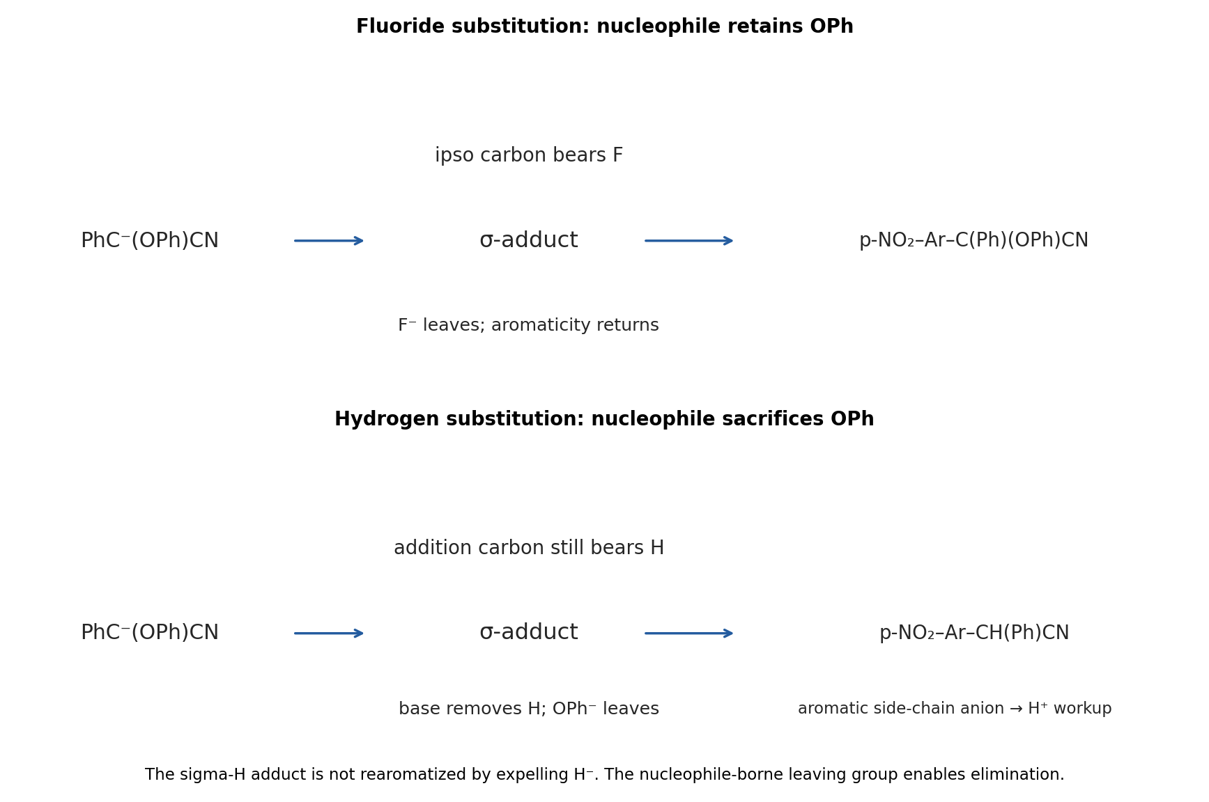
The same base first makes . This time addition occurs at a carbon bearing hydrogen para to the nitro group, giving a sigma-H adduct. The attacked carbon temporarily bears both its original H and the new carbon substituent. Negative charge is again delocalized through the ring toward the nitro oxygens.
A further base removes the hydrogen on that aromatic addition site. Its C–H electrons form an exocyclic bond toward the incoming carbon, while that carbon expels phenoxide. This is a base-induced elimination across the new ring–side-chain bond. Redistribution of the nitronate/quinoid pi system restores an aromatic ring and gives the side-chain carbanion . protonation during acidic workup yields .
This is vicarious nucleophilic substitution: the ring loses hydrogen while the nucleophile sacrifices its phenoxy leaving group. hydrogen is not expelled as hydride, and the product's side-chain hydrogen can be supplied on workup rather than moving directly from the ring in a compulsory hydride shift. Base and isotope experiments in the primary mechanistic work support reversible addition followed by base-induced elimination, while not uniquely choosing every E2/E1cb limit.[ref]
Reaction of organic anions. Part 108. On the mechanism of the vicarious nucleophilic substitution of hydrogen in nitroarenes by Mieczysław Mąkosza and Tomasz Glinka. 1983, pp. 3860–3861, isotope effects, base dependence and hydrogen/halogen competition
The displayed para product is consistent with access to the less crowded activated position.Hydroxide initially deprotonates a little thiophenol, generating the reactive thiolate . Number the extended conjugated system , where C1–C2–C3 is the ring enone and C4=C5 is the attached vinyl group. sulfur attacks the terminal delta carbon C5. The pi bonds relay toward oxygen, generating an extended enolate.
proton transfer at C4, either directly from thiophenol or through solvent-mediated enolization, gives the product retaining the conjugated ring enone. The product is 3-[2-(phenylsulfanyl)ethyl]cyclohex-2-en-1-one, drawn below. Its side chain is ; the ring C2=C3 double bond remains. This is net sulfur/hydrogen addition to the terminal part of the extended system, proceeding through 1,6-Conjugate addition and protonation of the extended enolate.
The terminal site is less crowded than the ring beta carbon, and the isolated product retains favorable carbonyl–alkene conjugation. Reversible proton transfers and conjugate addition under thiolate catalysis can favor that product over an unconjugated enone-derived alternative. Transfer of a proton from another PhSH molecule regenerates PhS, explaining why a catalytic quantity of hydroxide suffices. The net balance is one thiophenol added, ; carbonyl 1,2 addition would instead produce a different functional group and is not favored for this soft sulfur nucleophile.
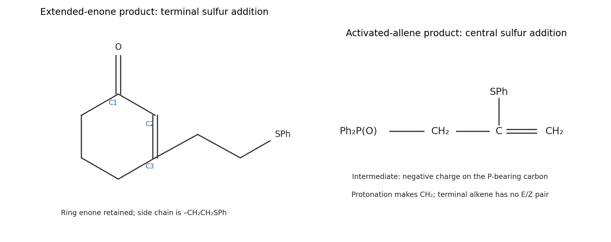
Base again generates catalytically. In , attack at the central allene carbon moves the adjacent C=C pi electrons onto the phosphorus-bearing carbon. This makes the stabilized alpha-phosphoryl carbanionprotonation at that carbon gives , the second product drawn above. The remaining double bond is terminal, so no E/Z pair is possible. The new sulfur bond is at the central carbon of the original allene, not at the terminal methylene.
This regiochemistry places the developing negative charge beside the electron-withdrawing phosphine-oxide group, instead of at a less stabilized vinylic position. It is an ionic addition/proton-transfer cycle, rather than radical thiol–ene chemistry. proton donation by another thiophenol molecule regenerates thiolate. One thiophenol is incorporated without loss of atoms, giving .
The depicted trans-fused is chiral; cis-fused is achiral as a configurational species. The unsaturated ring makes the two sides of the fused system constitutionally different. At the trans junctions of , the mirror image cannot be superposed by a proper rotation; there is no surviving mirror plane or inversion center that exchanges the two distinct rings correctly. A twofold rotational relationship is compatible with chirality and does not remove it. This is why simply importing the chirality conclusion for fully saturated trans-decalin would be unsafe.
For , interchange of the two junction ends relates the opposite local stereochemical senses. An individual folded cis conformer may look chiral, but ring inversion interconverts its mirror-related conformations without changing junction configuration. The configurations therefore describe a meso/achiral species rather than a persistent enantiomeric pair. The conformational sketches below distinguish the rigid trans arrangement from the two accessible cis folds; the alkene ring is represented schematically as a half-chair-like unit.
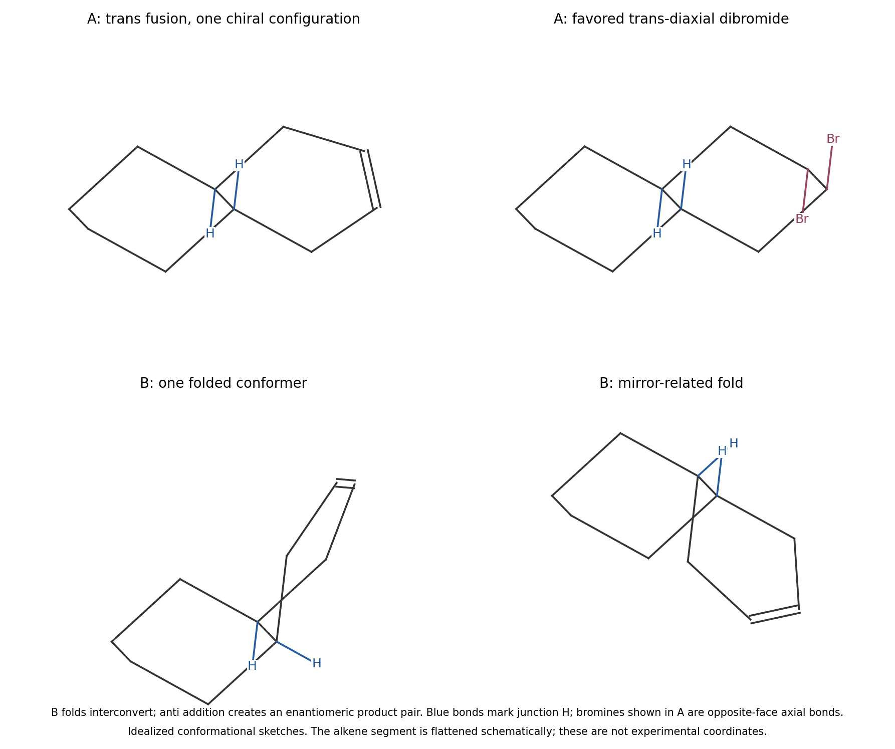
Fused-ring conformations and the stereochemical consequences of anti bromination
. Details. For both substrates, the alkene pi bond attacks bromine and polarizes/breaks Br–Br. A bromine lone pair completes a three-membered bromonium ion, and bromide subsequently attacks a carbon from the opposite face, cleaving that C–Br bridge bond. The two new C–Br bonds are anti. A free carbocation that rotates before capture is not needed.
In trans-fused , the fixed conformation makes the alternative anti-opening pathways diastereomeric. A chair-like, trans-diaxial opening is favored over a geometrically strained alternative that requires a twist/boat-like arrangement of the six-membered ring. The Fürst-Plattner rule describes this conformational preference. One depicted enantiomer of therefore gives essentially one dibromide diastereomer, with the two bromines occupying the trans-diaxial arrangement in its favored chair. If a racemic sample of were used, its other enantiomer would of course give the mirror product as well.
For cis-fused , the accessible mirror-related folds and the equivalent anti pathways produce mirror-related dibromides with equal rates in an achiral environment. Adding the two bromines destroys the symmetry that made the starting configuration achiral. gives a racemic dibromide pair. The racemate follows from equivalent enantiomer-forming pathways, not from a loss of anti stereospecificity.
Number the dioxane O1–C2–O3–C4–C5–C6, with methyl at C2 and tert-butyl at C5. Their cis relationship is maintained on ring inversion, but because they occupy opposite ring positions, one is axial and the other equatorial in either chair. The two alternatives are C2–Me equatorial/C5–tert-butyl axial, and C2–Me axial/C5–tert-butyl equatorial.
The key signal is U, the C5 methine, with only small vicinal couplings, 3.7 and 1.3 Hz. An axial H5 would have large trans-diaxial couplings to axial hydrogens on C4 and C6. Their absence identifies H5 as equatorial, hence the preferred conformer has axial tert-butyl and equatorial methyl. It is not sufficient to guess from tert-butyl bulk. The tert-butyl singlet itself supplies no vicinal H–H coupling information about its orientation.
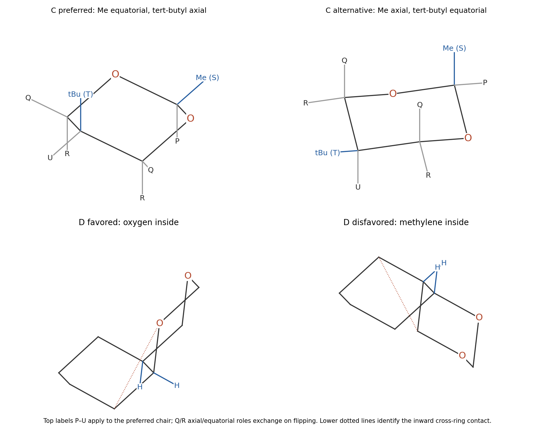
The axial penalties depend on the ring position. An axial methyl at C2 encounters the axial hydrogens on C4 and C6. The short C–O links and dioxane geometry make these 1,3-Diaxial interactions particularly unfavorable. In contrast, an axial substituent at C5 points toward ring oxygens O1 and O3 rather than toward axial C–H groups at those positions. Those oxygen sites present much less steric obstruction than axial hydrogens on carbon.
Avoiding the expensive axial C2 methyl outweighs the cost of axial tert-butyl at C5. Transferring cyclohexane tert-butyl preferences unchanged would therefore give the wrong answer. This is primarily a position-dependent steric comparison, not the usual anomeric preference for an axial electronegative substituent at an acetal carbon. Primary NMR work on substituted dioxanes documents this axial C5 tert-butyl conformer.[ref]
Carbon-13 magnetic resonance. Conformation in some tert-butyl-1,3-dioxanes by Alan J. Jones and others. 1971, pp. 4772–4777, discussion of cis-2-methyl-5-tert-butyl-1,3-dioxane
P is H2, the acetal methine between two oxygens; its only resolved coupling is to the attached methyl, giving the quartet. S is that C2 methyl, whose doublet has the same 6.9 Hz coupling. T is tert-butyl, integrating to nine equivalent hydrogens with no adjacent carbon-bound methine proton across its quaternary attachment.
U is equatorial H5, coupled to the four hydrogens at C4 and C6. The two equivalent equatorial methylene hydrogens are Q, each a doublet of doublets with a 12.3 Hz geminal coupling to its axial partner and a 1.3 Hz coupling to H5. The two equivalent axial methylene hydrogens are R, each with the same 12.3 Hz geminal coupling and a 3.7 Hz vicinal coupling to H5. Equivalence between C4 and C6 makes two pairs rather than four separately shifted methylene hydrogens.Coupling U to two equivalent R hydrogens and two equivalent Q hydrogens produces its triplet of triplets. The different small vicinal couplings are consistent with oblique H–C–C–H torsions for equatorial H5; the 12.3 Hz value is geminal and must not be mistaken for a large vicinal trans-diaxial coupling.
The cis fusion admits two chair–chair conformers, related by simultaneous ring inversion. The bridgehead hydrogens remain cis, while their axial/equatorial descriptions exchange. In one fold the oxygen next to the fusion occupies the concave, inside position; in the other, the corresponding inside position is occupied by a methylene group. Both chair-only structures are shown in the lower panels of the dioxane diagram.
The O-inside chair–chair conformer is strongly favored. In the unfavorable fold, the inward methylene and its C–H bonds create close nonbonded contacts with the other ring, analogous to costly 1,3-diaxial/gauche contacts in cis-decalin. Placing oxygen in that location removes the corresponding carbon-bound hydrogens and reduces the obstruction. The comparison is between the two conformers of the same cis configuration; making the fusion trans would be a configurational change, not a chair flip. Nor is it appropriate to flatten both six-membered rings into regular hexagons, which would hide the decisive inside contacts.
The alkene undergoes concerted oxygen transfer from the terminal peroxide oxygen of mCPBA. In the cyclic electron-flow description, the alkene pi bond begins one C–O bond, the peroxide oxygen supplies the second, O–O breaks, and proton transfer/carboxyl rearrangement gives m-chlorobenzoic acid. Both C–O bonds are installed from the same alkene face, so the alkene's relative substituent geometry is retained in the epoxide. There is no freely rotating carbocation intermediate.
The neighboring free OH hydrogen-bonds to the approaching peracid and favors a cyclic, organized transition-state geometry. oxygen is delivered from the OH-directed face, giving the indicated syn relationship between epoxide oxygen and the allylic hydroxyl. In the supplied projection, the epoxide C–O bonds and the original C–OH bond are on the rear/hashed face. Steric interactions still influence the preferred hydrogen-bonded conformer, but “attack from the least hindered face” alone misses the positive directing role of OH. The distant benzyl ether is not the directing free hydroxyl.
The classical experimental comparison of allylic alcohols and protected derivatives established this directing principle; it supports the mechanistic interpretation without itself measuring the selectivity of this acyclic substrate.[ref]
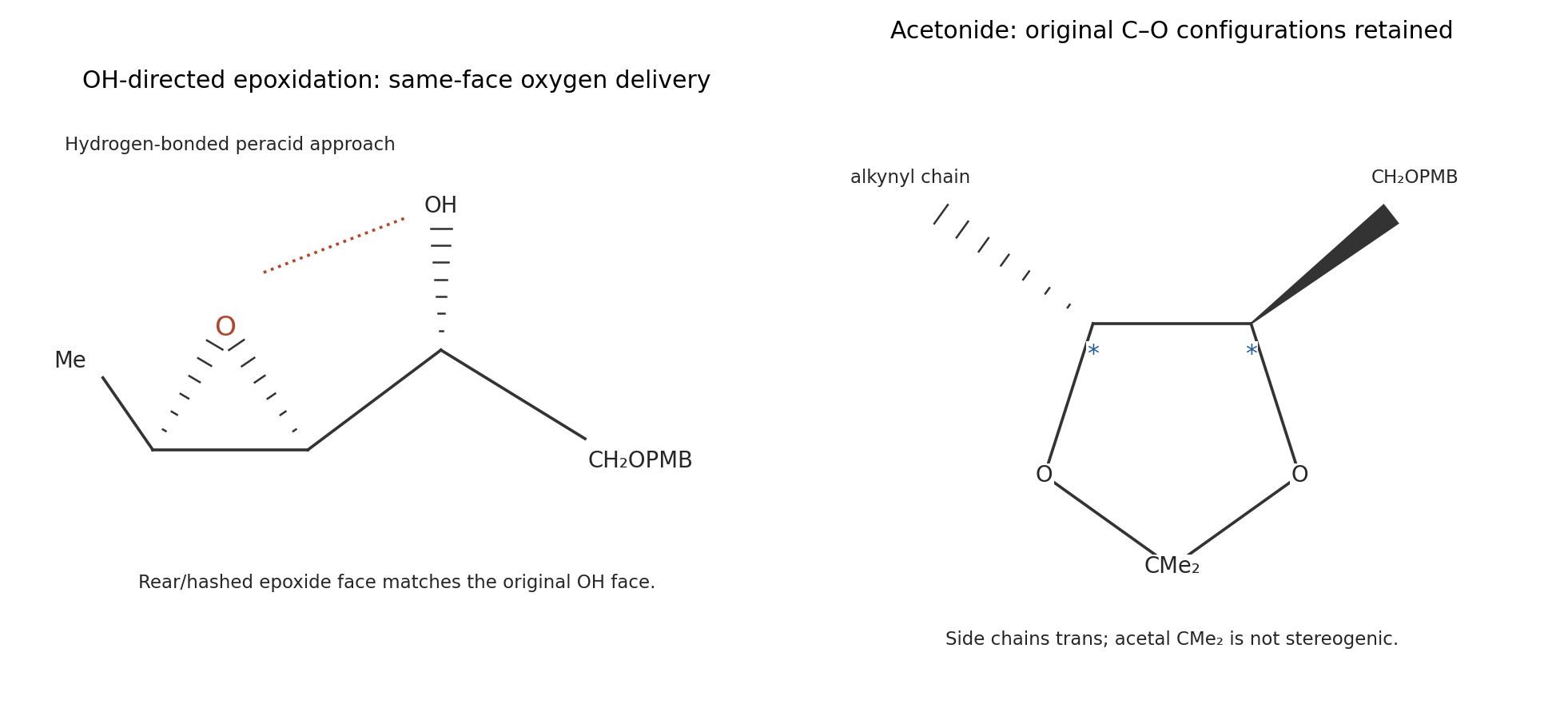
Acid protonates one methoxy group of 2,2-Dimethoxypropane. methanol departure gives the oxocarbenium species , which is captured by one diol oxygen. After proton transfer, protonation and loss of the remaining methoxy group generate a second oxocarbenium center. Intramolecular attack by the other diol oxygen closes the five-membered ring; deprotonation regenerates acid. The net transacetalization exchanges two methoxy groups for the two diol oxygens, releasing two methanol molecules.
The two starred carbon centers retain their original configurations, and the two carbon side chains on the acetonide are trans. No C–O bond at either original stereocenter is broken; reaction takes place at the reagent-derived CMe acetal carbon. The apparent change in wedge/dash presentation comes from redrawing the backbone into the five-membered ring, not inversion at a starred center. In the planar pentagon used in the diagram, draw the alkynyl-bearing side chain behind and the side chain in front. The unstarred methyl-bearing center also retains its configuration. The acetal carbon itself is not stereogenic because its two methyl substituents are identical.
The reacting alkene pi bond and two Os=O units undergo a concerted cycloaddition to give a five-membered cyclic osmate ester. Both new carbon–oxygen bonds form on the same face. Hydrolysis and proton transfer cleave the Os–O attachments, releasing the vicinal diol and a reduced osmium-containing species.
This is syn dihydroxylation of the terminal alkene. The terminal carbon becomes , and the adjacent carbon becomes ; the remote vinyl chloride remains as drawn. The protected region denoted by is a spectator for this local mechanism. No explanation of alkene chemoselectivity is required here. Since no terminal reoxidant is specified, do not invent a catalytic regeneration step for Os(VIII); osmium reduction and product release suffice for the stated transformation.
The free propargylic OH first reacts with an aluminium hydride, releasing hydrogen and forming an O–Al bond. Coordination of the nearby alkyne to this aluminium center organizes a five-membered O–C–C–C–Al unit. Hydride addition from the opposite side of the activated alkyne gives a cyclic alkenyl aluminate: aluminium bonds to the distal alkyne carbon, while hydride is delivered to the carbon nearer .
The key step is directed trans hydroalumination. The new C–H and C–Al bonds are anti. Aqueous workup protonolyzes C–Al with retention of its alkene geometry and hydrolyzes the O–Al attachment. Replacing that C–Al bond by C–H therefore produces the E allylic alcohol, with and the rest of the chain on opposite alkene sides. A generic syn hydroalumination drawing followed by simple protonation would predict the wrong geometry.
The free OH provides the directing tether; the silyl-protected oxygen is not equivalently available. Primary electrophile-trapping studies of Red-Al-treated propargylic alcohols support the alkenyl-alanate description.[ref]
trans-Hydroalumination/Alkylation: One-Pot Synthesis of Trisubstituted Allylic Alcohols by Neil F. Langille and Timothy F. Jamison. 2006, pp. 3761–3764, trans-hydroalumination and trapping of alkenyl alanates
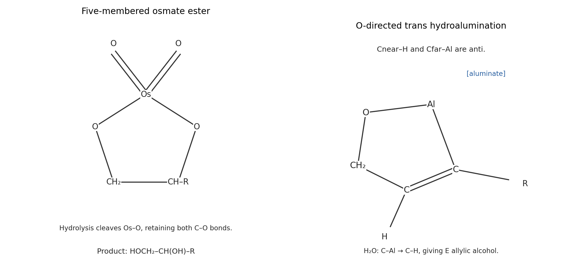
Cyclic intermediates in osmium dihydroxylation and directed trans hydroalumination
. Details. The alcohol oxygen attacks sulfur of methanesulfonic anhydride. Cleavage of the anhydride S–O bond releases methanesulfonate, and the amine base removes the O–H proton, giving the corresponding mesylate. This activation preserves the substrate C–O bond while replacing the poor hydroxide leaving group by .
The base then removes a hydrogen from the adjacent methylene on the side toward the vinyl chloride. In a favorable antiperiplanar arrangement, C–H electrons form the new C=C bond while the C–OMs bond breaks. An E2 elimination creates the alkene between the former alcohol carbon and that methylene. It links the two neighboring unsaturated regions into the extended conjugated triene segment, while the pre-existing carbon skeleton and vinyl chloride are retained.The left-hand polyenoate portion is unchanged. The base is diisopropylethylamine; it accepts acid during mesylation and promotes elimination. The product's E/Z mixture concerns the newly formed alkene, and no single geometry needs to be assigned.
A: aldehyde oxygen is the best acceptor; there is no conventional strong O–H or N–H donor. The N-methylpyrrole nitrogen contributes its lone pair to aromatic bonding and is not an ordinary amine acceptor. A formyl C–H or ring C–H can participate in weak contacts, but is not comparable to the donors in B and C.
B: pyridine nitrogen is the best acceptor and the phenolic OH is the best donor. Its oxygen can accept more weakly, but nitrogen provides the primary basic site in the depicted hydroxypyridine form.
C: lactam carbonyl oxygen is the best acceptor and the amide N–H is the donor. amide nitrogen is not a useful acceptor because its lone pair is delocalized toward the carbonyl.
D: the secondary amine nitrogen is the best acceptor, and its N–H is the only conventional donor, but a weak one here. Its ketone oxygen is also an acceptor, although weaker than the amine site in this comparison. These assignments distinguish functional-group identity and lone-pair availability, rather than counting heteroatoms.
4-Fluorophenol probes acceptor strength because it donates its OH hydrogen. The qualitative order is C D B A: the electron-rich lactam oxygen is the strongest acceptor here, the secondary amine is next, pyridine nitrogen is intermediate, and aldehyde oxygen is weakest. Resonance donation from amide nitrogen increases the lactam oxygen's basicity relative to an ordinary aldehyde or ketone oxygen. The N-methylpyrrole site in A does not add an independent amine-like acceptor.
N-Methylpyrrolidin-2-one is a carbonyl acceptor without N–H, so it instead probes the solute's donor ability. B C A and D: B's phenolic OH is a strong donor, the lactam N–H of C is appreciable, A lacks a conventional donor, and D's neutral secondary-amine N–H is weak. The amine can be a good acceptor and a poor donor at the same time; the two properties are not opposites on one scale.
These are association equilibria in cyclohexane, where solvent competition for hydrogen bonds is weak. The quoted constants should be compared within each probe series, not treated as solvent-independent measures. At 298 K, as an illustrative common temperature, a tenfold increase in corresponds to a binding free-energy advantage . This does not imply the experiment's temperature was specified, nor that every difference is the energy of one isolated hydrogen bond.
C is the best geometrical match for a strongly bound 1:1 complex with E. Both have an adjacent amide donor–acceptor edge. Arrange them head-to-tail so that the N–H of E donates to the carbonyl oxygen of C, and the N–H of C donates to the carbonyl oxygen of E. This closes the familiar two-hydrogen-bond cyclic amide motif, illustrated below.
C combines a strong acceptor with a useful donor in the correct spacing. B has a stronger individual donor, but its pyridine N and OH are separated around the ring and do not reproduce the compact adjacent amide edge of E. A lacks a matching donor, and D's N–H is weak and has the wrong geometry for the same amide–amide motif. Site strength and spatial complementarity must be considered together; multiplying two isolated probe constants is not a reliable prediction of the complete association equilibrium. Self-association of C or E can compete and must be included in a quantitative binding analysis.
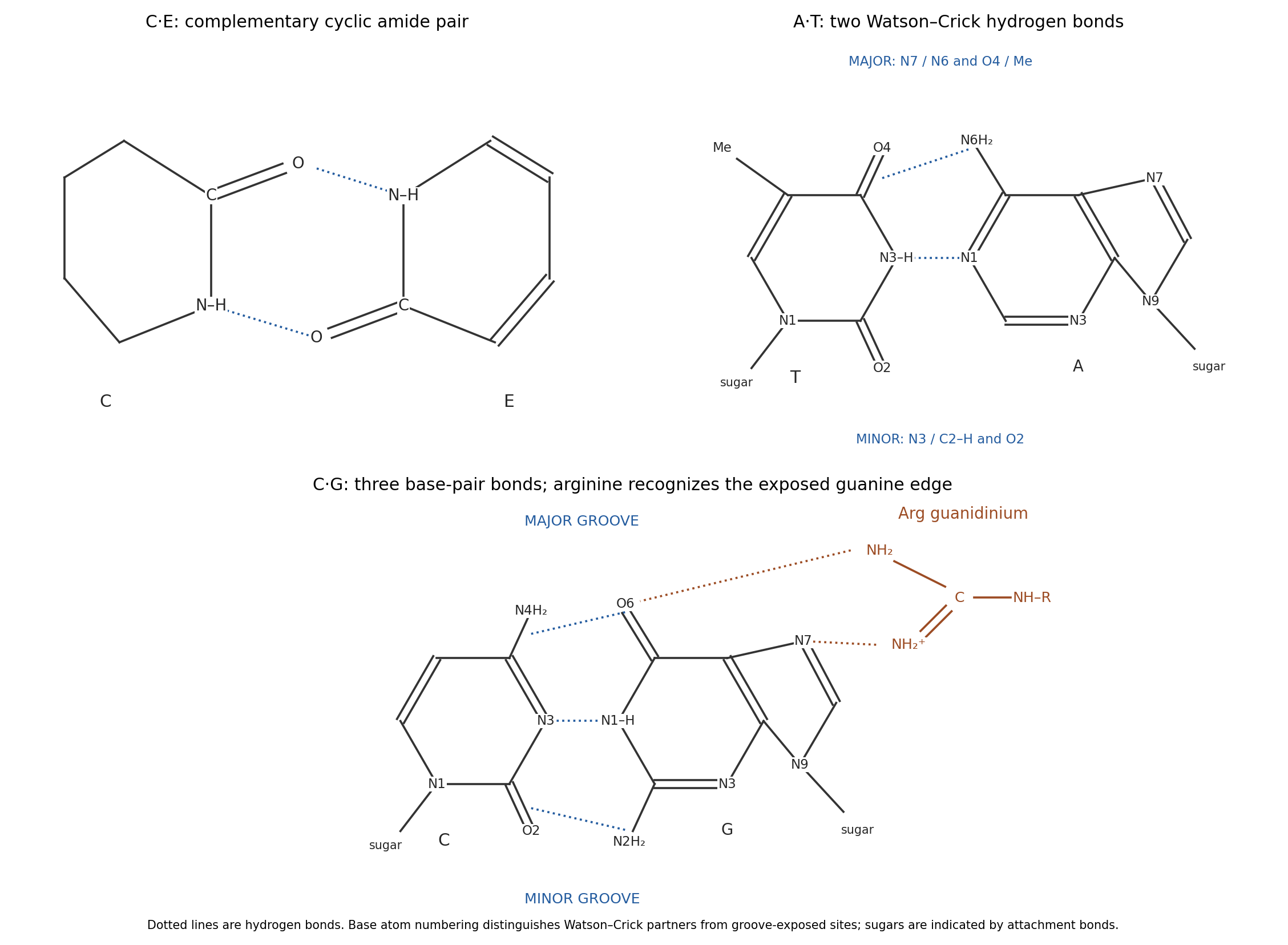
Use the standard Watson–Crick edges, with the glycosidic attachments at purine N9 and pyrimidine N1. The A–T pair has two hydrogen bonds: adenine N6–H donates to thymine O4, and thymine N3–H donates to adenine N1. The C–G pair has three: cytosine N4–H donates to guanine O6, guanine N1–H donates to cytosine N3, and guanine N2–H donates to cytosine O2. The middle and right panels above show these atoms and the sugar-attachment directions.
The major-groove side is the edge toward purine N7 and toward thymine O4/its methyl group or cytosine N4. The minor-groove side is toward purine N3 and adenine C2–H or guanine N2, and toward pyrimidine O2 and the sugar attachments. Groove labels refer to the exposed outer edges of the paired bases; the hydrogen bonds joining the bases are not themselves empty grooves. The resulting patterns differ even when the bases remain paired inside duplex DNA.
At ordinary physiological pH, the arginine side chain is predominantly guanidinium. Its N–H groups are donors, and its positive charge can reinforce favorable electrostatic interactions. guanine exposes adjacent O6 and N7 acceptors in the major groove, allowing two guanidinium N–H donors to make a paired interaction without breaking Watson–Crick pairing. The diagram places this arginine contact on the exposed guanine edge, not between the paired bases.
adenine also exposes N7, but at the corresponding neighboring position it has the exocyclic N6 amino donor rather than a carbonyl acceptor. It therefore cannot make the same donor-to-two-acceptors match with guanidinium. A single N7 contact would be less discriminating. The classic structural recognition analysis explicitly proposed paired arginine–guanine contacts in the major groove.[ref]
Sequence-specific recognition of double helical nucleic acids by proteins by Nadrian C. Seeman and others. 1976, pp. 804–808, major-groove arginine–guanine recognition model
protein geometry, hydration and phosphate contacts also affect sequence recognition; this local motif explains a preference rather than guaranteeing that every arginine-containing protein recognizes every G–C site.View the backbone along N–C to display in a Newman projection. The front amide nitrogen carries the preceding carbonyl and H, while the back C carries the next carbonyl, H and side chain. Rotation changes the contacts among these groups. A corresponding view along C–C displays . The peptide C–N bond itself is approximately planar and is not the freely rotating bond in either definition.
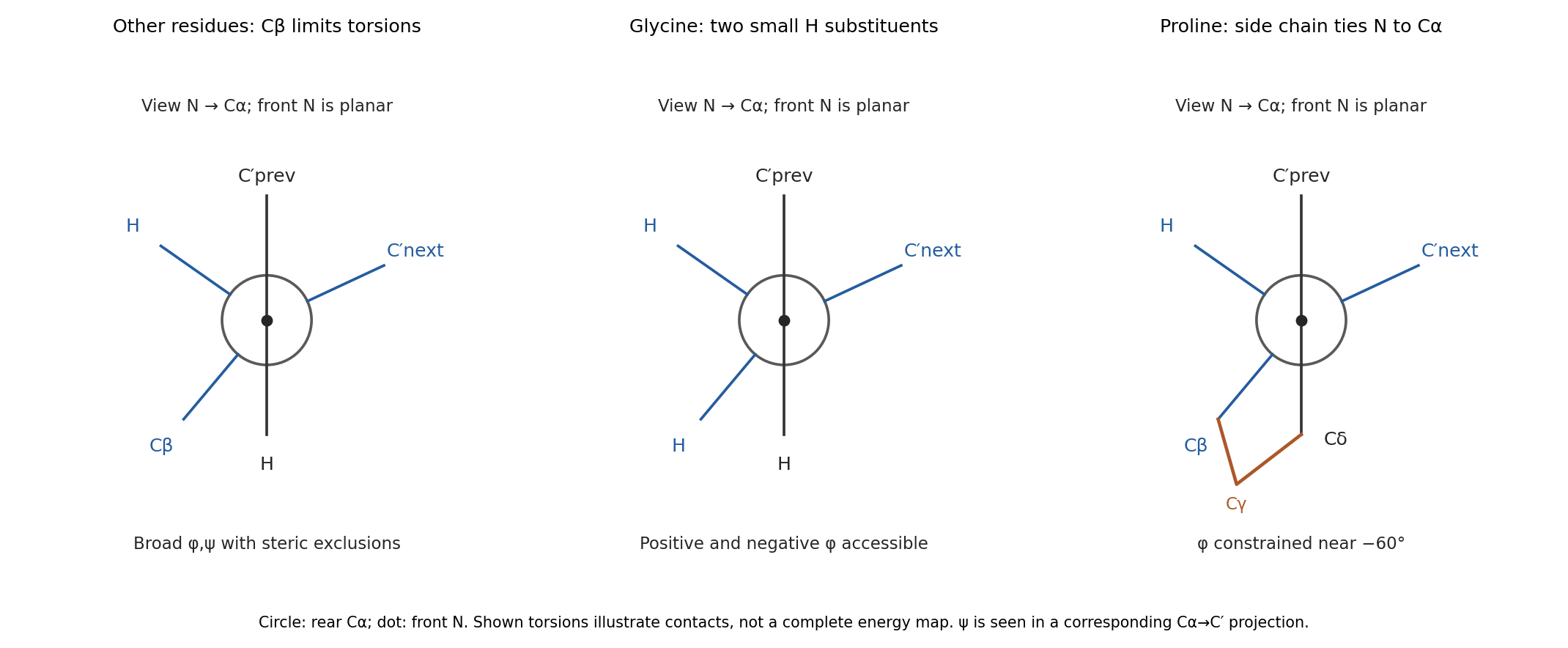
glycine replaces the side chain by a second hydrogen. Removing C removes many contacts that exclude backbone arrangements for other residues, so its allowed region is much broader and includes substantial positive- space. Its alpha carbon is also achiral, so the steric map is approximately mirror-symmetric between positive and negative torsions in the simple isolated-residue model.
proline closes a five-membered ring from C through three methylenes back to nitrogen. In the N–C projection, the nitrogen's side-chain arm and C's C arm are tied together, preventing unrestricted relative rotation. Its is consequently confined near the usual negative- region rather than exploring the broad range of an acyclic amino acid. proline also lacks a backbone N–H donor when incorporated into an ordinary internal peptide. Ring closure restricts strongly, but does not freeze every backbone or ring degree of freedom. These differences are naturally represented by distinct Ramachandran plots; the original torsion/steric analysis established the basis for such comparisons.[ref]
Stereochemistry of polypeptide chain configurations by G. N. Ramachandran and others. 1963, pp. 95–99, backbone torsions and steric restrictions
The carbon end of a carbonyl ligand has a filled molecular orbital with an outward-facing lobe. Its overlap with a vacant metal orbital of the same axial symmetry gives CO-to-metal sigma donation. Occupied metal orbitals can simultaneously overlap with the empty CO antibonding molecular orbitals, giving metal-to-CO pi backbonding. Two perpendicular pi channels are possible; the schematic represents one. Orbital signs indicate phase, not electric charge.
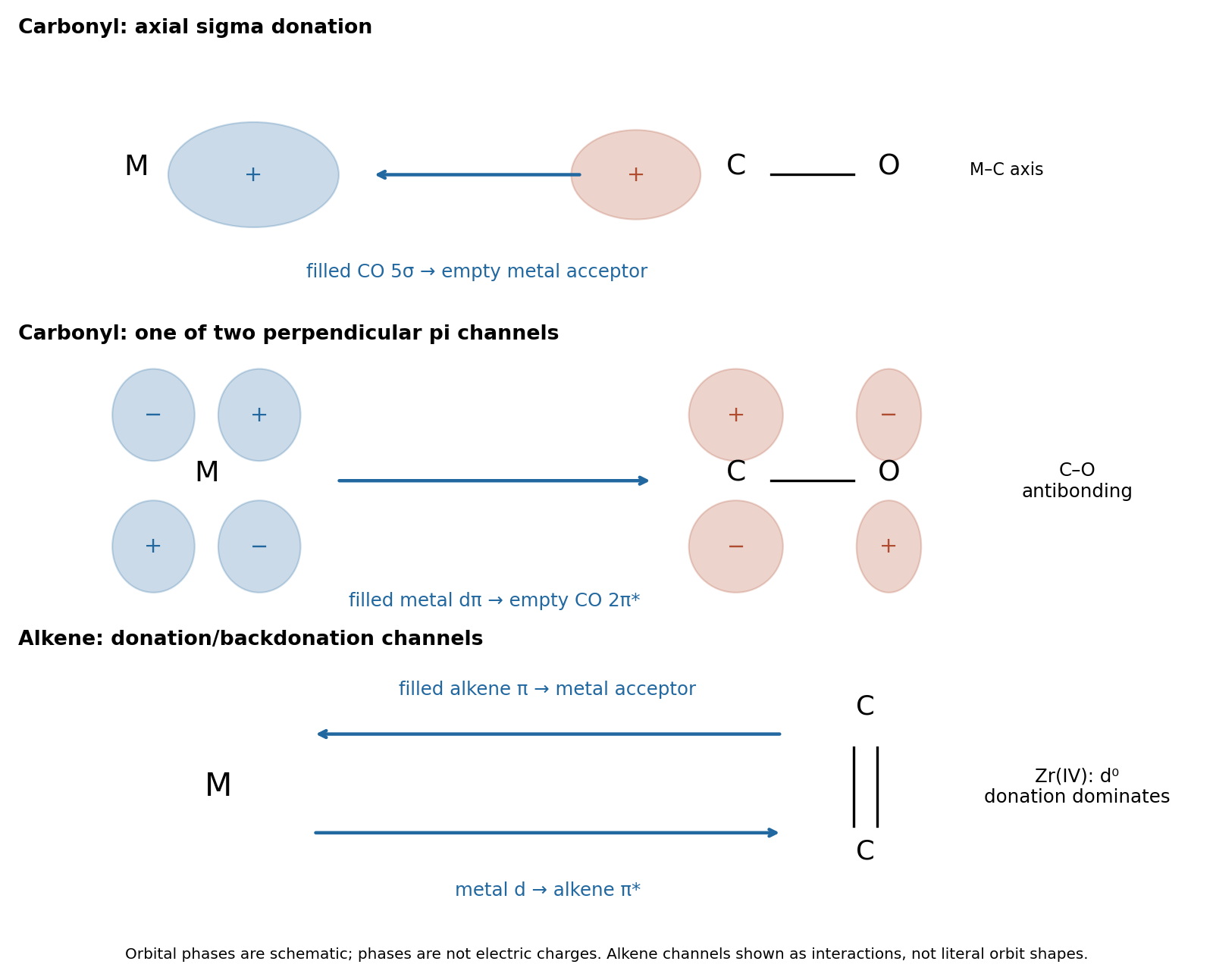
The two interactions reinforce one another: sigma donation transfers density toward the metal, and pi backbonding distributes it into the ligand acceptor orbitals. Both strengthen M–C attachment, while backbonding weakens C–O. Occupation of CO decreases the internal C–O bond order, generally increasing its bond length and lowering its stretching spectroscopic wavenumber. This is why a strongly backbonded carbonyl ligand can have a strong metal–carbon bond and a weakened internal carbon–oxygen bond at the same time.
Place four terminal trifluorophosphine donors on each cobalt and join the cobalt centers by a single metal-metal bond, as in the first panel. The P atoms bind to cobalt; the fluorines remain attached to phosphorus. Each neutral donor supplies two electrons. Neutral electron counting givesThe ionic description is Co(0), , with eight electrons from the four donors and one from the Co–Co bond. A Co–Co single bond supplies the missing electron at each center. No bridging PF is required by this count.
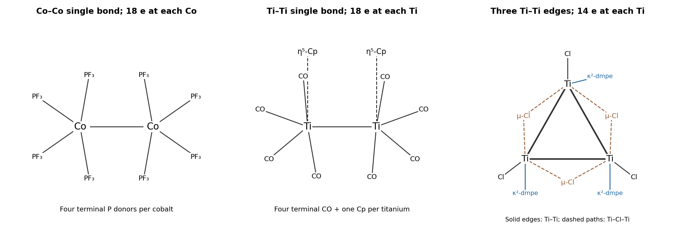
Use an cyclopentadienyl ligand and four terminal carbonyl ligands at each titanium, with a Ti–Ti single metal-metal bond. The middle panel shows this connectivity. Neutral electron counting assigns five electrons to each Cp and givesEquivalently, the two Cp anions require an average oxidation state Ti(I), hence ; ionic counting gives . The formal Ti–Ti bond order is one. The large coordination envelope can make this bond weak and long; reaching 18 electrons is bookkeeping, rather than proof of a short, strong bond. There is no need to invent extra CO bridges or a higher Ti–Ti bond order.
A suitable structure is a triangular Ti metal cluster with one 1,2-Bis(dimethylphosphino)ethane chelate and one terminal chloride at each titanium, and one bridging ligand -Cl spanning each edge. Include a direct Ti–Ti single metal-metal bond along each edge. The last panel distinguishes these direct bonds from the chloride bridges.
The six chlorides have total formal charge , so equivalent centers are Ti(II), . At each titanium the two phosphorus donors supply four electrons, the terminal chloride supplies two, and the two incident chloride bridges each donate a pair in ionic electron counting. Two incident Ti–Ti single bonds supply another two electrons:Each center can contribute one electron to each of its two edge bonds, making three bonds from six metal electrons overall. This is a 14-electron early-metal cluster, not an 18-electron complex. Forcing the 18-electron rule here would invent excessive bond orders. chloride bridges donate to more than one center, so summing local counts is not the same as counting distinct electrons of the whole cluster.
The principal electronic variable is the net population of metal–metal bonding molecular orbitals relative to antibonding molecular orbitals. For otherwise comparable complexes, a larger bond order generally gives a shorter metal-metal bond; removing an antibonding electron can shorten a bond even though the total electron count falls. Different orbital symmetries also matter: sigma, pi and delta overlaps respond differently to distance and rotation of the metal fragments.
metal size, oxidation state and radial orbital extent set the underlying distance scale. Comparing a first-row metal with a larger heavier congener does not isolate bond order. Increasing positive charge often contracts the metal orbitals, but can also change electron occupancy, ligand binding and electrostatic interactions. The ligand field and donor/acceptor properties of the surrounding ligands in coordination chemistry determine which metal orbitals remain available for direct bonding and how much density instead participates in pi backbonding.
Finally, the geometry can enforce or oppose the preferred separation. Bridging ligands impose bond-angle and bond-length constraints; bulky substituents produce steric hindrance; crystal packing can perturb the observed distance. A bond length is evidence for bonding only in its electronic and structural context. In particular, two bridges can hold metals close without proving a high direct bond order, while a sterically congested formal single bond can be unusually long.
For the doubly bridged tetracarbonyl, each iron has one terminal carbonyl ligand, two bridging carbonyl ligands and one cyclopentadienyl ligand. Neutral electron counting gives before direct bonding, so a single Fe–Fe bond gives 18 at each iron. For the triply bridged tricarbonyl, each iron instead has before direct bonding; formal completion to 18 requires a double Fe–Fe bond. Equivalently, the total fragment counts before metal–metal bonding are 34 and 32 respectively.The increased direct bond order explains the substantial contraction. The pentamethylcyclopentadienyl ligands also alter steric and electronic effects, and bridge geometry changes, so this is not a controlled measurement of bond order alone. Nevertheless, the markedly shorter distance in the less carbonyl-saturated dimer is consistent with stronger direct Fe–Fe bonding. The isolated triply bridged compound was characterized structurally and magnetically in the primary work.[ref]
Dinuclear, 18-electron species having a triplet ground state: isolation, characterization, and crystal structure of photogenerated (η5-C5Me5)2Fe2(μ-CO)3 by Josephine Paw Blaha and others. 1985, pp. 4561–4562, isolation, crystal structure and triplet assignment
The dimer has two unpaired electrons and a triplet ground state, . Its approximately threefold-symmetric Fe(CO) core supplies a pair of degenerate frontier molecular orbitals. The last two electrons occupy separate members with parallel spins, favored by exchange, rather than pairing in one. A useful qualitative picture is a sigma bond plus two perpendicular, singly occupied pi half-bonds: together these support a formal Fe–Fe double bond while retaining paramagnetism. The spin-only magnetic moment for is ; real moments can include orbital and spin–orbit effects.
The formal 18-electron count does not require every molecular orbital to be doubly occupied. It is therefore compatible with an open-shell dimer. The primary structural/magnetic study establishes this unusual triplet state; an isolated-atom high-spin diagram would not describe the coupled metal–ligand system.[ref]
In the ionic assignment, both tetrahedral anions would require Mn(VII), . Such a high oxidation state is strongly oxidizing and is best supported by strong bonding to oxygen. Short Mn–O bonds combine good orbital overlap with oxygen's resistance to further oxidation. This stabilizes permanganate relative to a hypothetical all-sulfide analogue.
sulfide is larger, more polarizable and much easier to oxidize than oxide. An attempted Mn(VII) sulfide would therefore combine an exceptionally oxidizing metal center with reducing ligands: internal redox can reduce manganese while oxidizing sulfur, for example toward S–S-bonded or elemental sulfur products. The larger sulfur donors also provide less favorable short multiple bonding to the small, highly charged manganese center. The oxygen compound supports Mn(VII); the sulfide combination is redox-incompatible. This predicts instability without requiring a specific unprovided decomposition product.
For an octahedral complex, strong axial sigma donation raises the metal–ligand antibonding -derived orbitals. A hydride is a very strong sigma donor: its occupied orbital overlaps effectively along a metal–ligand axis and has no filled perpendicular p orbitals to act as a conventional pi donor. It can therefore produce a large octahedral splitting by raising relative to .
Triphenylphosphine also gives sigma donation, but its useful acceptor orbitals additionally receive metal pi backbonding. This lowers the metal -derived levels and enlarges their separation from . The acceptors have substantial P–C antibonding character; invoking a dominant phosphorus acceptor is unnecessary. Thus hydride is strong-field principally through sigma bonding, while PPh combines sigma donation with pi acceptance. The spectrochemical series describes the resulting splitting, not just a ranking of ligand basicity.
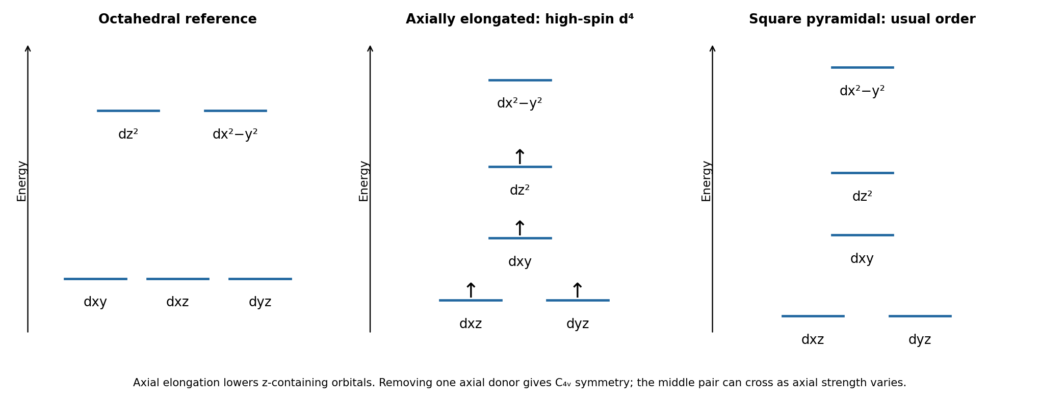
The metal is Cr(II), . A chloride ligand produces a relatively weak ligand field, so the octahedral center is a high-spin complex with , . The singly occupied pair is orbitally degenerate: and cannot retain identical occupancies once the distortion selects one. This is a strong setting for the Jahn-Teller effect.
On tetragonal elongation, the two axial Cr–Cl distances increase. Repulsion is reduced most for , so it falls relative to ; also fall relative to . The single electron occupies the lower -derived level. The electronic stabilization offsets an elastic cost until a distorted equilibrium geometry is reached. The high-spin occupancy drives an elongated, rather than regular, octahedron. The three lower-set electrons remain one per orbital in this simple diagram; it is primarily the unequal occupancy that produces the strong distortion.


The all-carbonyl hydride, HCo(CO), is more acidic. Loss of gives , whose negative charge is stabilized by pi backbonding into four carbonyl ligands. Replacing one CO by triphenyl phosphite reduces the net electron-withdrawing character of the ligand set relative to four carbonyls. The substituted hydride is more electron rich and its conjugate-base anion is less stabilized against protonation.Both conjugate bases have a conventional 18-electron count, so the difference is not a missing-electron argument. It is the relative stabilization of acid and base by the ligands in coordination chemistry. This concerns heterolytic deprotonation, not the homolytic Co–H bond-dissociation energy.
For comparable octahedral environments with the same donor set, the expected qualitative order isAcross comparable first-row ions, the increasing effective attraction and changing metal–ligand distances tend to increase octahedral splitting, giving Mn(II) below Fe(II). Raising the charge on the same metal gives Fe(III) above Fe(II), because the ligand interaction generally strengthens and metal–ligand distances contract. Moving from to more radially extended orbitals gives larger metal–ligand overlap, placing Mo(III) above Fe(III); Rh(III) lies further along the same series and typically gives the larger splitting. The ion Pt(IV) combines the heavier row with high charge and gives the largest splitting here.
This is a matched-environment trend, not an intrinsic list of constants for bare ions. Actual also depends strongly on donor identity, geometry and distance. It would be meaningless to compare an unspecified weak-field tetrahedral compound with a strong-field octahedral compound solely through this ordering.
Start from an octahedral molecular geometry with ligands on , with above . Remove the donor while keeping the other five positions. Loss of axial repulsion lowers substantially; still points directly at four basal donors and remains highest. Loss of an axial donor also lowers the orbitals with dependence relative to . The remaining fourfold axis keeps and degenerate.
The usual qualitative square-pyramidal molecular geometry diagram, shown in the last panel above, is thereforeThe corresponding labels are . Energies can be re-zeroed to the new orbital barycenter after ligand removal. Symmetry fixes the degeneracy and labels, but does not universally fix the separation of and : a weak or distant axial donor can reverse this middle pair on approach to the square-planar limit. The displayed ordering assumes an appreciable fifth axial interaction.
Articles were limited to the first 100 out of 148 total.
Articles by others on the same topic
There are currently no matching articles.