A: aldehyde oxygen is the best acceptor; there is no conventional strong O–H or N–H donor. The N-methylpyrrole nitrogen contributes its lone pair to aromatic bonding and is not an ordinary amine acceptor. A formyl C–H or ring C–H can participate in weak contacts, but is not comparable to the donors in B and C.
B: pyridine nitrogen is the best acceptor and the phenolic OH is the best donor. Its oxygen can accept more weakly, but nitrogen provides the primary basic site in the depicted hydroxypyridine form.
C: lactam carbonyl oxygen is the best acceptor and the amide N–H is the donor. amide nitrogen is not a useful acceptor because its lone pair is delocalized toward the carbonyl.
D: the secondary amine nitrogen is the best acceptor, and its N–H is the only conventional donor, but a weak one here. Its ketone oxygen is also an acceptor, although weaker than the amine site in this comparison. These assignments distinguish functional-group identity and lone-pair availability, rather than counting heteroatoms.
4-Fluorophenol probes acceptor strength because it donates its OH hydrogen. The qualitative order is C D B A: the electron-rich lactam oxygen is the strongest acceptor here, the secondary amine is next, pyridine nitrogen is intermediate, and aldehyde oxygen is weakest. Resonance donation from amide nitrogen increases the lactam oxygen's basicity relative to an ordinary aldehyde or ketone oxygen. The N-methylpyrrole site in A does not add an independent amine-like acceptor.
N-Methylpyrrolidin-2-one is a carbonyl acceptor without N–H, so it instead probes the solute's donor ability. B C A and D: B's phenolic OH is a strong donor, the lactam N–H of C is appreciable, A lacks a conventional donor, and D's neutral secondary-amine N–H is weak. The amine can be a good acceptor and a poor donor at the same time; the two properties are not opposites on one scale.
These are association equilibria in cyclohexane, where solvent competition for hydrogen bonds is weak. The quoted constants should be compared within each probe series, not treated as solvent-independent measures. At 298 K, as an illustrative common temperature, a tenfold increase in corresponds to a binding free-energy advantage . This does not imply the experiment's temperature was specified, nor that every difference is the energy of one isolated hydrogen bond.
C is the best geometrical match for a strongly bound 1:1 complex with E. Both have an adjacent amide donor–acceptor edge. Arrange them head-to-tail so that the N–H of E donates to the carbonyl oxygen of C, and the N–H of C donates to the carbonyl oxygen of E. This closes the familiar two-hydrogen-bond cyclic amide motif, illustrated below.
C combines a strong acceptor with a useful donor in the correct spacing. B has a stronger individual donor, but its pyridine N and OH are separated around the ring and do not reproduce the compact adjacent amide edge of E. A lacks a matching donor, and D's N–H is weak and has the wrong geometry for the same amide–amide motif. Site strength and spatial complementarity must be considered together; multiplying two isolated probe constants is not a reliable prediction of the complete association equilibrium. Self-association of C or E can compete and must be included in a quantitative binding analysis.
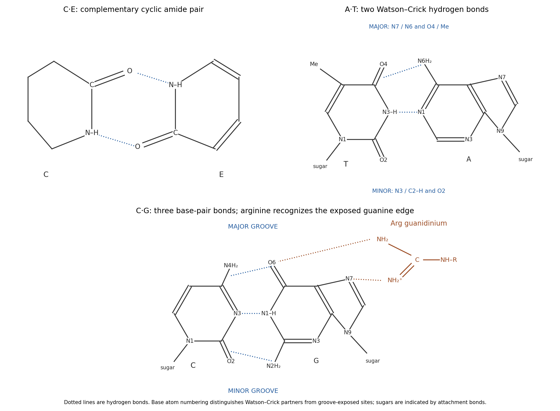
Use the standard Watson–Crick edges, with the glycosidic attachments at purine N9 and pyrimidine N1. The A–T pair has two hydrogen bonds: adenine N6–H donates to thymine O4, and thymine N3–H donates to adenine N1. The C–G pair has three: cytosine N4–H donates to guanine O6, guanine N1–H donates to cytosine N3, and guanine N2–H donates to cytosine O2. The middle and right panels above show these atoms and the sugar-attachment directions.
The major-groove side is the edge toward purine N7 and toward thymine O4/its methyl group or cytosine N4. The minor-groove side is toward purine N3 and adenine C2–H or guanine N2, and toward pyrimidine O2 and the sugar attachments. Groove labels refer to the exposed outer edges of the paired bases; the hydrogen bonds joining the bases are not themselves empty grooves. The resulting patterns differ even when the bases remain paired inside duplex DNA.
At ordinary physiological pH, the arginine side chain is predominantly guanidinium. Its N–H groups are donors, and its positive charge can reinforce favorable electrostatic interactions. guanine exposes adjacent O6 and N7 acceptors in the major groove, allowing two guanidinium N–H donors to make a paired interaction without breaking Watson–Crick pairing. The diagram places this arginine contact on the exposed guanine edge, not between the paired bases.
adenine also exposes N7, but at the corresponding neighboring position it has the exocyclic N6 amino donor rather than a carbonyl acceptor. It therefore cannot make the same donor-to-two-acceptors match with guanidinium. A single N7 contact would be less discriminating. The classic structural recognition analysis explicitly proposed paired arginine–guanine contacts in the major groove.[ref]
Sequence-specific recognition of double helical nucleic acids by proteins by Nadrian C. Seeman and others. 1976, pp. 804–808, major-groove arginine–guanine recognition model
protein geometry, hydration and phosphate contacts also affect sequence recognition; this local motif explains a preference rather than guaranteeing that every arginine-containing protein recognizes every G–C site.View the backbone along N–C to display in a Newman projection. The front amide nitrogen carries the preceding carbonyl and H, while the back C carries the next carbonyl, H and side chain. Rotation changes the contacts among these groups. A corresponding view along C–C displays . The peptide C–N bond itself is approximately planar and is not the freely rotating bond in either definition.
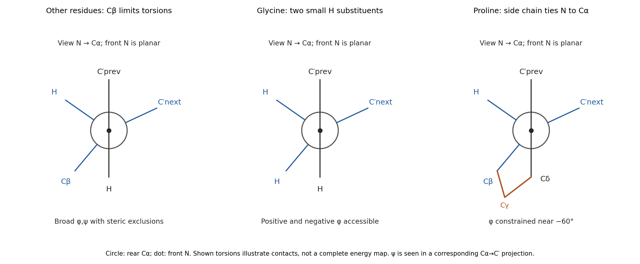


glycine replaces the side chain by a second hydrogen. Removing C removes many contacts that exclude backbone arrangements for other residues, so its allowed region is much broader and includes substantial positive- space. Its alpha carbon is also achiral, so the steric map is approximately mirror-symmetric between positive and negative torsions in the simple isolated-residue model.
proline closes a five-membered ring from C through three methylenes back to nitrogen. In the N–C projection, the nitrogen's side-chain arm and C's C arm are tied together, preventing unrestricted relative rotation. Its is consequently confined near the usual negative- region rather than exploring the broad range of an acyclic amino acid. proline also lacks a backbone N–H donor when incorporated into an ordinary internal peptide. Ring closure restricts strongly, but does not freeze every backbone or ring degree of freedom. These differences are naturally represented by distinct Ramachandran plots; the original torsion/steric analysis established the basis for such comparisons.[ref]
Stereochemistry of polypeptide chain configurations by G. N. Ramachandran and others. 1963, pp. 95–99, backbone torsions and steric restrictions
Articles by others on the same topic
There are currently no matching articles.