Western blot loading controls ask how much sample entered each lane; Western blot transfer controls ask what reached the receiving sheet; Western blot antibody specificity controls ask what the antibodies recognized. Each closes a different gap in Western blot band interpretation.
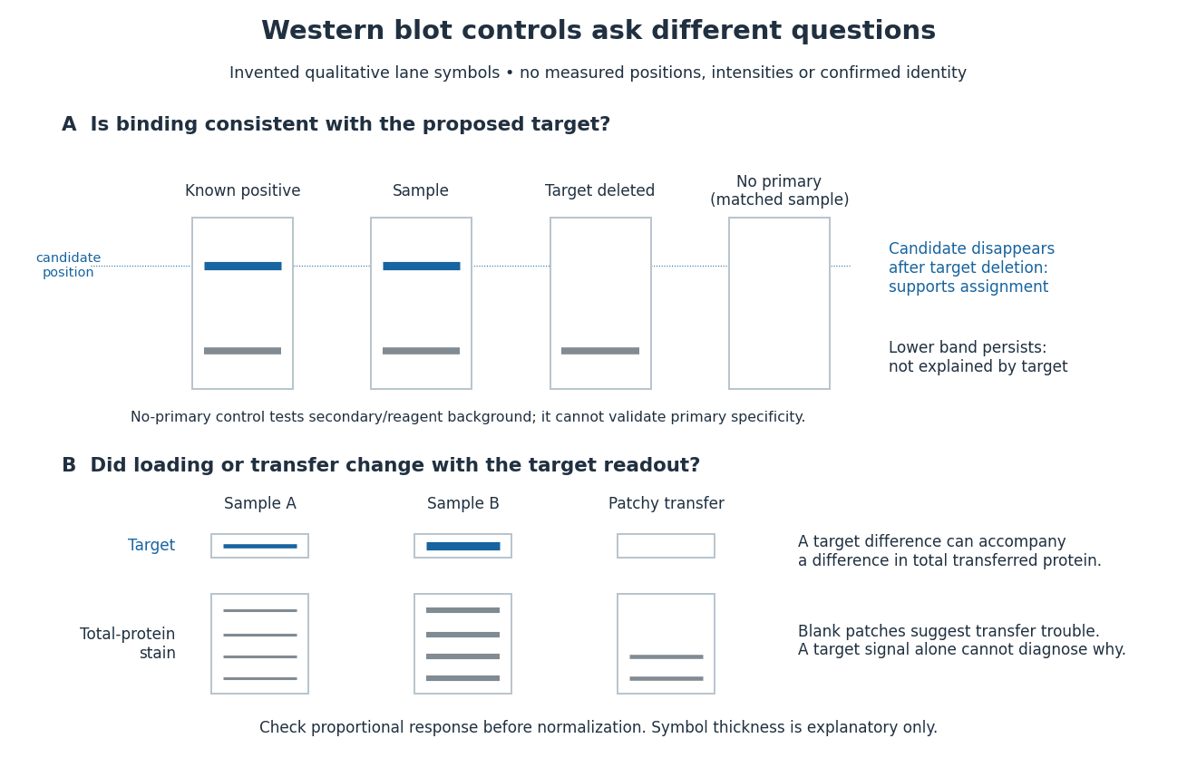
Conceptual lanes for Western blot controls
. Original schematic, with invented qualitative marks only. Candidate bands disappear in a target-deleted sample while a lower band persists; a separate panel shows how a loading or transfer difference can accompany a target-band difference. No lane, darkness, band position or relative amount represents a measurement, a specific real protein or confirmed identity. The no-primary comparison denotes a matched strip processed without primary antibody. Details.
A Western blot loading control estimates lane-to-lane sample differences. A reference protein, such as actin, must have stable abundance and detection responsive to amount. Equal saturated bands fail to establish equal loading. [ref]
Considerations when quantitating protein abundance by immunoblot by Alicia A. McDonough and others. McDonough and others, “Consider the Loading Controls”, Figure 4: sample doubling changes claudin detection but not the actin signal
A Western blot transfer control tests retention after Western blot transfer. Total-protein staining of the Western blot membrane can reveal uneven lanes or blank patches; staining the gel after transfer tests what remained behind. Transferred reference bands check selected positions, not recovery of every target. A pretransfer loading image alone cannot establish the amount retained on the receiving sheet.
A known target-containing sample is a positive comparison for Western blot antibody detection. A matched sample with gene deletion supplies a negative comparison: disappearance of the proposed band supports the assignment, while a persisting band indicates binding unrelated to the deleted target. Deletion must actually remove the recognized protein; changes in other proteins can complicate the comparison. Omitting the primary antibody checks binding or background from the secondary antibody and detection reagents, but cannot exclude binding of the primary antibody to an unintended protein. Independent antibodies recognizing different epitopes add evidence. [ref]
Considerations when quantitating protein abundance by immunoblot by Alicia A. McDonough and others. McDonough and others, “Consider the Specificity of the Antibody for the Target”, Figure 2: positive tissue, knockout tissue and antibodies to different termini
Articles by others on the same topic
There are currently no matching articles.