Fetal hemoglobin, commonly HbF, is a hemoglobin form built from two alpha-globin and two gamma-globin subunits, written . The major adult form, hemoglobin A, instead has the combination. Fetal identity concerns subunit composition; oxyhemoglobin and deoxyhemoglobin concern oxygen occupancy. The same fetal molecule can change its oxygenation state without becoming adult hemoglobin.
Primary structural evidence is available in the human fetal deoxyhemoglobin crystal structure 1FDH. Its experimental record identifies an alpha/gamma tetramer determined by X-ray diffraction at resolution.[ref]
Structure of human foetal deoxyhaemoglobin by J. A. Frier and M. F. Perutz. RCSB PDB 1FDH, Experimental Data Snapshot, Biological Assembly and Macromolecules
The related paper connects structural differences with weaker interaction with 2,3-Bisphosphoglycerate, an effector relevant to oxygen-binding behavior.[ref]Structure of human foetal deoxyhaemoglobin by J. A. Frier and M. F. Perutz. Indexed primary publisher abstract, diphosphoglycerate-site comparison
A structure constrains a mechanism, while an affinity curve requires a functional measurement under specified conditions.An original Hill equation model illustrates what higher oxygen affinity means. Let fractional occupancy at oxygen partial pressure beWith the same stipulated , compare midpoints and 30 in arbitrary pressure units. At , their occupancies areThe lower midpoint gives greater occupancy at the same pressure. It does not imply that the protein carries more than one oxygen molecule per available haem site. These midpoints and the exponent are teaching parameters, not measured fetal/adult reference values.
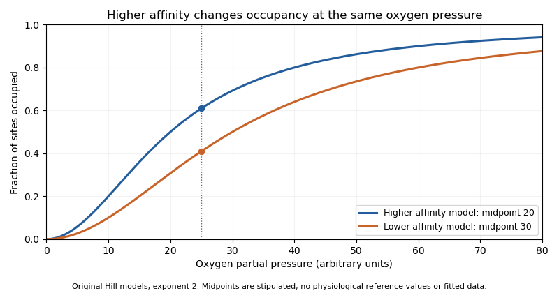
Original Hill models showing how a lower binding midpoint raises occupancy at the same oxygen pressure; parameters are illustrative
. Details. 
Subunit identity also matters analytically. A primary 1991 experiment measured fetal and adult hemoglobin derivative spectra over 450–1000 nm and found differences relevant to multicomponent analysis.[ref]
Absorption spectra of human fetal and adult oxyhemoglobin, de-oxyhemoglobin, carboxyhemoglobin, and methemoglobin by W. G. Zijlstra and others. Abstract
An optical model that distinguishes oxygenation states should therefore also justify which protein-form spectra it uses. Oxygen occupancy, subunit composition and total protein concentration are different quantities. Articles by others on the same topic
There are currently no matching articles.