Gamma-globin is the globin-chain class that pairs with alpha-globin in fetal hemoglobin, whose standard composition is . In humans, the HBG1 and HBG2 genes encode closely related gamma-chain products.[ref] The NCBI annotations distinguish A-gamma from G-gamma by alanine versus glycine at residue 136; gamma is therefore not necessarily a single sequence label.
The human fetal deoxyhemoglobin structure 1FDH provides a molecular example, identifying alpha and gamma entities within the tetramer.[ref]
Structure of human foetal deoxyhaemoglobin by J. A. Frier and M. F. Perutz. RCSB 1FDH, Macromolecules and Biological Assembly
The gamma chain occupies a protein-subunit role; it is not a gamma photon or a description of the molecule's oxygenation state.An original amount calculation makes the subunit distinction useful. Suppose an assay quantifies 120 pmol total gamma chains, with 90 pmol of one sequence form and 30 pmol of the other. Their chain fractions are 0.75 and 0.25. If every measured gamma chain belongs to fetal tetramers containing exactly two gamma chains, total fetal hemoglobin isIf 12 pmol of that measured gamma pool is instead free or belongs to other material, the same tetramer interpretation gives pmol. A total chain assay does not by itself prove where every chain was assembled.
A separate sequence-selective assay can establish which gamma forms are present, while a composition or functional measurement tests the assembled hemoglobin. Changes in HBG1 or HBG2 expression are questions about the producing system; changes in oxyhemoglobin versus deoxyhemoglobin are questions about oxygen binding. Keeping those levels separate prevents a molecular identity result from being mistaken for a complete physiological measurement.
Gamma-chain pairing bounds from bulk composition describe what a measured G-gamma globin/A-gamma globin chain ratio can establish about the assemblies containing those chains. Assume every measured gamma chain belongs to a standard fetal hemoglobin tetramer containing exactly two gamma chains. A bulk chain fraction then constrains, but generally does not determine, how the two forms are paired. The molecular distinction between the forms is experimentally established by glycine versus alanine at residue 136.[ref]
Evidence for multiple structural genes for the gamma chain of human fetal hemoglobin by W. A. Schroeder and others. Page 537, opening paragraph
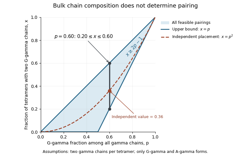
Let be the fraction of gamma chains that are G-gamma, and let be the fractions of tetramers containing respectively two G-gamma chains, one of each form, and two A-gamma chains. Counting assemblies and chains givesHence and . Requiring all three fractions to be nonnegative gives the sharp boundsEvery value in this interval yields nonnegative with the required bulk composition; a finite population additionally requires integral assembly counts.
For an original example, 100 tetramers containing 120 G-gamma and 80 A-gamma chains have . The endpoint compositions and give the same chain counts. Thus the fraction of two-G-gamma tetramers can range from to despite an exactly known bulk composition.
Independent random placement is an additional model. It selects the binomial distribution valuesAt , these are , one feasible point rather than a consequence of the measured chain ratio. The inequalities and show why the random-pairing value always lies within the bounds.
The argument does not infer actual biological pairing preferences. It identifies the information lost when an assay pools chains from many protein complexes. Free chains, other assemblies or unequal analytical responses would require a different input model. A change in bulk sequence proportions is therefore not automatically a measured change in every tetramer's composition or oxygen binding.

G-gamma globin is the human HBG2 gene product, a form of gamma-globin distinguished by glycine at residue 136. The corresponding A-gamma globin, encoded by HBG1, has alanine there. The letter G identifies that amino acid residue; it does not indicate a different oxygenation state or a gamma ray.[ref]
Two gamma chains and two alpha-globin chains form the standard fetal hemoglobin assembly, . Identifying a G-gamma chain is a sequence-level result. It does not by itself determine the identity of the other gamma chain in the same protein complex.
An original accounting example makes this distinction precise. Suppose 100 hypothetical fetal hemoglobin tetramers contain 120 G-gamma and 80 A-gamma chains. Let count tetramers with two G-gamma chains, one of each, and two A-gamma chains, respectively. ThenNonnegative counts imply , with and . Thus a G-gamma chain fraction of 0.60 is compatible with 20% to 60% of tetramers containing two G-gamma chains. Assuming independent placement would select , but independence is additional information, not a consequence of the chain counts.
Gamma-chain pairing bounds from bulk composition therefore matter whenever an assay breaks an assembly into its constituent chains before counting them. Sequence abundance, assembly composition and oxygen-binding behavior answer different questions.
The historical distinction emerged from direct chemical examination of fetal hemoglobin components. The 1968 primary report described glycine and alanine at position 136 in material from 17 additional infants, supporting gamma-chain heterogeneity.[ref]
Evidence for multiple structural genes for the gamma chain of human fetal hemoglobin by W. A. Schroeder and others. p.537, opening paragraph
Modern gene annotations associate the G-gamma form with HBG2; the earlier observation was a protein-sequence result rather than direct sequencing of that locus.A-gamma globin is the human HBG1 gene product, a form of gamma-globin with alanine at residue 136. Its name contrasts with G-gamma globin, whose corresponding residue is glycine. Both belong to the gamma-chain class used in fetal hemoglobin.[ref]
Human globin gene records in NCBI Gene. HBG1, Gene 3047, RefSeq Summary, alanine/glycine distinction at residue 136
A-gamma is not hemoglobin A: the latter is the adult assembly containing beta-globin.A single changed amino acid residue can supply a useful analytical signature without changing which general globin class is being measured. Replacing glycine with alanine adds the elemental composition , since their side chains are hydrogen and methyl. In a deliberately nominal-mass calculation this adds about 14 unified atomic mass units. If an otherwise identical diagnostic peptide produces ions with charge number , the expected separation in mass-to-charge ratio is approximatelyFor , that is about seven mass-to-charge units. This is a proposed analytical discrimination, not the experimental method used in the historical paper. Actual mass spectrometry must use the appropriate exact masses and account for other sequence changes, isotope peaks and modifications.
A sequence-selective amount measurement also requires a response model. In an original two-channel example, suppose one pmol of each form produces detector responses and in arbitrary signal units. Signals and correspond to amounts 20 and 60 pmol, giving an A-gamma fractionUsing the raw signal fraction would instead give 0.40. A distinct molecular signature identifies a channel; it does not guarantee equal analytical response between channels.
The 1968 primary study reported the additional alanine-containing form at position 136 in human fetal hemoglobin components.[ref]
Evidence for multiple structural genes for the gamma chain of human fetal hemoglobin by W. A. Schroeder and others. p.537, opening paragraph
That observation motivates distinguishing sequence forms experimentally, while HBG1 supplies their present gene-level identification. Neither an A-gamma amount nor its ratio to G-gamma alone establishes the oxygen-binding properties of every assembled tetramer. Articles by others on the same topic
There are currently no matching articles.