Immunoglobulin G is an antibody class whose conventional four-chain architecture supplies the example above.
An ordinary IgG has two matching immunoglobulin heavy chains and two matching immunoglobulin light chains, abbreviated . Its gamma-type immunoglobulin heavy chains contribute both to recognition and to interactions with immune partners.[ref]
Antibody Structure and Molecular Immunology by Gerald M. Edelman. Gerald M. Edelman, Antibody Structure and Molecular Immunology, printed pp. 35–37, Table 1 and Figure 3: gamma chains, four-chain architecture and separate functional regions
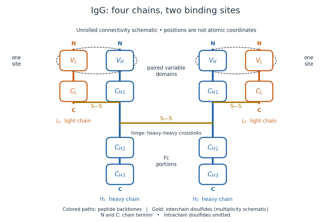
IgG chain continuity and protein disulfide bond connectivity
. Original chain-connectivity schematic. Gold crossbars denote interchain protein disulfide bonds; intrachain crosslinks are omitted. The drawn crossbar count and geometry illustrate connectivity, rather than atomic coordinates or a universal IgG subclass inventory. Details.
A useful way to read this picture is to follow a single polypeptide chain continuously from its N-terminus to its C-terminus, then examine the crosslinks separately. The same immunoglobulin heavy chain contributes to a Fab fragment and to the Fc fragment; these named fragments are not pre-existing separate chains. Conversely, one Fab fragment contains material from two different chains. The protein primary structure and the fragment inventory therefore answer different questions about the same protein.
Gerald M. Edelman reduced IgG disulfide bonds and used dissociating chemical solutions, including urea. Ultracentrifugal analysis showed a decrease in molar mass, supporting a multichain protein. In the comparison reproduced in his lecture, starch gel electrophoresis in urea separated serum IgG and a urinary protein from the same patient, alongside their reduced and alkylation-treated preparations. The untreated samples provided the treatment comparison; the urinary material provided a chain-identity comparison. The treated serum sample revealed heavy- and light-chain bands. Two-dimensional peptide mapping compared the urinary protein with the isolated immunoglobulin light chain. Together these readouts supported a shared chain identity.[ref]
Antibody Structure and Molecular Immunology by Gerald M. Edelman. Gerald M. Edelman, Antibody Structure and Molecular Immunology, Nobel Lecture dated 12 December 1972, printed pp. 33–34 and Figure 2a–b: dissociation, four comparison lanes and peptide maps
The controls address distinct alternatives. Treatment-dependent separation supports chains joined in the starting protein; a matching gel electrophoresis position alone would not uniquely identify a chain. Agreement of peptide mapping adds an independent chemical comparison. Neither readout gives atomic coordinates, and this patient's preparation does not establish a common protein primary structure for all antibodies.
Reduction targets protein disulfide bonds, while protein denaturation weakens the folded assembly. Neither operation is the proteolysis that cuts peptide bonds. This explains why chain separation and Papain digestion of IgG give different inventories: four intact chain backbones in the first case, three assembled fragments in the second.
The antibody hinge region is a flexible IgG immunoglobulin heavy chain segment between and containing inter-heavy-chain disulfide bonds.[ref]
The structure of a typical antibody molecule. Immunobiology: The Immune System in Health and Disease, 5th edition by Charles A. Janeway, Jr. and others; §3-3 and Figure 3.3: hinge and cleavage on the amino-terminal side of its inter-heavy-chain disulfides
Articles by others on the same topic
There are currently no matching articles.