Respiratory mechanics relates the movements of the thorax and respiratory diaphragm to pressure changes and airflow.
Peak airway pressure, , is the largest measured airway pressure over the specified breath or inspiratory interval. Its meaning includes the measurement location, reference pressure and temporal response. At the airway opening during positive-pressure inflation, the peak can include the pressure needed for flow through resistance as well as the pressure distending the respiratory system.[ref]
For a passive single-compartment model with constant airway resistance and respiratory-system compliance ,Here is volume above the end-expiratory state and is inspiratory flow. If constant flow persists to the end of inspiration, the largest volume and the resistive pressure occur together, givingWith varying flow, the peak is instead the maximum of the time-dependent sum; separate maxima cannot automatically be added.
An original example takes total end-expiratory pressure 5 cm HO, tidal volume 0.50 L, L/cm HO and L/s. For cm HO·s/L, peak pressure is cm HO. Doubling resistance raises the calculated peak to 25 cm HO while the equilibrated plateau airway pressure stays 15 cm HO. A larger peak can therefore occur without a larger static distending increment.
An end-inspiratory hold removes ongoing airway flow under appropriate conditions and helps separate these contributions. Active respiratory effort, leaks, nonlinear resistance or delayed redistribution complicate that separation. Peak pressure alone neither partitions lung and chest-wall loading nor establishes a unique cause of a pressure change.
Respiratory driving pressure, , is the difference between plateau airway pressure and total end-expiratory pressure when measured under suitable passive conditions:The total baseline includes intrinsic positive end-expiratory pressure when present; substituting only the externally imposed baseline can overestimate this tidal pressure difference.[ref]
For the linear static model, respiratory-system compliance is . An original calculation with tidal volume 0.45 L, plateau pressure 23 cm HO and total baseline 8 cm HO gives cm HO and L/cm HO. If the external baseline alone were 5 cm HO, incorrectly subtracting it would give 18 cm HO and a different apparent compliance.
This pressure difference loads both the lung and chest wall. At static endpoints, transpulmonary pressure gives the partitionThus a stipulated airway increment of 12 cm HO corresponds to a lung increment of 4 when the pleural increment is 8, but to a lung increment of 10 when the pleural increment is 2. Equal airway driving pressures do not establish equal lung distension without information about the chest-wall contribution.
The measurement method matters as much as the subtraction. A dynamic estimate during flow can include resistance; an end-inspiratory hold and an end-expiratory occlusion address different endpoints. The primary pilot study explicitly compared static and dynamic ways of obtaining whole-respiratory-system compliance and driving pressure.[ref]
Static and Dynamic Measurements of Compliance and Driving Pressure: A Pilot Study by Pierre Tawfik and others. Introduction, Figure 1 and Study Procedures
These distinctions explain the quantity's mechanical meaning without turning an illustrative number into an individual ventilation recommendation.Positive end-expiratory pressure, PEEP, is airway pressure maintained above atmospheric pressure at the end of exhalation. It specifies an end-expiratory pressure reference, not a tidal volume or a pressure rise measured from the beginning to the end of inspiration. A set external pressure and the total pressure assessed during an appropriate end-expiratory hold need not be identical.[ref]
How to measure driving pressure by Hamilton Medical. Measurement on Hamilton Medical ventilators; End-expiratory hold
One reason is incomplete emptying. In an original passive single-compartment model, let be excess volume above equilibrium with the prescribed outlet pressure. With airway resistance and respiratory-system compliance ,The remaining elastic pressure above the outlet is . Stipulated cm HO·s/L and L/cm HO give a time constant of 1 s. Starting with 0.50 L excess volume, after 0.5 s the model retains about 0.303 L, corresponding to 6.07 cm HO above the outlet pressure. These are constructed mechanics, not ventilator settings for a person.
If the prescribed outlet reference is 5 cm HO, the modeled retained elastic pressure raises the equilibrated total to about 11.07 cm HO. This illustrates intrinsic positive end-expiratory pressure: a pressure contribution that cannot be identified from the external setting alone. The result assumes a communicating passive compartment; active effort, leakage and heterogeneous time constants require further interpretation.
Respiratory driving pressure compares plateau airway pressure with total PEEP, rather than silently subtracting an external setting that may omit retained pressure. The manufacturer's primary measurement account states that distinction and the required passive, leak-free conditions.[ref]
How to measure driving pressure by Hamilton Medical. Driving-pressure definition and measurement conditions
Naming the pressure reference is therefore part of defining the reported mechanical quantity.An end-expiratory hold is an interruption of airflow at the end of exhalation during which airway pressure is observed while the respiratory system is occluded. In an appropriate passive, leak-free measurement it can assess total positive end-expiratory pressure, including intrinsic positive end-expiratory pressure. The set external pressure alone need not equal this measured total.[ref]
How to measure driving pressure by Hamilton Medical. End-expiratory hold and measurement conditions
The mechanical reason is easiest to see in one communicating compartment. With airway resistance , let relate airflow to the difference between alveolar pressure and measured airway pressure. Once flow vanishes and pressure has equilibrated through the communicating airway, this model gives . Zero flow removes the resistive pressure drop; it does not mean that either pressure is zero.
For stipulated pressures, an external setting of and an equilibrated total of differ by . If plateau airway pressure is , respiratory driving pressure is , whereas subtracting the external setting would give 18. These constructed values illustrate reference selection, not patient settings.
Occlusion changes the boundary condition rather than directly opening every lung region to the sensor. A compartment isolated by airway closure cannot be assumed to equilibrate with the measured opening. Active muscular effort, a leak or a pressure trace that has not settled also defeats the simple passive-compartment inference. The observed airway-pressure plateau and the inferred regional alveolar pressure must therefore remain distinct.
Intrinsic positive end-expiratory pressure is the residual alveolar pressure above the externally imposed end-expiratory pressure, associated with incomplete lung emptying. In a communicating passive compartment,The manufacturer's technical account makes this distinction and explains why a fitted whole-breath estimate can differ from the pressure obtained during an end-expiratory hold.[ref]
Measurement of AutoPEEP and total PEEP by Hamilton Medical. Opening definition; Measuring AutoPEEP; Calculations
An original repeated-breath model isolates the timing mechanism. Let be excess volume above equilibrium with the outlet just before inspiration, and let each inspiration add tidal volume . During exhalation, assume a passive linear compartment with time constant . After expiratory time ,The steady excess volume satisfies , henceFor stipulated L, respiratory-system compliance L/cm HO, s and s, , L and intrinsic pressure is about 4.66 cm HO. Giving this same ideal compartment two seconds to empty yields about 0.063 L and 1.25 cm HO. These are consequences of a model, not patient-specific timing instructions.
A nonzero external baseline does not by itself imply incomplete emptying. Conversely, setting an external baseline to zero does not prove that alveolar pressure has returned to zero. Measurements require attention to communication between regions, leakage and active muscle pressure; a single airway value need not reveal every trapped regional pressure. The recurrence explains why resistance, compliance and available time matter together rather than assigning the phenomenon to one pressure setting alone.
Plateau airway pressure, , is the airway pressure assessed during an end-inspiratory interval with no flow and sufficiently stable conditions. An end-inspiratory hold can allow the airway and communicating alveolar gas pressures to equilibrate when the respiratory system is passive and the circuit is not leaking.[ref]
Measured values for Ppeak and Pplateau by Hamilton Medical. Definition of airway pressures; Principles of measuring plateau pressures
[ref]The distinction from peak airway pressure follows a simple single-compartment model:During the modeled hold, , leavingHere is respiratory-system compliance, is airway resistance, and total positive end-expiratory pressure supplies the starting reference. The measured difference is the corresponding respiratory driving pressure.
In an original example, total PEEP 8 cm HO, tidal volume 0.40 L and compliance 0.04 L/cm HO give cm HO. If flow immediately beforehand is 0.5 L/s and resistance is 12 cm HO·s/L, the model's airway pressure during that flow is 24 cm HO. The 6 cm HO difference is the modeled resistive contribution, not an extra static distending pressure.
A plateau does not directly measure pleural pressure or isolate the lung from the chest wall. For the same stipulated alveolar pressure 18 cm HO, pleural pressures of 12 and 2 cm HO imply transpulmonary pressures of 6 and 16 cm HO. The airway value alone cannot distinguish those states.
Finally, a stable displayed number is not automatically the same operation as an actual hold. The manufacturer distinguishes measured holds, rapid occlusion and calculated monitoring estimates.[ref]
Measured values for Ppeak and Pplateau by Hamilton Medical. Principles of measuring plateau pressures; Monitoring parameter Pplateau
Reporting the method and its conditions connects plateau airway pressure to the mechanical question it can answer.An end-inspiratory hold is a pause after inflation during which the airway is occluded and external flow is interrupted. Under passive, leak-free conditions, the resulting pressure trace helps distinguish flow-related airway resistance from the pressure remaining at approximately fixed lung volume. A stable endpoint provides a plateau airway pressure for interpreting respiratory-system compliance.[ref]
How to measure driving pressure by Hamilton Medical. End-inspiratory hold; passive and leak-free measurement conditions
Stopping flow does not imply that every compartment instantaneously reaches a common pressure. Stress relaxation, redistribution between lung regions and the apparatus response can produce a continuing pressure change after the rapid resistive drop. The measured value should therefore identify the observation interval, rather than treating the first post-interruption sample as universally equilibrated.
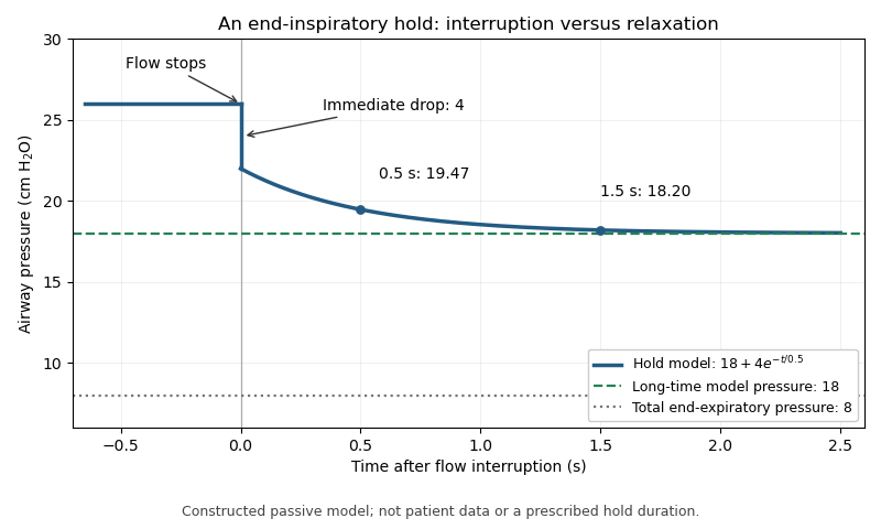
For an original illustrative trace, suppose flowing peak pressure is 26 cm HO and stopping flow immediately removes 4 cm HO. Let the subsequent relaxation beThe initial hold value is 22 cm HO, about 19.47 remains after 0.5 s, and about 18.20 after 1.5 s. The model's long-time value is 18. The change from 26 to 22 and the later change from 22 toward 18 represent different mechanisms in this constructed example.
With total end-expiratory pressure 8 cm HO, using the initial hold value gives a nominal respiratory driving pressure of 14, whereas using the asymptotic value gives 10 cm HO. The difference demonstrates the importance of time and model assumptions; it does not prescribe a hold duration. Continued muscle activity, leakage or airway closure can invalidate the simple static interpretation. The experimental pilot study compares static and dynamic measurement methods rather than assuming all displayed pressure estimates are interchangeable.[ref]
Static and Dynamic Measurements of Compliance and Driving Pressure: A Pilot Study by Pierre Tawfik and others. Figure 1 and Study Procedures

An esophageal balloon is a thin-walled pressure-sensing balloon placed in the oesophagus and connected to a pressure-measurement system. Its mechanical purpose is to couple surrounding pressure to the gas inside it. An esophageal balloon catheter supplies the pneumatic connection; esophageal pressure measurement concerns the interpretation of the resulting signal as evidence about pleural pressure.
A useful idealization separates the contributions:where balloon tension, tissue reaction and an instrumental offset can affect the reading. This additive model is bookkeeping, not a universal constitutive law for the tissue or balloon. A pressure change can be transmitted well even when the baseline has an offset.
The primary physiological study by Francesco Mojoli and collaborators changed balloon filling and compared pressure transmission with baseline contributions from the esophageal wall. Its observations demonstrate that favorable transmission of pressure swings does not by itself remove the esophageal balloon filling artifact.[ref]
In vivo calibration of esophageal pressure in the mechanically ventilated patient makes measurements reliable by Francesco Mojoli and others. Abstract and Results: filling, transmitted changes and wall-pressure contribution
For a constructed example, suppose surrounding pressure changes from 6 to while a fixed added pressure is . The balloon reads 8 and 12: it reproduces the change exactly but overestimates both absolute values by two. Consequently, agreement of a change measurement is a different validation claim from agreement of absolute pressure. Neither the balloon's name nor its presence inside the chest makes those claims automatic.
Esophageal pressure measurement records pressure in the oesophagus, commonly with an esophageal balloon, to obtain information about respiratory mechanics and pleural pressure. The measured signal is a local surrogate. It is not a direct map of pressure everywhere in the pleural cavity.
A primary validation experiment by Ahmet Baydur and collaborators compared changes in esophageal and mouth pressure during end-expiratory airway occlusion and spontaneous efforts. Ten subjects were studied in different postures; some discrepancies changed when the balloon was repositioned.[ref]
A Simple Method for Assessing the Validity of the Esophageal Balloon Technique by Ahmet Baydur and others. Abstract: subjects, posture, occlusion and repositioning
The esophageal balloon occlusion test therefore evaluates pressure-change transmission under specified conditions, rather than proving all absolute-pressure values correct.In an ideal local model, write . If and remain fixed during an effort, thenwhen mouth and pleural pressure changes coincide under the test's assumptions. A ratio near one supports transmission gain near one, but leaves unidentified. Even perfect change agreement cannot determine that constant baseline offset.
The consequence matters when calculating transpulmonary pressure . For an illustrative alveolar pressure of 15 and true local pleural pressure of , the difference is eight. Substituting an esophageal reading of nine with an uncorrected offset of two gives six instead. The arithmetic identifies an error mechanism; it does not supply a clinical correction or assume that all pleural regions have pressure seven.
Instrument calibration, balloon coupling, body position and the physiological assumptions of the comparison address separate sources of error. Reporting the waveform without those conditions can conceal which inference was actually tested.
An esophageal balloon catheter is the tube-and-balloon instrument used to transmit a pressure signal from a balloon in the oesophagus to a pressure sensor. The esophageal balloon is the coupling element; the catheter also introduces a gas-filled line, connections and an external measurement chain. Those components can affect both baseline pressure and dynamic response.
The physiological filling study by Francesco Mojoli and collaborators shows why treating the assembly as an ideal pressure wire is inadequate: changing balloon filling altered pressure-change transmission and wall-related baseline pressure.[ref]
In vivo calibration of esophageal pressure in the mechanically ventilated patient makes measurements reliable by Francesco Mojoli and others. Abstract, Methods and Results
This establishes a measurement dependence, not one universal filling volume for every catheter and surrounding pressure.A minimal model of pneumatic response lag of an esophageal catheter iswhere is a varying source pressure and the sensor output. For a step from to , the response isWith a stipulated , 63.2 percent of the step appears after 0.10 s and 95.0 percent after 0.30 s. These are model calculations, not specifications of a clinical device.
This dynamic error differs from a fixed offset: waiting longer removes the modeled step-response lag but not a baseline bias. A comparison against a known pressure waveform can test the instrument's response, while an esophageal balloon occlusion test addresses transmission in the measurement setting. Neither validation alone proves that esophageal pressure equals regional pleural pressure throughout the lung.
The esophageal balloon occlusion test checks how well changes measured by an esophageal balloon catheter track changes in pleural pressure. During an inspiratory effort against an occluded airway at end-expiration, the comparison is between the pressure change in the balloon and that at the mouth. The original study by Ahmet Baydur and colleagues tested ten subjects in sitting, supine and lateral positions. Repositioning the balloon could improve agreement when a supine measurement initially differed.[ref]
A Simple Method for Assessing the Validity of the Esophageal Balloon Technique by Ahmet Baydur and others. Abstract, end-expiratory occlusion protocol and positional findings
The mechanical rationale is a change comparison. With no external flow and approximately fixed lung volume, the change in transpulmonary pressure is small in the simple static model. Consequently,for a balloon that transmits pleural changes appropriately. The reasoning assumes communicating airways and a suitable measurement system; it does not establish equality merely because flow at one sensor is zero.
An original example uses simultaneous mouth-pressure changes cm HO and balloon changes cm HO. Their proportional response is 0.9. Repeating several efforts makes that relation more visible than interpreting one small pressure fluctuation in isolation.
Importantly, a successful change test does not determine the absolute offset. If a hypothetical sensor obeys , the ratio of changes assesses , while the constant cancels. A true pleural change from to recorded as 0 to has unit change ratio despite a 5 cm HO offset. Esophageal pressure measurement therefore needs a clear distinction between tracking respiratory swings and assigning an absolute pressure for a transpulmonary calculation. The test interrogates pressure transmission, not every possible source of measurement error.
Articles by others on the same topic
There are currently no matching articles.