
Base pairing is the recognition and association of compatible nucleobases. In canonical Watson–Crick geometry, an adenine–thymine base pair has two hydrogen bonds, while a guanine–cytosine base pair has three. These counts refer to those specific geometries and usual chemical forms; they are not a definition of every possible base pair. RNA can also contain other pairings, and canonical adenine–uracil pairing replaces adenine–thymine pairing. See Clark, Douglas and Choi, Biology 2e: DNA structure.
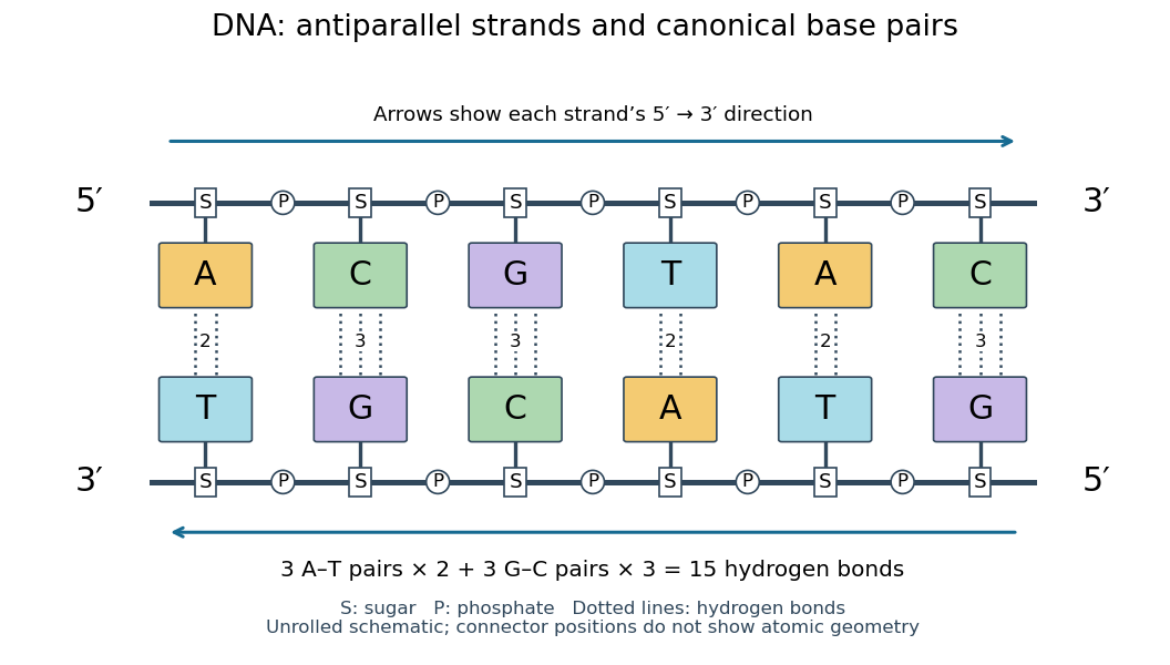
Six canonical Watson–Crick DNA base pairs: opposite backbone directions and two or three hydrogen bonds per pair
. Details. 
For this illustrative duplex, the six base pairs are A–T, C–G, G–C, T–A, A–T, and C–G. Thus three contribute two hydrogen bonds each and three contribute three each:Each dotted connector in the diagram counts one hydrogen bond; coloured boxes denote whole nucleobases, not individual chemical atoms. The solid rails denote the covalent bond scaffold. Separating the two strands disrupts base pairing and base stacking without requiring the backbone to be cut.
The count of fifteen hydrogen bonds is not fifteen identical contributions to a duplex's Gibbs free energy. Base stacking, interactions with water and ions, temperature, and the order of neighbouring base pairs also matter. Knowing the fraction of guanine–cytosine base pairs alone does not fix a nucleic acid double helix's shape or stability. See Brown, Genomes: pairing and stacking interactions. What makes base pairing especially useful is its selectivity: an exposed DNA strand constrains the partner's nucleic acid sequence, giving DNA replication a chemical recognition rule.
New to topics? Read the docs here!