
D. R. Enzmann 2026-11-05
D. R. Enzmann is the radiologist credited under that byline as first author of Normal flow patterns of intracranial and spinal cerebrospinal fluid defined with phase-contrast cine MR imaging by D. R. Enzmann and others. With N. J. Pelc, Enzmann reported measurements of moving cerebrospinal fluid, using magnetic resonance imaging to investigate physiology as well as anatomical appearance.[ref]
Normal flow patterns of intracranial and spinal cerebrospinal fluid defined with phase-contrast cine MR imaging by D. R. Enzmann and others. Radiology 178 (1991), 467–474; verified byline and abstract.
The study examined ten healthy volunteers and four patients whose conventional MR images were normal. Peripheral gating produced sixteen quantitative flow-encoded images over the cardiac cycle. Measurements distinguished the timing of fluid motion in ventricular and subarachnoid spaces, demonstrating why static anatomy alone does not describe pulsatile movement.[ref]
Normal flow patterns of intracranial and spinal cerebrospinal fluid defined with phase-contrast cine MR imaging by D. R. Enzmann and others. Abstract: participant groups, peripheral gating and regional flow timing.
The methodological contribution is an example of turning image contrast into a physiological measurement. Direction-sensitive signals and repeated sampling across a heartbeat distinguish oscillatory motion from a compartment that simply looks fluid-filled. The reported display convention assigned opposite intensities to opposite craniocaudal directions; that convention belongs to the acquisition and is not a universal rule for brightness in every MR image.
Interventricular foramina (neuroanatomy) 2026-11-05
The interventricular foramina are the paired openings through which the lateral ventricles communicate with the third ventricle. Each opening connects its own side's lateral ventricle to the shared midline cavity. This arrangement provides a route for cerebrospinal fluid to reach the rest of the ventricular system.[ref]
Lateral Ventricles in MeSH. Definition, each lateral cavity communicating with the third ventricle
Both sides reach the shared third ventricle, which joins the fourth ventricle through the cerebral aqueduct.
Measurements add a temporal picture to this connection map. In a 1991 study, phase-contrast magnetic resonance imaging encoded the direction of cerebrospinal fluid movement across the cardiac cycle. The ventricular measurements contained systolic and diastolic flow components, with essentially simultaneous timing at the interventricular opening and cerebral aqueduct. Anatomical continuity and measured pulsatile transport describe complementary features of the same pathway.[ref]
Normal flow patterns of intracranial and spinal cerebrospinal fluid defined with phase-contrast cine MR imaging by D. R. Enzmann and others. abstract, flow encoding and timing at ventricular passages
Lateral ventricle 2026-11-05
A lateral ventricle is one of the two large cerebrospinal fluid-filled cavities within the cerebral hemispheres. Each connects with the third ventricle through an interventricular foramen. Its location inside a hemisphere makes it a useful landmark when interpreting sections or images of the brain.[ref]
Lateral Ventricles in MeSH. Definition, paired cerebral cavities and communication with the third ventricle
The choroid plexus projects into ventricular cavities and contributes to cerebrospinal fluid production; the ependyma lines the ventricular wall. The resulting environment joins a secretory interface, a fluid compartment and neighbouring neural tissue. These components have different roles even though they appear close together in an anatomical section.[ref]
Anatomy and Physiology 2e by J. Gordon Betts and others. §12.2, ependymal cells; §13.3, cerebrospinal fluid and choroid plexuses
The connections below provide a simplified route through the ventricular system. Actual transport also has a time dependence: phase-contrast magnetic resonance imaging has resolved cerebrospinal fluid movement over the cardiac cycle, while work on the third ventricle has revealed structured local flows driven by motile cilia.[ref]
Normal flow patterns of intracranial and spinal cerebrospinal fluid defined with phase-contrast cine MR imaging by D. R. Enzmann and others. abstract, cardiac-cycle-dependent flow encoding
[ref]Cilia-based flow network in the brain ventricles by Regina Faubel and others. abstract, ciliary modules and ventricular flow networks
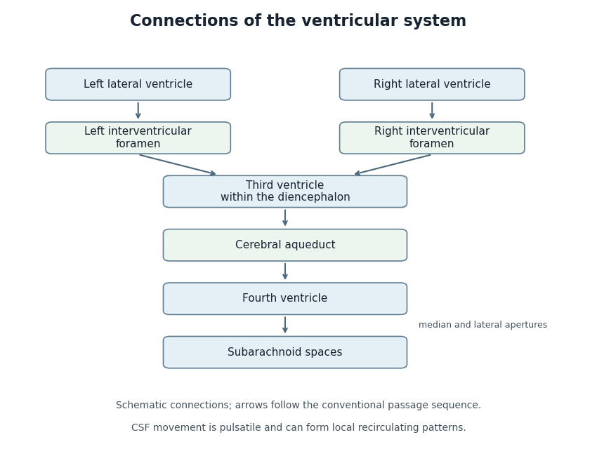
Anatomical connections of the ventricular system
. Original schematic showing paired lateral ventricles, the interventricular foramina, third ventricle, cerebral aqueduct, fourth ventricle and subarachnoid spaces. Arrows follow the conventional passage sequence; cerebrospinal fluid also undergoes pulsatile movement and local recirculation.[ref]Neuroscience, 2nd edition edited by D. Purves and others. The Ventricular System, Figure 1.17, anatomical connections
Details.
N. J. Pelc 2026-11-05
N. J. Pelc is a coauthor with D. R. Enzmann of a 1991 study that used phase-contrast magnetic resonance imaging to measure cerebrospinal fluid motion through the cardiac cycle. The work illustrates how magnetic resonance imaging can provide a time-dependent physiological measurement rather than only an anatomical picture.[ref]
Normal flow patterns of intracranial and spinal cerebrospinal fluid defined with phase-contrast cine MR imaging by D. R. Enzmann and others. Radiology178,467–474(1991), originating byline and abstract.
The investigators studied ten healthy volunteers and four patients whose conventional magnetic-resonance images were normal. Peripheral cardiac gating divided the measurement into 16 flow-encoded images per cycle. The flow encoding distinguished opposite directions of cerebrospinal fluid motion, and measurements at several locations showed that the timing of the motion differed between the ventricular system and surrounding spaces.[ref]
Normal flow patterns of intracranial and spinal cerebrospinal fluid defined with phase-contrast cine MR imaging by D. R. Enzmann and others. Abstract: participant groups,16 cardiac phases, directional encoding and regional timing.
For an illustrative one-second cycle, 16 equally spaced phases would separate samples by seconds. The paper's number of cardiac phases therefore matters: an average over the entire cycle could hide changes of direction within it. Conversely, sixteen phases are a temporal sampling scheme, not evidence of sixteen independent subjects. This example explains the measurement design without assigning an unreported individual contribution to either coauthor.
Phase-contrast magnetic resonance imaging 2026-11-05
Phase-contrast magnetic resonance imaging estimates motion from differences in the phase of nuclear magnetic resonance signals acquired with controlled magnetic field gradients. The encoding measures a component of velocity, with its sign set by the gradient and reconstruction convention. A 1982 paper described an early flow-sensitive phase-encoding approach.[ref]
For a constant-velocity teaching model, write position along the encoding direction as . If is the effective gradient, including refocusing signs, thenHere is the gyromagnetic ratio in radians per second per tesla, and is the gradient first moment. With balanced zeroth moments, two encodings giveThese equations are an explicit idealized derivation, not a claim that every measured voxel contains one uniform velocity.
Define the velocity encoding limit by . If it is , a corrected phase difference of corresponds to under the chosen sign convention. Phase wrapping modulo causes an ambiguity beyond the encoding range, related to aliasing.
A 1991 gated study measured differently timed cerebrospinal fluid motion in ventricular and subarachnoid compartments over the cardiac cycle.[ref]
Normal flow patterns of intracranial and spinal cerebrospinal fluid defined with phase-contrast cine MR imaging by D. R. Enzmann and others. Abstract; 16 flow-encoded images per cardiac cycle
Opposite velocities during equal parts of a cycle can average to zero despite substantial oscillatory motion. A volumetric flow rate additionally requires integrating the normal velocity across a defined cross-section; peak speed alone does not supply transported volume. Radiologist 2026-11-05
A radiologist is a physician specializing in diagnosis and treatment using medical imaging. The role includes choosing or advising on suitable imaging methods, interpreting measurements in anatomical and clinical context, and communicating their significance. Some radiologists also perform procedures under imaging guidance through interventional radiology.[ref]
What does a radiologist do? by RadiologyInfo.org. Professional-society account of physician interpretation, collaboration and minimally invasive procedures.
The distinction from image acquisition matters. A radiologic technologist often operates the acquisition equipment, while a radiologist interprets the resulting images and relates them to the question being investigated. The work is collaborative; neither the instrument's output nor a single visual feature automatically supplies a diagnosis.[ref]
What does a radiologist do? by RadiologyInfo.org. Your Radiologist and the description of the healthcare team.
A scientific example is phase-encoded imaging of cerebrospinal fluid. D. R. Enzmann and collaborators used a sequence synchronized to the cardiac cycle to distinguish motion in different fluid spaces. Interpretation required more than recognizing their outlines: opposite flow directions and different timing provided information about fluid dynamics.[ref]
Velocity encoding limit 2026-11-05
The velocity encoding limit, commonly written , specifies the velocity scale represented by a phase difference of magnitude in a chosen phase-contrast magnetic resonance imaging encoding. With equal gradient zeroth moments and first-moment difference , an ideal constant-velocity model givesThe gyromagnetic ratio is angular, in radians per second per tesla, and is the difference of gradient first moments. A sign convention must be specified separately. A 1982 original paper described obtaining motion information through phase-sensitive nuclear magnetic resonance encoding.[ref]
A flow velocity zeugmatographic interlace for NMR imaging in humans by P. R. Moran. Abstract; phase encoding of flow information; displayed relations are the stated model
Choose the convention . If , then produces . But produces , which wraps to when phase is represented in . A reconstruction that ignores this aliasing would report . The limit describes an encoding ambiguity, not a physical maximum speed of the fluid.
For a fixed phase uncertainty in this simple model, propagation gives . Reducing improves velocity sensitivity under that assumption while narrowing the unambiguous range. For example, radians and give . Real measurements must also account for background phase, intravoxel variation and timing. Gated measurements of cerebrospinal fluid show why timing within the cardiac cycle matters even when net displacement is small.[ref]