Imaging based on spatial encoding of nuclear magnetic resonance signals. Acquisition and reconstruction choices determine which physical contrasts can be measured.
Magnetic resonance velocity encoding makes a component of motion measurable through the phase of a nuclear magnetic resonance signal. Controlled magnetic field gradients give different phase shifts to spins with different trajectories. Phase-contrast magnetic resonance imaging compares encodings to extract a velocity component, rather than interpreting ordinary image brightness as speed.[ref]
A flow velocity zeugmatographic interlace for NMR imaging in humans by P. R. Moran. Primary abstract: phase-sensitive flow encoding and reconstructed motion information
In an original idealized model, assume constant velocity along one encoding axis and effective gradients with equal gradient zeroth moments. Then the encoding difference obeyswhere is the angular gyromagnetic ratio and is the gradient first moment difference. For , and , this gives . These stipulated values illustrate the conversion, rather than a particular scanner experiment.
Reversing the moment difference reverses the phase sign under this convention. Reducing its magnitude decreases sensitivity to a given velocity while increasing the velocity encoding limit. A measured voxel can contain a range of velocities, and background phase or phase aliasing can complicate reconstruction.
The encoded component also differs from volumetric flow rate: estimating transported volume requires combining the appropriate normal velocity with area and time, not merely recording one peak speed.
Phase-contrast magnetic resonance imaging estimates motion from differences in the phase of nuclear magnetic resonance signals acquired with controlled magnetic field gradients. The encoding measures a component of velocity, with its sign set by the gradient and reconstruction convention. A 1982 paper described an early flow-sensitive phase-encoding approach.[ref]
For a constant-velocity teaching model, write position along the encoding direction as . If is the effective gradient, including refocusing signs, thenHere is the gyromagnetic ratio in radians per second per tesla, and is the gradient first moment. With balanced zeroth moments, two encodings giveThese equations are an explicit idealized derivation, not a claim that every measured voxel contains one uniform velocity.
Define the velocity encoding limit by . If it is , a corrected phase difference of corresponds to under the chosen sign convention. Phase wrapping modulo causes an ambiguity beyond the encoding range, related to aliasing.
A 1991 gated study measured differently timed cerebrospinal fluid motion in ventricular and subarachnoid compartments over the cardiac cycle.[ref]
Normal flow patterns of intracranial and spinal cerebrospinal fluid defined with phase-contrast cine MR imaging by D. R. Enzmann and others. Abstract; 16 flow-encoded images per cardiac cycle
Opposite velocities during equal parts of a cycle can average to zero despite substantial oscillatory motion. A volumetric flow rate additionally requires integrating the normal velocity across a defined cross-section; peak speed alone does not supply transported volume.A bipolar gradient is a magnetic field gradient waveform with lobes of opposite sign. In nuclear magnetic resonance, such waveforms can control sensitivity to position and motion. Opposite signs alone do not guarantee equal signed areas, and an applied waveform must be distinguished from its effective encoding waveform when refocusing pulses change phase evolution.
For an original counterexample, apply for and then for . The gradient zeroth moment isThe waveform is bipolar but unbalanced.
If the two durations are instead equal to , and the effective lobes are contiguous, from time 0 to and from to , then but the gradient first moment is . Thus cancellation of stationary-position phase can coexist with sensitivity to constant velocity. The sign follows this stated ordering.
Bipolar pulses also appear outside flow imaging. A 1989 stimulated echo study addressed background gradients that distort diffusion measurements. Alternating pulses could cancel a cross term under specified conditions; tests used proton signals from homogeneous and granular samples.[ref]
Pulsed field gradient stimulated echo methods for improved NMR diffusion measurements in heterogeneous systems by R. M. Cotts and others. Primary abstract: background-gradient reduction, conditional cross-term cancellation and measurements
This is a sequence-specific result, not a promise that every bipolar pair removes every imaging error.The gradient zeroth moment is the signed time integral of an effective magnetic field gradient waveform along a specified encoding axis:Its units are tesla seconds per metre. Effective means that the sign convention accounts for any refocusing pulses in the nuclear magnetic resonance sequence; it is not necessarily the unsigned physical pulse area.
For a stationary spin at position , choose the phase convention . Then , where is the angular gyromagnetic ratio. A balanced waveform with cancels this position-dependent contribution within the model. It does not assert that every possible background contribution to phase vanishes.
As an original rectangular-pulse example, for has . Adding for cancels it. Equal areas, rather than equal pulse heights, produce the balance.
For a moving spin with , the remaining contribution can be , involving the gradient first moment. That separation explains how magnetic resonance velocity encoding can suppress sensitivity to stationary position while preserving motion information. Early phase-sensitive flow imaging supplied the experimental motivation for extracting such information from resonance phase.[ref]
A flow velocity zeugmatographic interlace for NMR imaging in humans by P. R. Moran. Primary abstract; displayed moment relations are explicit idealized derivations
The velocity encoding limit, commonly written , specifies the velocity scale represented by a phase difference of magnitude in a chosen phase-contrast magnetic resonance imaging encoding. With equal gradient zeroth moments and first-moment difference , an ideal constant-velocity model givesThe gyromagnetic ratio is angular, in radians per second per tesla, and is the difference of gradient first moments. A sign convention must be specified separately. A 1982 original paper described obtaining motion information through phase-sensitive nuclear magnetic resonance encoding.[ref]
A flow velocity zeugmatographic interlace for NMR imaging in humans by P. R. Moran. Abstract; phase encoding of flow information; displayed relations are the stated model
Choose the convention . If , then produces . But produces , which wraps to when phase is represented in . A reconstruction that ignores this aliasing would report . The limit describes an encoding ambiguity, not a physical maximum speed of the fluid.
For a fixed phase uncertainty in this simple model, propagation gives . Reducing improves velocity sensitivity under that assumption while narrowing the unambiguous range. For example, radians and give . Real measurements must also account for background phase, intravoxel variation and timing. Gated measurements of cerebrospinal fluid show why timing within the cardiac cycle matters even when net displacement is small.[ref]
The gradient first moment of an effective magnetic field gradient waveform is its time-weighted integral,Here effective includes the sign changes caused by refocusing pulses. Its units are when is in tesla per metre. The gradient zeroth moment is . For position with constant velocity, the accumulated nuclear magnetic resonance phase iswhere is the angular gyromagnetic ratio. Thus removes the dependence on initial position while can retain sensitivity to velocity, the basis of an idealized phase-contrast magnetic resonance imaging encoding.[ref]
A flow velocity zeugmatographic interlace for NMR imaging in humans by P. R. Moran. Original abstract; motion-sensitive phase encoding; equations here are an explicit constant-velocity derivation
For a teaching bipolar gradient, let the first lobe have signed area and time centroid , and the second have signed area and centroid . ThenEqual and opposite lobe areas cancel stationary-spin phase, but their time separation prevents cancellation of the constant-velocity phase. For and , the first moment is . These are illustrative waveform values, not measurements from the source.
The time origin is part of the definition: replacing by gives . Balanced makes the first moment independent of that choice. Two encodings with equal zeroth moments use their first-moment difference , rather than either individual value, to infer velocity.
An imaging approach describing direction-dependent water diffusion using a tensor model. In white matter it provides indirect information about tissue organisation; crossing fibres, motion and model limitations affect interpretation.
Diffusion MRI gradient sampling is the choice of magnetic field gradient directions, strengths and repetitions used to acquire diffusion magnetic resonance imaging measurements. Under a Gaussian diffusion tensor model, different directions probe different values of a quadratic form. Acquiring many measurements in an uninformative set of directions cannot determine every tensor coefficient.
For a unit gradient direction , a separately measured matched baseline and fixed positive diffusion weighting b-value , the ideal signal model isThe symmetric matrix has six independent coefficients. Opposite directions and give the same model prediction. Thus counting orientations without considering their independent information can overstate diffusion tensor identifiability.
A 2003 Monte Carlo simulation study controlled the observation budget: one baseline plus60 diffusion-weighted observations. Six distinct orientations repeated ten times and30 distinct orientations repeated twice each use60 diffusion-weighted observations. The comparison asks how angular coverage and repetition affect estimated orientation, rather than simply comparing a longer acquisition with a shorter one.[ref]
The Effect of Gradient Sampling Scheme on Estimates of Fiber Orientation: Implications for Fiber Tractography by D. K. Jones. Methods and Table1: observation budget, directions and repetitions
The subsequent 2004 study compared direction schemes through estimated orientation, trace and fractional anisotropy at . These are distinct outputs of the same tensor fit. An acquisition suited to one readout need not minimize uncertainty in another.[ref]
The effect of gradient sampling schemes on measures derived from diffusion tensor MRI: A Monte Carlo study by Derek K. Jones. Primary Abstract: Monte Carlo design and tensor-derived measurements
An original design comparison separates two questions. Repeating the three coordinate axes may reduce random variation in their measured attenuations, but it adds no equations for the three off-diagonal tensor entries. Adding suitable mixed directions can supply those equations. Full matrix rank establishes uniqueness in the ideal model; practical measurement uncertainty, motion and model mismatch still require assessment.
Diffusion tensor identifiability asks whether the measurements determine a unique diffusion tensor within a stated model. For a constant symmetric matrix , known matched baseline and known , defineFor unit directions , these equations give a linear system with rowAll six coefficients are uniquely determined in this unrestricted noise-free linear model exactly when . The condition concerns independent equations, not merely the number of acquired images. Direction design and uncertainty in tensor-derived measurements were examined experimentally through controlled simulations by Derek Jones.[ref]
The effect of gradient sampling schemes on measures derived from diffusion tensor MRI: A Monte Carlo study by Derek K. Jones. Primary Abstract: sampling comparisons; displayed rank criterion is an explicit linear-algebra derivation
For a counterexample, use the six directions . Each antipodal pair repeats a row, so the matrix rank is three. Only are determined. Even restricting to physically positive tensors does not generally repair this: the identity matrix and a matrix with unit diagonal and both fit those data for any , while having different off-diagonal entries.
Replace the negative axes by , and . The axes first give the diagonal entries, thenand similarly for and . This directly proves uniqueness for the new design. If , and , all in units of , then in those units.
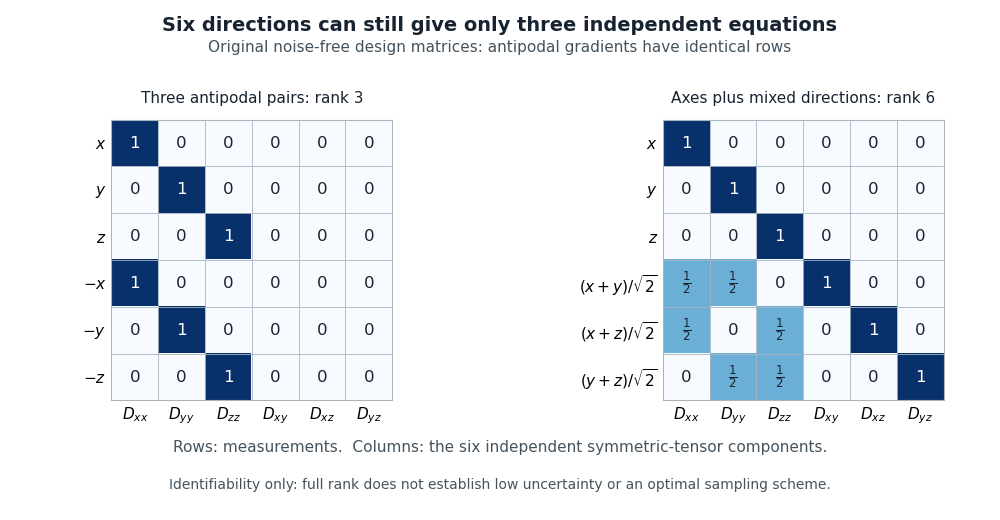
Original design-matrix example: antipodal axes give rank three, while axes and mixed directions give rank six
. Reused algebra illustration, not a reconstruction of a published acquisition or simulation. Rows represent measurements; columns represent the six independent tensor coefficients. Details.Full matrix rank is not a guarantee of a stable estimate in noisy data. If must also be fitted, its logarithm is another unknown; the augmented signal model needs a corresponding independent baseline constraint. The six-coefficient argument above explicitly assumes is separately known. Likewise, tractography requires additional interpretation beyond uniqueness of a local tensor.
A dimensionless measure of dispersion among the diffusion tensor eigenvalues. With eigenvalues and mean , when the denominator is nonzero. It is not a direct measure of axon number or functional necessity.
Magnetic-resonance measurements sensitive to water diffusion. Models of directional diffusion can support tractography, subject to limitations such as crossing fibers, partial volume and modeling assumptions.
Estimation of plausible pathways from diffusion measurements and modeling choices. Reconstructed streamlines are not a direct count of axons and do not by themselves determine synaptic direction, signal flow or functional necessity.
Imaging task-related or spontaneous changes in magnetic-resonance signals associated with physiology, commonly the BOLD signal. Hemodynamic timing and spatial averaging constrain inferences about neural firing and cell-level computations.
A magnetic-resonance signal sensitive to blood oxygenation and related vascular changes. It is an indirect correlate of local neural processing, with slower dynamics than individual spikes.
An ideal resonant inversion pulse reverses the longitudinal magnetization. Subsequent spin-lattice relaxation gives the displayed recovery law. Observation at a chosen inversion time provides contrast or nulling for a specified relaxation time; an RF inversion alone does not amplify the initial polarization magnitude.
Magnetic resonance imaging with a prepared nuclear population imbalance exceeding ordinary thermal polarization under the imaging conditions. Dynamic nuclear polarization, optical spin transfer and spin-order transfer offer different preparation routes. Preparation and acquisition must account for spin-lattice relaxation, which erases the excess polarization.
The spatial Fourier-frequency coordinates sampled by a magnetic-resonance acquisition. With gyromagnetic ratio in radians per time per field, the displayed expression is in cycles per length. Signal phase, gradient history and sequence refocusing determine the trajectory; the samples require reconstruction to form a spatial image.
Articles by others on the same topic

Magnetic Resonance Imaging (MRI) is a medical imaging technique used to visualize detailed internal structures of the body. It employs strong magnetic fields, radio waves, and a computer to produce images of organs, soft tissues, bones, and other internal body structures. Key features of MRI include: 1. **Non-Invasive**: MRI is a non-invasive procedure that does not involve ionizing radiation, making it safer compared to techniques like X-rays or CT scans.


How MRI Works Part 1 by thePIRL (2018)
Source. - youtu.be/TQegSF4ZiIQ?t=326 the magnet is normally always on for the entire lifetime of the equipment!
- youtu.be/TQegSF4ZiIQ?t=465 usage of non-ionizing radiation (only radio frequencies) means that it is very safe to use. The only dangerous part is the magnetic field interacting with metallic objects.
Tomorrow's World gave audiences a true world first as Dr Peter Mansfield of the University of Nottingham demonstrated the first full body prototype device for Magnetic resonance imaging (MRI), allowing us to see inside the human body without the use of X-rays.