A strongly moving lung target is a relative contraindication to a particular proton therapyplan if respiratory motion and changing air–tissue paths make robust target coverage and normal-tissue constraints unattainable. A changing water-equivalent path changes the particle range; pencil-beam scanning can also interact with respiratory motion. A photon radiotherapyplan may then offer more reliable coverage despite its greater exit dose. This is a conditional planning contraindication, not a blanket claim that lung cancer cannot be treated with protons: gating, breath-hold, rescanning and adaptive planning can change the decision. If a robust comparative plan shows no meaningful benefit, the additional cost and access burden also weigh against the protonplan.[ref]
A localized paediatric intracranial tumour is a representative indication where proton therapy can reduce unnecessary absorbed dose to the developing brain and other normal tissue. The finite particle range can reduce exit and integral dose compared with many photon radiotherapyplans, which may reduce late effects and secondary cancerrisk over a long expected survival. It remains necessary to maintain target coverage, account for changing anatomy and keep uncertain distal-edge relative biological effectiveness away from sensitive structures. Travel, cost and daily positioning are practical disadvantages. The relevant comparison is a robust individual treatment plan and clinical endpoint, rather than the label “proton” alone.[ref]
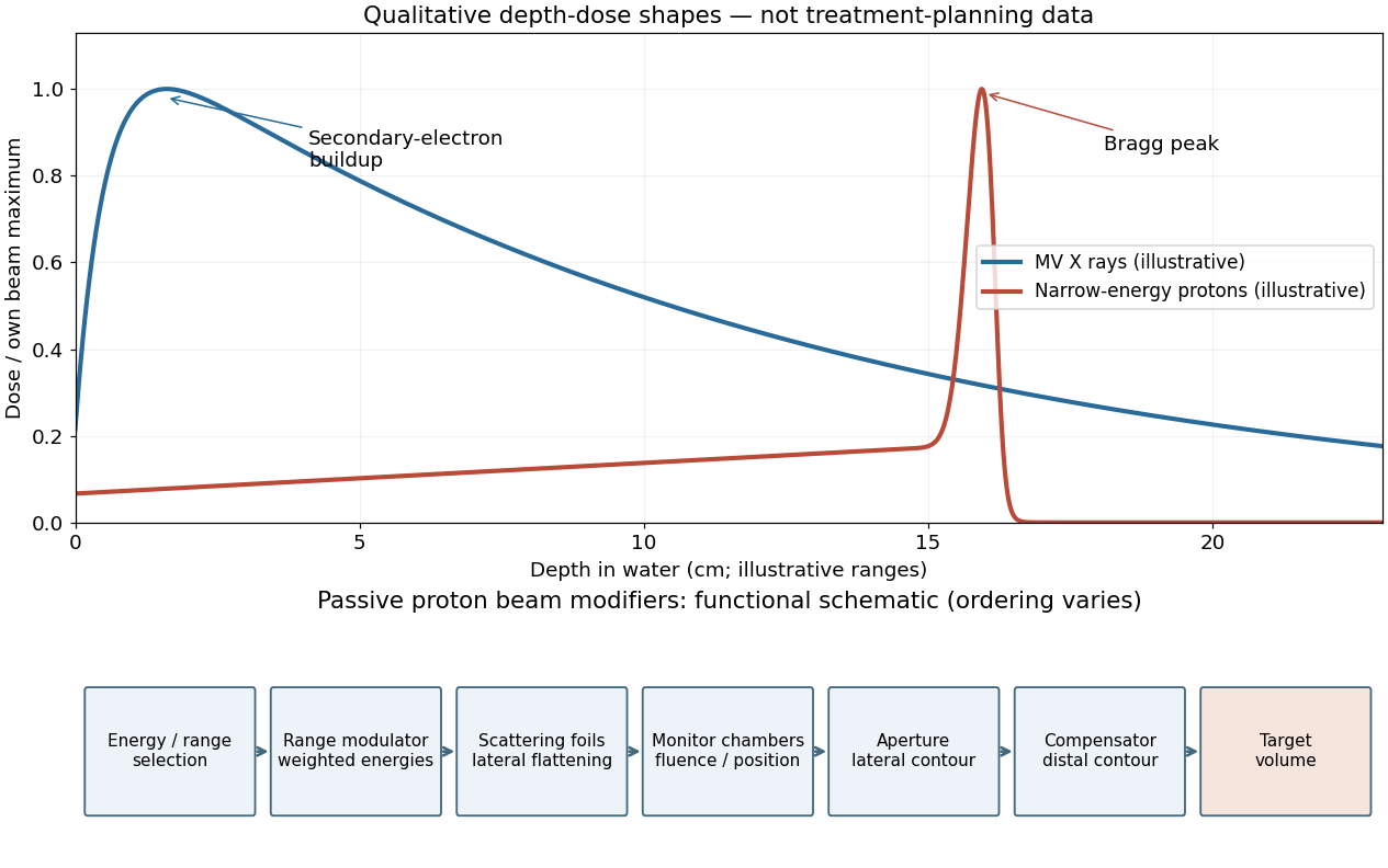
A nearly monoenergetic proton beam deposits a moderate entrance dose, then a sharp Bragg peak near the end of its range. As nonrelativistic protons slow, their electronic stopping power rises approximately as 1/v2 over the relevant part of the track. Once the protons stop, the dose falls steeply. Range straggling and energy spread broaden the peak; nuclear interactions add a smaller nonideal tail, so an exact hard cutoff is an idealization. Superposing appropriate incident energies produces a spread-out Bragg peak covering an extended target.[ref]
A passively scattered proton therapy beam uses distinct devices to control lateral spread, range and distal shape. The lower panel of the figure gives afunctional schematic; the exact ordering varies among beamlines. An energy selector or range shifter sets the maximum range. A stepped range-modulation wheel or ridge filter gives a weighted collection of protonenergies, forming the spread-out Bragg peak. One or more scattering foils broaden the narrow beam; a double-scattering system can improve lateral uniformity. Ionization chambers monitor delivered fluence and position. Apatient-specificaperture or collimator blocks unwanted lateral regions. A shaped range compensator makes the remaining range vary across the field, matching the distal target boundary. Their effects must be combined: lateral collimation alone cannot flatten the depth distribution, and an energy modulator alone cannot set the lateral contour.[ref]