An ionization chamber measures ionizing radiation by collecting the electric charge that it creates in a gas. Two electrodes establish an electric field; liberated electrons and positive ions move toward opposite electrodes. In the chamber's intended operating regime the field collects the initial charge with negligible electron avalanche multiplication, distinguishing it from a proportional counter.
Varying the applied electric potential difference while holding the incident radiation fixed produces a collection curve. Too weak a field allows electron-ion recombination before collection; an approximately flat region indicates near-complete collection under those conditions. Increasing the field far enough can instead cause gas multiplication. A 2013 synchrotron radiation experiment compared free-air chambers with different electrode gaps and chopped the X-ray beam, showing that collection depends on both geometry and the temporal delivery of radiation.[ref]
Current saturation in free-air ionization chambers with chopped synchrotron radiation by Nobuteru Nariyama. abstract and sections 2–3: chamber gaps, chopped irradiation and recombination
For a simple counting model, let each interacting particle create on average electron-ion pairs and let be their collected fraction. An interaction rate then gives mean electric current , where is the elementary charge. This accounting explains why loss of charge can mimic a reduced incident particle flux; converting a current to absorbed dose requires additional information about energy deposition and the irradiated mass.
Detector volume averaging is the spatial averaging of a nonuniform radiation field over a detector's finite sensitive region. A reading associated with the detector centre need not equal the local absorbed dose at that point. This limits spatial resolution even if the detector position and electronic readout are exact.
For an idealized linear response, let be the absorbed dose field in the undisturbed medium and let be a normalized spatial weighting function, with . Model the spatially averaged quantity byThe model separates averaging from the change in the radiation field caused by inserting the detector. A real ionization chamber can require both effects to be considered. The IAEA definition compares a point dose with the average over the detector's sensitive volume, both evaluated in water with the detector absent.[ref]
Dosimetry of Small Static Fields Used in External Beam Radiotherapy by the International Atomic Energy Agency. §3.1.4, printed p. 37, definition and spatial weighting in equation (5)
For an original one-dimensional example, suppose the detector averages uniformly over a full width . ThenAn ideal sharp edge , where is the Heaviside step function, is recorded asThus a detector gives a distance between the 20-percent and 80-percent response points, although the stipulated field edge has zero transition width. A detector gives in the same model.
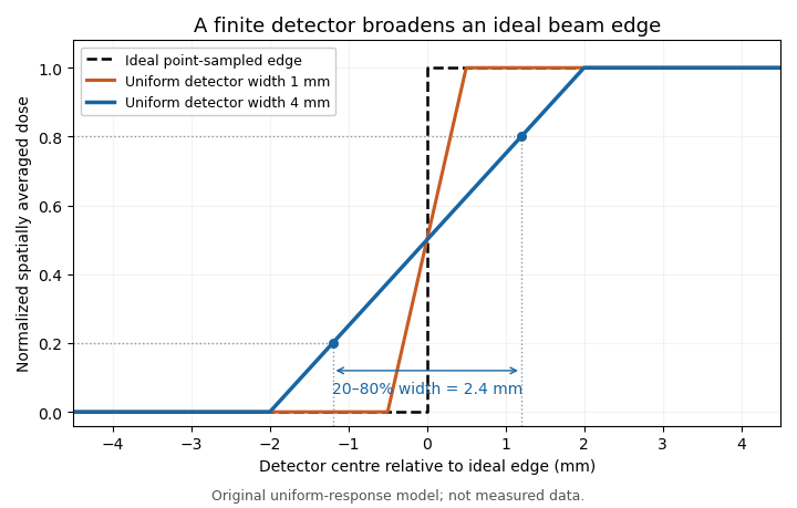
Uniform finite-detector averaging turns a sharp radiation-field edge into a ramp. Original model calculation; the indicated 2.4 mm width belongs to the 4 mm detector, not to an intrinsic field transition.
Details. For a smooth field, a Taylor expansion under the symmetric uniform weighting gives . A constant slope averages to its centre value; curvature produces the leading local discrepancy. A detector-volume-averaging correction factor therefore depends on the field shape and detector geometry, rather than only on the detector's nominal volume.
A proposed apparatus test scans a stable radiation field with detectors of different effective widths or orientations, keeping depth and irradiation geometry fixed. Changes in the recorded edge width test spatial response, while a uniform region checks relative sensitivity. The IAEA's geometric weighting examples show why a projected detector area may need nonuniform weighting; finer scan steps alone cannot remove averaging within each reading.[ref]
A detector-volume-averaging correction factor converts a specified spatially averaged absorbed dose into the point value associated with that measurement. It depends on the radiation field and the detector geometry. The IAEA defines this comparison in undisturbed water, with the detector absent, so the factor isolates detector volume averaging from perturbations caused by inserting an ionization chamber.[ref]
For a normalized nonnegative spatial weighting function , letassuming the denominator is positive. Multiplication by then recovers the point value in this response model. The averaging law is developed in detector volume averaging; the correction requires additional information about the spatial field, not just one scalar detector reading.
For an original exact example, take a uniformly sensitive one-dimensional detector of full width centred at a smooth peak. Over its footprint stipulate with and . Direct integration givesWith and , the average is and . A small local under-response becomes a factor greater than one.
At a valley with and the same parameters, the factor is instead . Thus an averaging correction need not exceed one. A uniform field gives exactly one. Reusing a peak-derived factor at a different location can therefore introduce an error.
In a proposed characterization, obtain a sufficiently resolved beam profile, account for the profiling detector's own spatial resolution, and integrate that profile with the intended detector's geometric weighting. The IAEA's geometric models explicitly distinguish different weightings across the projected detector area.[ref]
Dosimetry of Small Static Fields Used in External Beam Radiotherapy by the International Atomic Energy Agency. Appendix I.2.3, printed pp. 147–150, equations (52)–(53) and weighting models
A factor derived this way addresses spatial averaging under the stated field conditions; it does not independently correct energy response, recombination or an erroneous measurement calibration.Electron-ion recombination combines a free electron with a positive ion, reducing the number of free charge carriers. The released binding energy must be carried away, for example by a photon or another particle.
In an ionization chamber, recombination before charge collection reduces the measured signal. A simplified local model has loss rate , with coefficient depending on the species and conditions. Increasing the collection field can reduce the time available for recombination, but complete collection must be established experimentally rather than assumed from the applied voltage alone.
Articles by others on the same topic


An ionization chamber is a type of radiation detector that measures ionizing radiation by collecting and measuring the charge created when radiation interacts with a gas within the chamber. It consists of a gas-filled chamber with two electrodes: an anode (positive electrode) and a cathode (negative electrode). When ionizing radiation, such as alpha particles, beta particles, or gamma rays, passes through the gas, it ionizes the gas molecules, creating positive ions and free electrons.