Secretory protein routing connects entry into the endoplasmic reticulum, protein folding and sorting, transfer to the Golgi apparatus, and delivery onward. In the conventional soluble-cargo route, transport vesicles carry lumenal contents between compartments; exocytosis releases cargo outside the cell membrane. Resident endoplasmic reticulum proteins can be retained or retrieved, and defective cargo can be held back. Many cytosol proteins never enter this route.[ref]
The Endoplasmic Reticulum. The Cell: A Molecular Approach, 2nd edition by Geoffrey M. Cooper; Protein Folding and Processing in the ER, Figure 9.14; Export of Proteins and Lipids from the ER, Figures 9.20 and 9.21
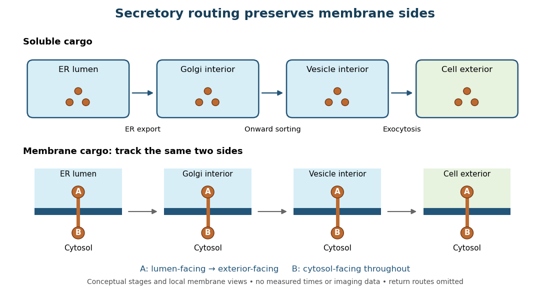
Secretory protein routing preserves membrane sides
. Original conceptual route for soluble cargo and a membrane protein. Intermediate compartments and return routes are omitted. Arrows show connectivity, not measured timing, fluorescence or electron microscopy. Details.
The useful geometric rule is preservation of membrane sides: during budding and fusion, a lumen-facing region of a membrane protein stays lumen-facing, then faces the exterior after exocytosis; its cytosol-facing region stays cytosolic.[ref]
Work through the schematic using two distinguishable labels A and B on a stipulated membrane protein. Start with A facing the ER lumen and B facing the cytosol. At each budding or fusion step, carry the surrounding membrane together with the labels; do not turn the protein over. The resulting pair is A facing the Golgi apparatus interior and B facing the cytosol, then A facing a transport vesicle interior and B facing the cytosol, and finally A facing the exterior and B facing the cytosol. This is a stepwise argument about the depicted operations, not a claim that the compartments have identical chemical contents. Likewise, once soluble cargo is inside the ER lumen, this schematic reaches the exterior with zero further crossings through a lipid bilayer: enclosed spaces join when membranes fuse. Delivery therefore need not mean repeatedly pushing a folded protein through a membrane.
In 1967, James D. Jamieson and George E. Palade incubated guinea pig pancreas slices, following a three-minute pulse of tritium-labeled leucine with excess unlabeled leucine. This pulse-chase analysis marked newly made proteins while reducing subsequent incorporation. Fixed, Epon-embedded thin sections were mounted on coated grids, covered with Ilford L-4 photographic emulsion, developed and stained for electron microscopy. Autoradiography located photographic grains relative to pancreatic acinar cell structures.[ref]
Intracellular Transport of Secretory Proteins in the Pancreatic Exocrine Cell. II. Transport to Condensing Vacuoles and Zymogen Granules by James D. Jamieson and George E. Palade. Jamieson and Palade, Intracellular Transport of Secretory Proteins in the Pancreatic Exocrine Cell. II. Transport to Condensing Vacuoles and Zymogen Granules (1967), p. 598 Methods; Figure 10: three-minute pulse
Pulse-end samples provided a temporal reference: label lay mainly over rough endoplasmic reticulum; later samples shifted toward peripheral Golgi apparatus elements, condensing vacuoles and secretory granules. As a contamination check, isolated granule pellets were also examined by autoradiography: early pellet labeling chiefly marked condensing vacuoles. Grids from the compared pellet samples were coated together to control photographic processing. This supported transport of a previously labeled cohort rather than separate synthesis at each location. Fixed samples did not track individual proteins; localization was finite, and labeling was not absolutely exclusive to secretory cargo. The result concerns this preparation, not every protein in every biological cell.[ref]
Intracellular Transport of Secretory Proteins in the Pancreatic Exocrine Cell. II. Transport to Condensing Vacuoles and Zymogen Granules by James D. Jamieson and George E. Palade. pp. 603–606 and Figures 10–13: changing localization; pp. 610–611 and Figures 14–15, Table III: pellet check and matched emulsion processing; p. 611 General Premises: nonexportable-protein labeling
Articles by others on the same topic
There are currently no matching articles.