An organelle is a specialized subcellular structure. Usage varies over whether non-membrane-bounded structures such as ribosomes count as organelles, so explicitly say membrane-bounded organelles when comparing eukaryotic compartmentalization with prokaryotic organization. Mitochondria and chloroplasts are membrane-bounded examples. See OpenStax Microbiology, 3.4: cellular compartments.
A membrane-bounded organelle with pathways including oxidative metabolism. Many matrix proteins are imported after cytosolic synthesis, including folded substrates.
The maintained combination of membrane composition, resident proteins, lumenal conditions, geometry and trafficking functions that distinguishes a compartment. Its molecules can turn over while this functional organization persists.
A microtubule-organizing structure in relevant animal cells. Positioning centrosomes relative to a niche can prepare the orientation of a later mitotic spindle. Budding yeast instead has a spindle pole body.
A highly reduced mitochondrion-related organelle retaining selected metabolic functions rather than oxidative ATP production. Functions differ among lineages, including iron-sulfur cluster maturation in Giardia and sulfate activation in Entamoeba histolytica.[ref]
A mitochondrion-related organelle class associated with anaerobic metabolism and, in typical examples, hydrogen and ATP production. Substrate-level phosphorylation can supply energy without a conventional oxygen-terminated respiratory chain.[ref]
The endoplasmic reticulum is a membrane-enclosed network of tubules and flattened sacs in a typical eukaryotic biological cell. Its continuous boundary encloses the ER lumen, separate from the cytosol, and connects with the outer nuclear envelope. Rough endoplasmic reticulum and smooth endoplasmic reticulum are regions of this network.[ref]
The Endoplasmic Reticulum. The Cell: A Molecular Approach, 2nd edition by Geoffrey M. Cooper; The Endoplasmic Reticulum, opening paragraph and Figure 9.1: continuous membrane and nuclear-envelope connection
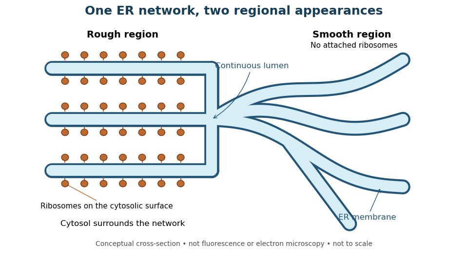
One endoplasmic reticulum network with rough and smooth regions
. Original conceptual cross-section: blue interiors connect, while attached ribosomes distinguish rough regions. Shapes and dimensions are illustrative, not measured fluorescence or electron microscopy. Details.The sarcoplasmic reticulum is the specialized endoplasmic reticulum of muscle cells that stores and releases calcium. Pump uptake, channel-mediated release and passive leak exchange calcium with the cytoplasm; these internal transfers cancel in the appropriately volume-weighted total-calcium balance. They can nevertheless change cytoplasmic calcium rapidly and control contraction.
endoplasmic reticulum stress arises when ER demands exceed its processing or homeostatic capacity. It can alter folding, degradation and transport, confounding interpretation of a supposedly selective secretion defect.
The endoplasmic reticulum lumen is the aqueous space enclosed by the ER membrane. A soluble lumenal cargo cannot directly contact a cytoplasmic coat across an intact membrane.
An endoplasmic reticulum exit site is a region organizing export machinery and cargo departure from the ER. Local coat assembly, cargo concentration and membrane supply can be coordinated there.
The ER lumen is the internal aqueous compartment of the endoplasmic reticulum. A ribosome attached to its membrane sits on the cytosol side, not inside the ER lumen. This distinction separates the machinery making a protein from the compartment receiving it.[ref]
The Endoplasmic Reticulum. Molecular Biology of the Cell, 4th edition by Bruce Alberts and others; Membrane-bound Ribosomes Define the Rough ER, Figure 12-36: cytosolic ribosomes and lumenal delivery
Rough endoplasmic reticulum bears attached ribosomes on its cytosol surface. In a common mammalian route, protein targeting delivers a nascent chain into the ER lumen during biological translation. Other proteins enter the membrane. Free and attached ribosomes share the same basic machinery; targeting depends on their cargo.[ref]
The Endoplasmic Reticulum. Molecular Biology of the Cell, 4th edition by Bruce Alberts and others; Membrane-bound Ribosomes Define the Rough ER, Figures 12-36 and 12-37; Translocation Across the ER Membrane Does Not Always Require Ongoing Polypeptide Chain Elongation
An ER signal sequence is polypeptide chain targeting information recognized by machinery delivering a protein to the endoplasmic reticulum. Many soluble cargoes have a removable N-terminus signal; membrane insertion can use internal signals. An ER signal sequence alone does not specify the final destination.[ref]
The Endoplasmic Reticulum. Molecular Biology of the Cell, 4th edition by Bruce Alberts and others; The ER Signal Sequence Is Removed from Most Soluble Proteins After Translocation; In Single-Pass Transmembrane Proteins, a Single Internal ER Signal Sequence Remains in the Lipid Bilayer as a Membrane-spanning α Helix
For an original counting example, stipulate an unbranched precursor polypeptide chain of amino acid residues whose first amino acid residues form a cleaved ER signal sequence. With no further processing, the remaining chain has amino acid residues and peptide bonds. The initial chain had peptide bonds; the detached signal has , so remain after one bond is cut. The check counts adjacent positions: an unbranched chain of amino acid residues has peptide bonds. These stipulated lengths are not a natural sequence, a measured size or evidence that a particular sequence functions as an ER signal sequence.
Smooth endoplasmic reticulum lacks attached ribosomes. Specialized regions support lipid metabolism and processing of foreign compounds; the endoplasmic reticulum also stores and releases calcium ions. Regional specialization does not split the network into two separate organelles, nor make every smooth region perform every function.[ref]
The Endoplasmic Reticulum. Molecular Biology of the Cell, 4th edition by Bruce Alberts and others; Smooth ER Is Abundant in Some Specialized Cells, Figures 12-38A and 12-38B and calcium-storage paragraphs
Secretory protein routing connects entry into the endoplasmic reticulum, protein folding and sorting, transfer to the Golgi apparatus, and delivery onward. In the conventional soluble-cargo route, transport vesicles carry lumenal contents between compartments; exocytosis releases cargo outside the cell membrane. Resident endoplasmic reticulum proteins can be retained or retrieved, and defective cargo can be held back. Many cytosol proteins never enter this route.[ref]
The Endoplasmic Reticulum. The Cell: A Molecular Approach, 2nd edition by Geoffrey M. Cooper; Protein Folding and Processing in the ER, Figure 9.14; Export of Proteins and Lipids from the ER, Figures 9.20 and 9.21
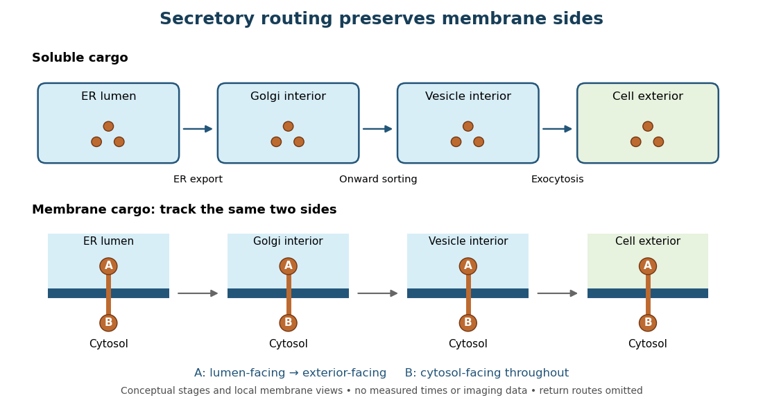
Secretory protein routing preserves membrane sides
. Original conceptual route for soluble cargo and a membrane protein. Intermediate compartments and return routes are omitted. Arrows show connectivity, not measured timing, fluorescence or electron microscopy. Details.The useful geometric rule is preservation of membrane sides: during budding and fusion, a lumen-facing region of a membrane protein stays lumen-facing, then faces the exterior after exocytosis; its cytosol-facing region stays cytosolic.[ref]
Work through the schematic using two distinguishable labels A and B on a stipulated membrane protein. Start with A facing the ER lumen and B facing the cytosol. At each budding or fusion step, carry the surrounding membrane together with the labels; do not turn the protein over. The resulting pair is A facing the Golgi apparatus interior and B facing the cytosol, then A facing a transport vesicle interior and B facing the cytosol, and finally A facing the exterior and B facing the cytosol. This is a stepwise argument about the depicted operations, not a claim that the compartments have identical chemical contents. Likewise, once soluble cargo is inside the ER lumen, this schematic reaches the exterior with zero further crossings through a lipid bilayer: enclosed spaces join when membranes fuse. Delivery therefore need not mean repeatedly pushing a folded protein through a membrane.
In 1967, James D. Jamieson and George E. Palade incubated guinea pig pancreas slices, following a three-minute pulse of tritium-labeled leucine with excess unlabeled leucine. This pulse-chase analysis marked newly made proteins while reducing subsequent incorporation. Fixed, Epon-embedded thin sections were mounted on coated grids, covered with Ilford L-4 photographic emulsion, developed and stained for electron microscopy. Autoradiography located photographic grains relative to pancreatic acinar cell structures.[ref]
Intracellular Transport of Secretory Proteins in the Pancreatic Exocrine Cell. II. Transport to Condensing Vacuoles and Zymogen Granules by James D. Jamieson and George E. Palade. Jamieson and Palade, Intracellular Transport of Secretory Proteins in the Pancreatic Exocrine Cell. II. Transport to Condensing Vacuoles and Zymogen Granules (1967), p. 598 Methods; Figure 10: three-minute pulse
Pulse-end samples provided a temporal reference: label lay mainly over rough endoplasmic reticulum; later samples shifted toward peripheral Golgi apparatus elements, condensing vacuoles and secretory granules. As a contamination check, isolated granule pellets were also examined by autoradiography: early pellet labeling chiefly marked condensing vacuoles. Grids from the compared pellet samples were coated together to control photographic processing. This supported transport of a previously labeled cohort rather than separate synthesis at each location. Fixed samples did not track individual proteins; localization was finite, and labeling was not absolutely exclusive to secretory cargo. The result concerns this preparation, not every protein in every biological cell.[ref]
Intracellular Transport of Secretory Proteins in the Pancreatic Exocrine Cell. II. Transport to Condensing Vacuoles and Zymogen Granules by James D. Jamieson and George E. Palade. pp. 603–606 and Figures 10–13: changing localization; pp. 610–611 and Figures 14–15, Table III: pellet check and matched emulsion processing; p. 611 General Premises: nonexportable-protein labeling
A microsome is a closed lipid bilayer compartment produced during disruption of a biological tissue; rough endoplasmic reticulum-derived microsomes provide a sealed compartment for protein targeting experiments.
A plastid is an organelle of the lineage that includes chloroplasts. A chloroplast is a photosynthetic plastid; the terms are not interchangeable because some plastids are nonphotosynthetic. Endosymbiotic theory connects plastid origins to cyanobacteria, while endosymbiotic gene transfer and organelle protein import explain genetic and functional integration with the host.
An apicoplast is a reduced nonphotosynthetic plastid in many apicomplexan parasites. Retained biosynthetic outputs can make it essential after photosystems and photosynthetic carbon fixation are lost.
A mitochondrion is an organelle with an outer mitochondrial membrane and an inner mitochondrial membrane. The latter encloses the mitochondrial matrix and folds into cristae; the intermembrane space lies on its opposite side. This architecture separates a chemical workspace from a membrane that couples electron transport to ATP formation.[ref]
4.3 Eukaryotic Cells. Biology 2e by Mary Ann Clark and others; §4.3, Mitochondria and Figure 4.14: two membranes, matrix and cristae
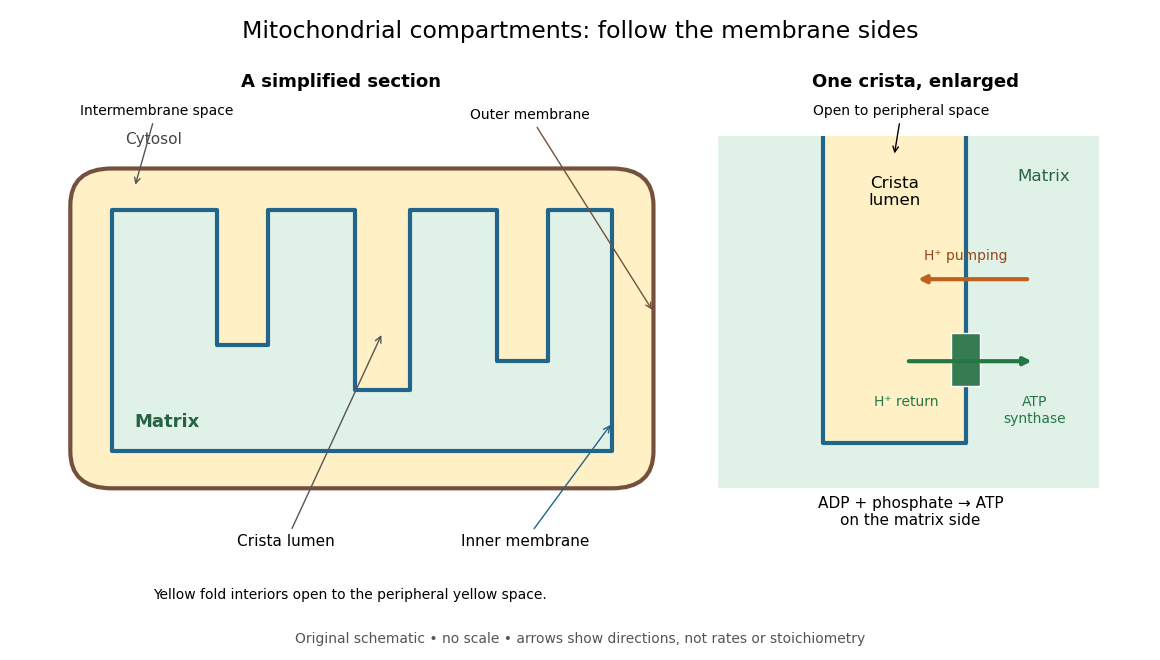
For an original location exercise, place soluble label A in the mitochondrial matrix and soluble label B in the interior of a crista. In the depicted geometry, B can reach the peripheral intermembrane space through the fold's opening without crossing a lipid bilayer. A must cross the inner mitochondrial membrane to reach either location. A fold projecting into the mitochondrial matrix therefore does not make its enclosed fluid part of the mitochondrial matrix. Following a continuous path through the colored regions proves the distinction; merely asking whether a label is “inside the mitochondrion” loses it.
The functional connection is equally spatial. The mitochondrial respiratory chain pumps protons from the mitochondrial matrix side across the inner mitochondrial membrane. Their return through ATP synthase can drive ATP formation. Oxidative phosphorylation develops the mechanism and chemiosmosis gives its free energy bookkeeping. Compartmentalization is useful because it lets a shared electrochemical gradient couple different proteins without direct transfer of a chemical intermediate between each pair.
Under aerobic conditions, mitochondria supply much of the ATP in many eukaryotic biological cells; the contribution varies with biological tissue, demand and oxygen supply. Glycolysis also makes ATP in the cytosol. Mature mammalian red blood cells lack mitochondria, and bacteria have no mitochondria at all. Neither all ATP production nor all biological cells fit the familiar mitochondrial picture.[ref]
Biology 2e by Mary Ann Clark and others. §4.3, Mitochondria; §7.2, glycolysis; §40.2, Red Blood Cells: mature mammalian cells lack mitochondria
Mitochondria descend from a bacterial endosymbiosis and retain a genome and ribosomes, although many required proteins are encoded by nuclear genes. Endosymbiotic theory develops the ancestry argument; organelle protein import explains delivery of nuclear-encoded proteins. A functioning isolated mitochondrion demonstrates biochemical autonomy under supplied conditions, rather than independently establishing that ancestry.
Dynamic remodeling of a mitochondrial population through division and merger. Fusion permits content mixing, while fission supports distribution and can segregate portions for mitophagy.
The inner mitochondrial membrane separates mitochondrial matrix from intermembrane space. Its selective permeability permits distinct chemical conditions, while embedded mitochondrial respiratory chain proteins and ATP synthase support oxidative phosphorylation. Cristae increase its surface area.[ref]
Molecular Biology of the Cell, 4th edition by Bruce Alberts and others. Chapter 14, The Mitochondrion Contains an Outer Membrane, an Inner Membrane, and Two Internal Compartments; Figure 14-8
A selective boundary and a productive enzyme have different jobs. Even active respiratory enzymes cannot sustain a useful electrochemical gradient if protons return freely across the boundary. Conversely, an intact boundary without an electron donor does not supply the energy for continued pumping. The isolated mitochondria respiration experiment separates some of these dependencies by additions to the same preparation.
The electric potential difference across the inner mitochondrial membrane, contributing to the proton-motive force. A change in potential can alter both useful phosphorylation and leak behavior.
A transport protein supplying phosphate across the inner mitochondrial membrane for processes including ATP synthesis.
A carrier exchanging adenine nucleotides across the inner mitochondrial membrane, supplying matrix ADP and exporting synthesized ATP during ordinary oxidative phosphorylation.
A transport protein exchanging suitable dicarboxylates, including succinate and malate, with counter-substrates such as phosphate. Its relevance to respiration depends on the fuel and preparation.
A crista is a fold of the inner mitochondrial membrane into the mitochondrial matrix. Its membrane remains continuous with the surrounding inner mitochondrial membrane; it is not a separately inserted sac like a thylakoid.[ref]
Molecular Biology of the Cell, 4th edition by Bruce Alberts and others. Chapter 12, The Transport of Proteins into Mitochondria and Chloroplasts, Figure 12-22: continuity of cristae and contrast with thylakoids
The diagram idealizes access through the fold's opening. Connectivity alone does not imply instant mixing or identical concentrations throughout the intermembrane space. Narrow geometry and molecular interactions can matter to transport. Increasing surface area also does not, by itself, prove a proportional increase in ATP production: the supply of substrates, available oxygen and the state of the coupling machinery must also support the flux.
The mitochondrial matrix is the compartment enclosed by the inner mitochondrial membrane, containing mitochondrial DNA, ribosomes and many enzymes. Most citric acid cycle steps occur here; succinate dehydrogenase is membrane-associated. The citric acid cycle supplies NADH for the mitochondrial respiratory chain.[ref]
Molecular Biology of the Cell, 4th edition by Bruce Alberts and others. Chapter 14, The Mitochondrion Contains an Outer Membrane, an Inner Membrane, and Two Internal Compartments; Figure 14-8, matrix enzymes and inner-membrane succinate dehydrogenase
Distinguish the location of a chemical reaction from the location of its detection. An external oxygen electrode can report consumption by an intact preparation without entering the mitochondrial matrix. That signal does not locate every intermediate, establish protein targeting or directly count newly formed ATP.
In a mitochondrion, the intermembrane space lies between the outer mitochondrial membrane and inner mitochondrial membrane, extending into the interiors of cristae. It includes the side of the inner mitochondrial membrane from which protons return through ATP synthase to the mitochondrial matrix.
A useful sidedness check is to follow a single arrow: outward pumping goes from green to yellow in the diagram, while coupled return goes from yellow to green. These are opposing movements of protons, not a circuit in which electrons return through ATP synthase. The mitochondrial respiratory chain and chemiosmosis articles distinguish the two ledgers.
The outer mitochondrial membrane separates the surrounding cytosol from the intermembrane space. Channels permit passage of many small molecules, while the inner mitochondrial membrane supplies the main selective barrier between them and the mitochondrial matrix.[ref]
Molecular Biology of the Cell, 4th edition by Bruce Alberts and others. Chapter 14, The Mitochondrion Contains an Outer Membrane, an Inner Membrane, and Two Internal Compartments: outer-membrane channels and inner-membrane selectivity
Small-solute access does not imply unrestricted access for proteins. The cytochrome c addition in the isolated mitochondria respiration experiment tests a consequence of damage to this boundary. Organelle protein import describes purposeful delivery across mitochondrial boundaries.
Respiratory control can be investigated outside an intact biological cell by supplying isolated mitochondria with substrates and ADP. This makes the boundary between oxygen consumption and ATP formation experimentally accessible. The historical mitochondrial oxygen electrode assay describes the 1955 experiment; the following authored protocol adds modern preparation and inhibitor controls.
In Functional Assessment of Isolated Mitochondria In Vitro (2009), Ian R. Lanza and K. Sreekumaran Nair describe human skeletal-muscle biopsy mincing, cold protease treatment, homogenization and differential centrifugation. A low-speed spin removes coarse material; subsequent spins collect and wash mitochondria. A stirred, closed Oxygraph-2k chamber at uses a calibrated polarographic oxygen electrode. Its signal gives dissolved oxygen concentration; the negative time derivative gives consumption. Instrument corrections include electrode consumption and oxygen leakage.[ref]
Functional Assessment of Isolated Mitochondria In Vitro. Methods in Enzymology, volume 457: Mitochondrial Function, Part B; Tissue homogenization; Separation of mitochondria by differential centrifugation; Mitochondrial respiration, Overview; An oxygraph protocol for mitochondrial assessment, steps 1–2
Their additions include glutamate plus malate, an ADP pulse, then saturating ADP. Added cytochrome c tests outer-membrane damage: stimulation suggests loss of endogenous cytochrome c. Later oligomycin inhibits ATP synthase; FCCP produces respiratory uncoupling; antimycin A inhibits respiratory complex III to estimate residual background. Figure 6 includes intervening succinate dehydrogenase and respiratory complex I interventions; the modeled traces below simplify that sequence. Isolation can damage or select mitochondria, and saturating assay conditions differ from those in living biological tissue.[ref]
The control logic matters more than a single steep slope. If ADP accelerates consumption and a finite pulse is followed by slowing, demand is affecting respiratory flux. If oligomycin slows consumption, but subsequent FCCP accelerates it, the electron transport chain can still turn over while ATP synthase remains inhibited. That contrast separates restricted coupled turnover from a completely inactive chain. Failure of FCCP to restore a rate is less specific: substrate limitation, damage, low oxygen or excessive uncoupler can also restrict respiration.
Neither the cytochrome c test alone nor a large respiratory control ratio establishes every aspect of mitochondrial integrity. Likewise, oxygen consumption is not a direct ATP assay: respiratory uncoupling provides a concrete counterexample. The preparation, additions and normalization denominator determine what comparison the experiment can support.
The following original model chooses piecewise constant oxygen consumption rates in a closed, well-mixed suspension. It assumes no gas exchange, negligible addition volume, a corrected sensor and sufficient oxygen throughout. The curves are invented teaching examples, not measured traces, fitted data or a reconstruction of Figure 6.
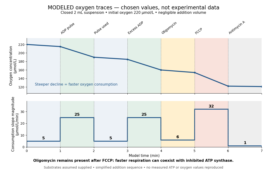
Start at . On successive one-minute intervals, choose positive slope magnitudes . Integration gives the endpoint concentrations . This is just the recurrence , with ; the step plot shows . The trace remains above zero by construction.
During the second ADP interval, the total consumption rate isWith a stipulated mitochondrial protein mass of , that is . Using a denominator twice as large would halve this reported rate without changing the raw oxygen trace. Comparisons therefore need both loading and the chosen denominator.
For a second bookkeeping check, the modeled post-antimycin A slope of corresponds to background. If that background is additive and unchanged in earlier windows, subtraction gives , and for excess ADP, oligomycin and FCCP, respectively. The oligomycin-sensitive difference is . It is a difference in oxygen consumption under the chosen interventions, not an ATP production rate. Converting it into one requires additional coupling assumptions or an independent ATP measurement; the FCCP interval shows why slope alone cannot supply that conversion.
Across the whole seven-minute example, consumption is , also the sum of the seven interval consumptions. This equality checks the rate-to-amount accounting independently of how the intervals are named. Actual records require attention to calibration, mixing transients, background stability and decreasing oxygen availability; a line that steepens or flattens alone does not identify the cause.
The Golgi apparatus modifies and sorts cargo arriving from the endoplasmic reticulum for delivery to other cellular destinations. The eponym refers to Camillo Golgi.
Its usual stacked organization is polarized: cargo enters on the cis side, encounters medial processing regions, and leaves through the trans side and trans-Golgi network. Golgi cisternae enclose a Golgi lumen separated from the surrounding cytosol. Processing changes cargo; sorting selects its destination.[ref]
The Golgi Apparatus. The Cell: A Molecular Approach, 2nd edition by Geoffrey M. Cooper; Organization of the Golgi, Figures 9.22–9.23; Protein Sorting and Export from the Golgi Apparatus
This division of labor explains why reaching the Golgi apparatus does not imply being secreted: cargo can take different onward routes, while resident enzymes continue working there. The conventional route discussed here is part of secretory protein routing; many cytosol proteins never enter it.
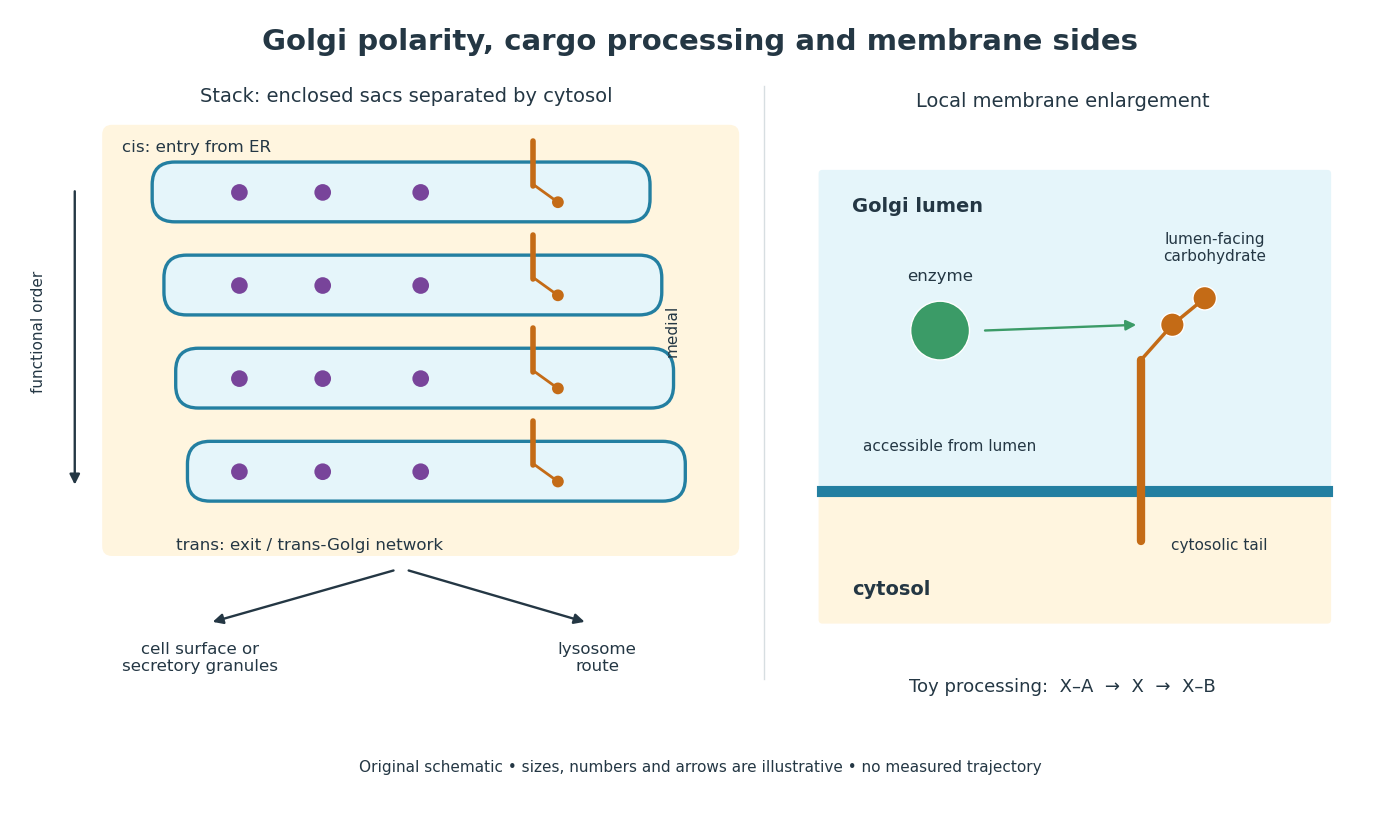
Golgi apparatus polarity, processing and membrane sides
. Original schematic of a stack and a local membrane enlargement. The spaces between Golgi cisternae contain cytosol, while each enclosed sac contains Golgi lumen. Purple cargo is soluble; the orange membrane protein has a lumen-facing carbohydrate chain and a cytosolic tail. Arrows show functional ordering, not a measured trajectory or a universal carrier mechanism. The chain-processing symbols are a constructed example. Details.A Golgi cisterna is a flattened membrane-enclosed sac. Stacking several sacs does not make their interiors one open space: membrane separates Golgi lumen from intervening cytosol.[ref]
The Golgi Apparatus. The Cell: A Molecular Approach, 2nd edition by Geoffrey M. Cooper; Organization of the Golgi, Figure 9.22: membrane-enclosed cisternae
The Golgi lumen is the enclosed interior of a Golgi cisterna, distinct from the surrounding cytosol.
The membrane-sidedness rule in secretory protein routing still applies within the Golgi apparatus. For a local worked example, stipulate a membrane protein with a carbohydrate chain exposed to Golgi lumen and a tail exposed to cytosol. An enzyme confined to Golgi lumen can contact that chain, but cannot directly contact the cytosolic tail without a boundary-crossing step. A change in which Golgi cisterna contains the protein need not turn the protein over. Thus chemical accessibility and cargo location are different questions: moving cargo onward preserves its membrane sides under the budding and fusion operations described in secretory protein routing.
Golgi cargo processing includes enzyme-mediated remodeling of attached oligosaccharides. Glycosylation can involve addition of sugars, while processing can also remove them. Different Golgi apparatus regions have different processing enzymes; their substrate requirements constrain the order of possible chemical reactions.[ref]
Transport from the ER through the Golgi Apparatus. Molecular Biology of the Cell, 4th edition by Bruce Alberts and others; Oligosaccharide Chains Are Processed in the Golgi Apparatus; The Golgi Cisternae Are Organized as a Series of Processing Compartments, Figure 13-28
An original model makes the importance of ordering explicit. Write a toy carbohydrate chain as , read with its terminal symbol on the right. Define a trimming function that removes a terminal , and an extension function that adds only when the terminal symbol is . In every other case the corresponding function leaves the chain unchanged. ThenIn particular, but : the two functions do not commute. Each result follows by inspecting the terminal symbol before applying the stipulated rule. These symbols are artificial labels, rather than a natural oligosaccharide sequence or measured enzyme pathway. The example explains why “two processing steps occurred” does not determine the product unless their order and substrate conditions are also specified. In the Golgi apparatus, spatial organization can help arrange successive opportunities for processing.
The trans-Golgi network is the sorting and export region at the trans side of the Golgi apparatus. Onward traffic can supply the cell membrane, secretory granules, or the lysosome route.[ref]
The Golgi Apparatus. The Cell: A Molecular Approach, 2nd edition by Geoffrey M. Cooper; Protein Sorting and Export from the Golgi Apparatus, Figure 9.27 and lysosomal targeting discussion
Processing and sorting answer different questions. In a constructed example, two cargo proteins could receive the same carbohydrate modification yet carry different targeting information and leave in different transport vesicles. Conversely, two chemically different cargo proteins could share a destination. A processing readout alone therefore cannot establish the final address; an experiment must also identify cargo location or delivery. Return traffic and retention likewise matter: the Golgi apparatus is a working compartment, not merely a one-way queue that empties all of its enzymes into outgoing carriers.
In cisternal maturation, a cargo-containing Golgi cisterna changes from early to later identity as its resident proteins change; retrieval of resident enzymes toward earlier compartments helps maintain Golgi apparatus polarity. In the contrasting stable-compartment model, cargo moves between persistent Golgi cisternae in transport vesicles. These mechanisms need not be mutually exclusive.[ref]
Transport from the ER through the Golgi Apparatus. Molecular Biology of the Cell, 4th edition by Bruce Alberts and others; Transport Through the Golgi Apparatus May Occur by Vesicular Transport or Cisternal Maturation, Figure 13-30 and concluding comparison
The distinction is between following cargo and following its container. A cargo label encountered first in an early processing environment and later in a late one is compatible with either changing containers or a container changing identity. Finding transport vesicles nearby does not by itself establish which cargo they carry or their direction of travel. Testing cisternal maturation therefore requires evidence about compartment identity and continuity as well as cargo position; a single mechanism should not be assumed for every cargo and biological cell.
The pancreatic secretory-protein pulse-chase experiment of James D. Jamieson and George E. Palade used guinea pig pancreas slices and a short pulse of tritium-labeled leucine followed by a chase. Thin sections on coated grids received Ilford L-4 emulsion; developed grains were read against structures by electron microscopy. Their Table II localized label to peripheral Golgi apparatus vesicles and condensing vacuoles, without significant labeling of the piled flattened Golgi cisternae.[ref]
Intracellular Transport of Secretory Proteins in the Pancreatic Exocrine Cell. II. Transport to Condensing Vacuoles and Zymogen Granules by James D. Jamieson and George E. Palade. p. 598 Methods; p. 609 Table II and its footnote
An important comparison checked the isolated granule fraction by autoradiography and electron microscopy: early labeling came substantially from contaminating condensing vacuoles, rather than mature secretory granules. Thus a fraction's name did not uniquely identify the labeled structure. This supported transport into the Golgi apparatus's condensing region and onward storage, while fixed samples and finite grain localization limited interpretation. It did not establish a resolved cis-to-trans itinerary across every Golgi cisterna. The preparation's labeling also did not mark secretory cargo exclusively.[ref]
Intracellular Transport of Secretory Proteins in the Pancreatic Exocrine Cell. II. Transport to Condensing Vacuoles and Zymogen Granules by James D. Jamieson and George E. Palade. p. 609 Figure 14; p. 613, Transport of Labeled Proteins to Condensing Vacuoles, Zymogen Granules, and the Acinar Lumen: comparison of fraction labeling with microscopy; p. 611, General Premises
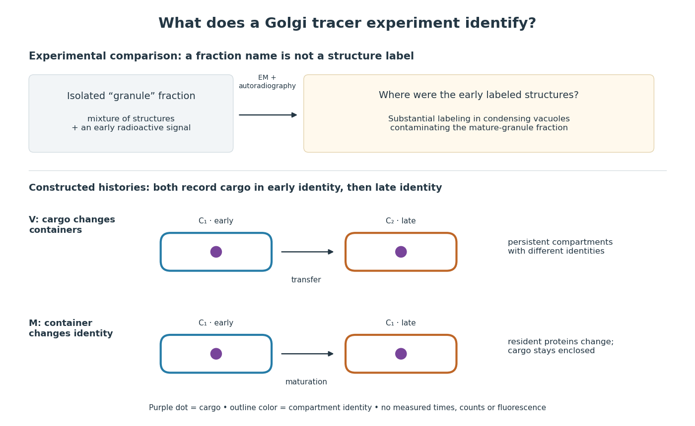
For a minimal worked inference, record only two outcomes for one labeled cargo cohort: early identity first, late identity later. Model V transfers cargo from container to container ; model M keeps it in while changes identity. Both produce the same recorded outcomes, so those outcomes alone cannot distinguish V from M. The proof is constructive: exhibit both histories and discard container identity from each record. More informative tracking would retain both cargo and resident-compartment markers, while resolving whether the same compartment persists. This is why pulse-chase analysis and compartment tracking answer complementary questions about the Golgi apparatus.
A transport vesicle is a membrane-bounded carrier that moves cargo between cellular compartments. Budding and fusion preserve the distinction between lumen-facing and cytosol-facing membrane surfaces.
A membrane-enclosed particle released by a cell into the extracellular environment. Isolation methods and cargo characterisation determine what signalling or transfer claims can be made.
A lysosome is an acidic digestive and recycling organelle: one surrounding lipid bilayer encloses a lysosomal lumen containing acid hydrolases. These enzymes break down proteins, nucleic acids, carbohydrates and lipids by hydrolysis. Cargo can originate outside the biological cell, entering through endocytosis, or within its own cytoplasm, delivered through autophagy. Digestion therefore serves both disposal and reuse.[ref]
Lysosomes. The Cell: A Molecular Approach, 2nd edition by Geoffrey M. Cooper; Cooper, The Cell, 2nd edition (2000), Lysosomes: opening paragraph; Lysosomal Acid Hydrolases; Phagocytosis and Autophagy, Figure 9.37
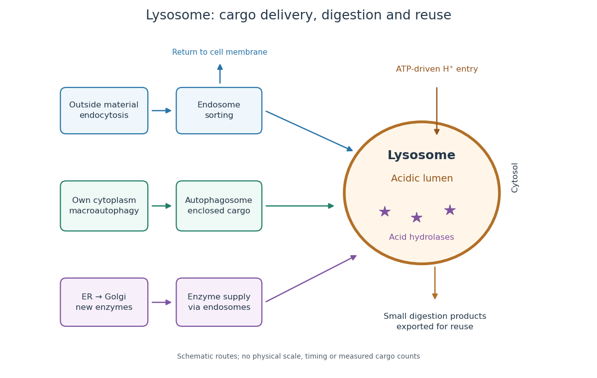
An endosome sorts endocytosed cargo: some returns to the cell membrane, while some proceeds toward lysosomes. An autophagosome instead encloses material already in the cytoplasm. Both routes bring cargo into contact with digestive enzymes; neither implies that every internalized molecule is destroyed.[ref]
Transport from the Trans Golgi Network to Lysosomes. Alberts and others, Molecular Biology of the Cell, 4th edition (2002), Multiple Pathways Deliver Materials to Lysosomes, Figure 13-35
Enzyme supply is a different itinerary from cargo disposal. Secretory protein routing and protein targeting deliver newly made digestive enzymes through the endoplasmic reticulum, Golgi apparatus and trans-Golgi network toward lysosomes.[ref]
The Golgi Apparatus. The Cell: A Molecular Approach, 2nd edition by Geoffrey M. Cooper; Protein Sorting and Export from the Golgi Apparatus, lysosomal enzyme targeting
The existing membrane transport and differential centrifugation articles explain the boundary-crossing and separation principles used below.A proteasome is a different digestive machine: a protein complex without a surrounding lipid bilayer, acting on individual proteins in the cytosol and cell nucleus. Many targets carry ubiquitin chains. A lysosome accepts a much broader range of cargo, including material delivered inside an autophagosome. Thus an unwanted soluble protein and an unwanted mitochondrion pose different disposal problems; a proteasome does not swallow an intact mitochondrion.[ref]
Protein Degradation. The Cell: A Molecular Approach, 2nd edition by Geoffrey M. Cooper; Cooper, The Cell, 2nd edition (2000), The Ubiquitin-Proteasome Pathway, Figure 7.39; Lysosomal Proteolysis, Figure 7.41
The lysosomal lumen is the aqueous interior of a lysosome, separated from cytosol by its surrounding lipid bilayer. A membrane pump couples ATP hydrolysis to inward transport of hydrogen ions, maintaining an acidic environment for acid hydrolases. Membrane transport proteins export digestion products, including amino acids and sugars, for reuse in metabolism. Compartmentalization combines restricted access with controlled exchange.[ref]
Transport from the Trans Golgi Network to Lysosomes. Molecular Biology of the Cell, 4th edition by Bruce Alberts and others; Lysosomes Are the Principal Sites of Intracellular Digestion, Figure 13-31: ATP-driven acidification and export of digestion products
Autophagy delivers a biological cell's own material for digestion and recycling in lysosomes. Macroautophagy is the enclosing-carrier route illustrated here; other forms deliver cargo differently. The cargo's internal origin distinguishes this route from uptake by endocytosis.[ref]
Protein Degradation. The Cell: A Molecular Approach, 2nd edition by Geoffrey M. Cooper; Lysosomal Proteolysis, Figure 7.41 and subsequent selective-uptake paragraph: enclosed cargo and alternative uptake
Selective turnover of mitochondria or mitochondrial portions through autophagy. Mitochondrial fragmentation alone is not proof of successful turnover.
In macroautophagy, a portion of cytoplasm, potentially including an organelle, is enclosed in an autophagosome before delivery to a lysosome. Sequestration precedes digestion.[ref]
Transport from the Trans Golgi Network to Lysosomes. Molecular Biology of the Cell, 4th edition by Bruce Alberts and others; Multiple Pathways Deliver Materials to Lysosomes, autophagy paragraph and Figure 13-35
An autophagosome is the membrane-enclosed carrier formed around macroautophagy cargo. Enclosing a mitochondrion does not immediately digest it: subsequent delivery must expose the cargo to digestive enzymes in lysosomes.[ref]
Lysosomal enzyme latency is the difference between the enzyme activity accessible to an added substrate before disruption of the surrounding membrane and the activity revealed after disruption. Low accessible activity can reflect separation of enzyme from substrate, rather than absence of enzyme. This is an assay-accessibility statement, not a claim that the intact lysosome is inactive in the living biological cell.[ref]
Exploring Cells with a Centrifuge by Christian de Duve. Christian de Duve, Exploring Cells with a Centrifuge (Nobel Lecture, 12 December 1974), printed pp. 157–159, enzyme-latency discussion and Figure 6
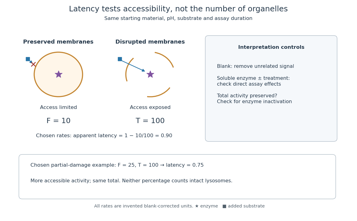
Lysosomal enzyme latency: matched assay conditions and disruption controls
. Original assay-logic diagram and explicitly chosen activity example. The values , and are arbitrary blank-corrected rate units, not historical observations. The soluble-enzyme comparison is an additional control proposed for the schematic, not an asserted measurement from the rat-liver acid-phosphatase latency experiment. Details.Let be the blank-corrected initial rate measured with a membrane-preserving preparation and the rate after a treatment that exposes all assayable enzyme. Use matched amounts of starting material, pH, substrate and measurement time. Define the apparent latent fraction byThis definition requires more than a large treated signal. Disruption must preserve enzyme activity, and the treatment must not independently alter the readout. A soluble-enzyme comparison with and without treatment helps test direct activation or inhibition; an appropriate blank tests signal unrelated to enzyme-catalysed hydrolysis. These are controls for interpreting the constructed assay, rather than additional historical observations.
For a deliberately chosen example, take and arbitrary rate units. Then : ninety percent of the assayable activity was initially inaccessible. After hypothetical partial damage, with unchanged gives . The preparation has lost latency without gaining total assayable activity. These percentages describe activity accessibility; they do not count intact lysosomes. Unequal enzyme contents, partial leakage and mixed particle populations prevent that identification. If , the formula is negative, flagging failure of this simple accessibility interpretation rather than a negative population of hidden enzymes.
To check the logic, divide the same uniformly mixed preparation into two equal portions. If dilution halves both measured rates, , so is unchanged. In contrast, comparing an undiluted untreated portion with a diluted treated portion changes the ratio artificially. Normalization to the same amount of original material is therefore part of the definition, not an optional numerical correction.
In the 1951 study by Jacques Berthet and Christian de Duve, liver from fasted rats was immediately chilled in an ice-cold isotonic chemical solution. The tissue was generally pressed through wire mesh; the weighed pulp was homogenized in cold sucrose using a glass Potter-Elvehjem homogenizer. Their acid phosphatase test concerned activity toward added glycerophosphate at pH : the readout was substrate hydrolysis, rather than an image of a particle.[ref]
Tissue fractionation studies. 1. The existence of a mitochondria-linked, enzymically inactive form of acid phosphatase in rat-liver tissue by Jacques Berthet and Christian de Duve. Berthet and de Duve (1951), Tissue fractionation studies. 1, printed p. 174, introduction and Methods: Fractionation of tissue constituents; inspected first-page text
Christian de Duve's retrospective describes successive centrifugation steps, separating centrifugation pellets and supernatants, and comparing enzyme distributions across the recovered fractions. Membrane disruption exposed previously inaccessible acid phosphatase activity, providing the crucial comparison with the membrane-preserving preparation. Joint fractionation patterns and release behavior of several acid hydrolases supported a shared population of digestive particles. By 1955, this evidence supported the lysosome concept.[ref]
The inference was biochemical: a barrier restricted access to active enzymes. Co-sedimentation could confuse distinct particles; a crude mitochondrial fraction did not establish residence inside mitochondria. Lysosomal enzyme latency neither images an organelle nor determines its shape. Electron microscopy of enriched fractions supplied separate morphological evidence.[ref]
Exploring Cells with a Centrifuge by Christian de Duve. Printed pp. 153–157: mixed populations and distribution patterns; pp. 161–164: incomplete separation and subsequent electron microscopy of lysosome-rich fractions
An endosome is a membrane-enclosed sorting compartment in the endocytosis pathway. Cargo may return to the cell membrane or proceed toward lysosomes; entry therefore does not by itself establish a digestive fate.[ref]
Progressive changes in endosomal membrane identity and lumenal processing, accompanied by sorting and retrieval rather than uniform conversion of all endocytosed cargo into a degradative route.
A maturing degradative compartment distinguished from early sorting endosomes by changes in proteins, lipids, lumenal conditions and fusion competence.
An early endosome is an early sorting compartment for internalized material. Delivery to it, recycling from it and onward trafficking are experimentally distinct from uptake across the plasma membrane.