The dominant factor raises the energy loss as a charged projectile slows, while the logarithm moderates that rise. This produces a Bragg peak close to the end of its particle range. At still lower velocity, changing effective charge, shell effects and nuclear stopping invalidate extrapolation of the elementary Bethe stopping formula to zero energy. A finite initial energy gives a finite continuous-slowing-down range,
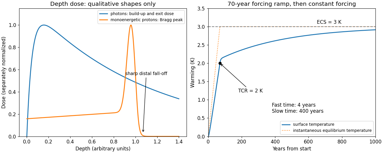
A higher initial energy increases the particle range; greater electron density increases stopping power per unit length. Absorber and also affect the depth. Multiple Coulomb scattering, energy-loss straggling, nuclear reactions and the incident energy spread broaden the measured peak and distal fall-off; nuclear secondaries can produce a small tail. Superposing many incident energies makes a spread-out Bragg peak for an extended target. The schematic curve below illustrates shapes only and is not patient radiation dosimetry.
Figure 1.
Qualitative depth-dose curves and a two-timescale climate response; depth is in arbitrary units
. Details.
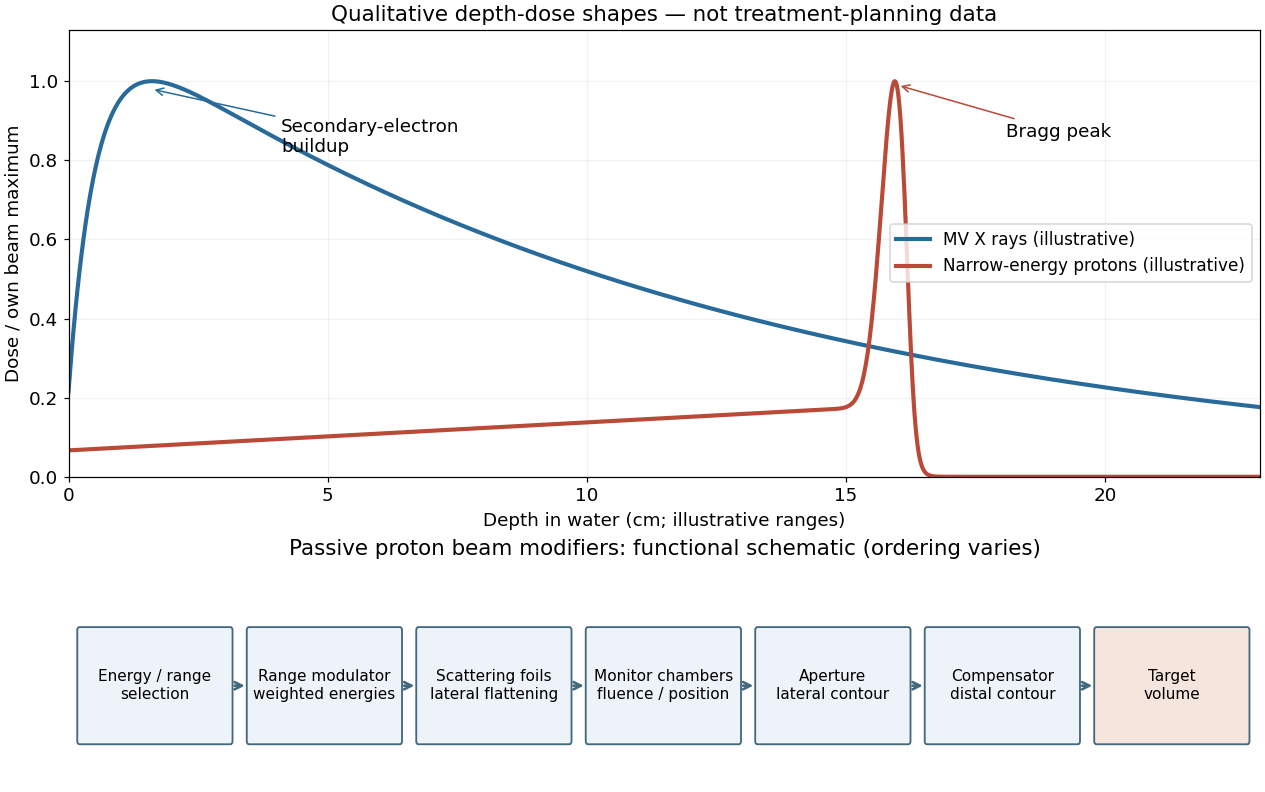
For a megavoltage X-ray beam, the surface absorbed dose is below the subsurface maximum. Photons generate energetic secondary electrons, mainly through Compton scattering; the downstream electron fluence initially builds up until approximate charged-particle equilibrium is established. Beyond this buildup region, decreasing primary photon fluence, scattering and geometric spreading make the depth-dose curve fall gradually. A penetrating photon beam has a substantial exit dose.
A nearly monoenergetic proton beam deposits a moderate entrance dose, then a sharp Bragg peak near the end of its range. As nonrelativistic protons slow, their electronic stopping power rises approximately as over the relevant part of the track. Once the protons stop, the dose falls steeply. Range straggling and energy spread broaden the peak; nuclear interactions add a smaller nonideal tail, so an exact hard cutoff is an idealization. Superposing appropriate incident energies produces a spread-out Bragg peak covering an extended target.[ref]
The label MV for an X-ray treatment beam specifies the nominal accelerating potential of the electron accelerator. Its bremsstrahlung beam is a broad photon energy spectrum with an endpoint of order , not a monoenergetic beam whose individual photons all carry the quoted numerical energy. Proton therapy beam energies can instead be specified directly in MeV per incident proton.
Figure 1.
Qualitative photon and proton depth-dose profiles and functions of passive proton beam modifiers
. Details.
A passively scattered proton therapy beam uses distinct devices to control lateral spread, range and distal shape. The lower panel of the figure gives a functional schematic; the exact ordering varies among beamlines. An energy selector or range shifter sets the maximum range. A stepped range-modulation wheel or ridge filter gives a weighted collection of proton energies, forming the spread-out Bragg peak. One or more scattering foils broaden the narrow beam; a double-scattering system can improve lateral uniformity. Ionization chambers monitor delivered fluence and position. A patient-specific aperture or collimator blocks unwanted lateral regions. A shaped range compensator makes the remaining range vary across the field, matching the distal target boundary. Their effects must be combined: lateral collimation alone cannot flatten the depth distribution, and an energy modulator alone cannot set the lateral contour.[ref]