Boris Komrakov could refer to various individuals or concepts depending on the context, but he is most likely recognized as a mathematician known for his work in areas such as optimization theory and other mathematical fields.
As of my last update in October 2023, Boris Rufimovich Vainberg does not appear to be a widely recognized public figure, historical personality, or a prominent character in popular culture. It's possible that he may be a private individual or a lesser-known figure.
Konstantin Andreev could refer to multiple individuals, as it is a relatively common name in Russian-speaking regions. Without specific context, it is difficult to determine which Konstantin Andreev you are referring to. If you have particular details about the person or their achievements, such as their profession (e.g.
Nikolai Kapitonovich Nikolski was a prominent Soviet and Russian mathematician known for his contributions to various fields, including analysis, function theory, and complex analysis. Born on April 29, 1898, and passing away on October 20, 1979, Nikolski's work particularly focused on topics such as approximation theory, operator theory, and the study of analytic functions.
Tatyana Velikanova is a prominent figure in the field of education, particularly recognized for her work in developing innovative teaching methodologies. She has made significant contributions to pedagogical practices, promoting the use of technology and interactive learning approaches in the classroom. Her work often focuses on enhancing student engagement and improving educational outcomes.
Victor Ginzburg is not a widely recognized individual in general knowledge up to date. There may be several people with that name in various fields, but without more specific context—such as their profession or notable achievements—it's difficult to provide detailed information.
Soviet women physicists refers to female scientists from the Soviet Union who made significant contributions to the field of physics. Despite facing various societal and institutional challenges, many women in the Soviet Union excelled in science and played important roles in research and education. During the Soviet era, especially from the mid-20th century onwards, there was a strong emphasis on higher education and the participation of women in science.
Vladimir Mironenko does not appear to be a widely recognized figure in historical or contemporary contexts, as of my last knowledge update in October 2023. It's possible that he could be a private individual, a figure in a specific niche, or perhaps someone who has gained prominence after that date. If you can provide more context or specify the field (e.g.
Andrei Slavnov is a notable Russian theoretical physicist, best known for his contributions to quantum field theory and statistical mechanics. He is particularly recognized for his role in the development of the Slavnov-Taylor identities, which are important for ensuring the consistency of gauge theories in particle physics. His work has had significant implications in the field of quantum gravity and other areas of theoretical physics.
As of my last knowledge update in October 2023, there is no widely known or notable figure by the name of Bogdan Voitsekhovsky. It is possible that he is a private individual, a lesser-known person in a specific field, or a fictional character. If you have more context or specific details about him, I may be able to provide more information. Please let me know!
Evsei Rabinovich may refer to a specific individual, but without additional context, it's difficult to provide a definitive answer, as there could be multiple people with that name or references to it in different fields, such as academia, literature, or other domains.
Gennadi Zakharov is a name that could refer to various individuals, but one notable person by that name is a Russian-born scientist and researcher known for his work in the field of physics, specifically in quantum physics and photonics. He has made significant contributions to research involving light-matter interactions, as well as the development of advanced optical technologies.
Georgy Flyorov refers to a notable Russian physicist and his contributions to science. Georgy Nikolaevich Flyorov (1913-1990) was an influential figure in the field of nuclear physics, best known for his role in the discovery of the phenomenon of nuclear fission and his work on heavy elements. He was a prominent advocate for the peaceful use of nuclear energy and played a significant role in the Soviet nuclear program.
As of my last update in October 2023, Gregory Garibian does not appear to be a widely recognized figure in public domains such as politics, entertainment, or academia. It's possible that he could be a private individual or a figure who has gained prominence after my last update.
Acta Astronautica is a peer-reviewed scientific journal that focuses on the field of astronautics and space exploration. It covers a wide range of topics related to space science and technology, including but not limited to spacecraft design, space missions, astrobiology, and various aspects of human and robotic spaceflight. The journal serves as a platform for researchers, practitioners, and engineers to publish their findings, share insights, and discuss advancements in the field of space exploration.
Leonid Yatsenko does not appear to be a widely recognized public figure or concept as of my last knowledge update in October 2023. It is possible that he may be an individual known in specific local contexts or in certain professional fields, but there may not be significant information available about him in broader public discourse.
Pinned article: Introduction to the OurBigBook Project
Welcome to the OurBigBook Project! Our goal is to create the perfect publishing platform for STEM subjects, and get university-level students to write the best free STEM tutorials ever.
Everyone is welcome to create an account and play with the site: ourbigbook.com/go/register. We belive that students themselves can write amazing tutorials, but teachers are welcome too. You can write about anything you want, it doesn't have to be STEM or even educational. Silly test content is very welcome and you won't be penalized in any way. Just keep it legal!
Intro to OurBigBook
. Source. We have two killer features:
- topics: topics group articles by different users with the same title, e.g. here is the topic for the "Fundamental Theorem of Calculus" ourbigbook.com/go/topic/fundamental-theorem-of-calculusArticles of different users are sorted by upvote within each article page. This feature is a bit like:
- a Wikipedia where each user can have their own version of each article
- a Q&A website like Stack Overflow, where multiple people can give their views on a given topic, and the best ones are sorted by upvote. Except you don't need to wait for someone to ask first, and any topic goes, no matter how narrow or broad
This feature makes it possible for readers to find better explanations of any topic created by other writers. And it allows writers to create an explanation in a place that readers might actually find it.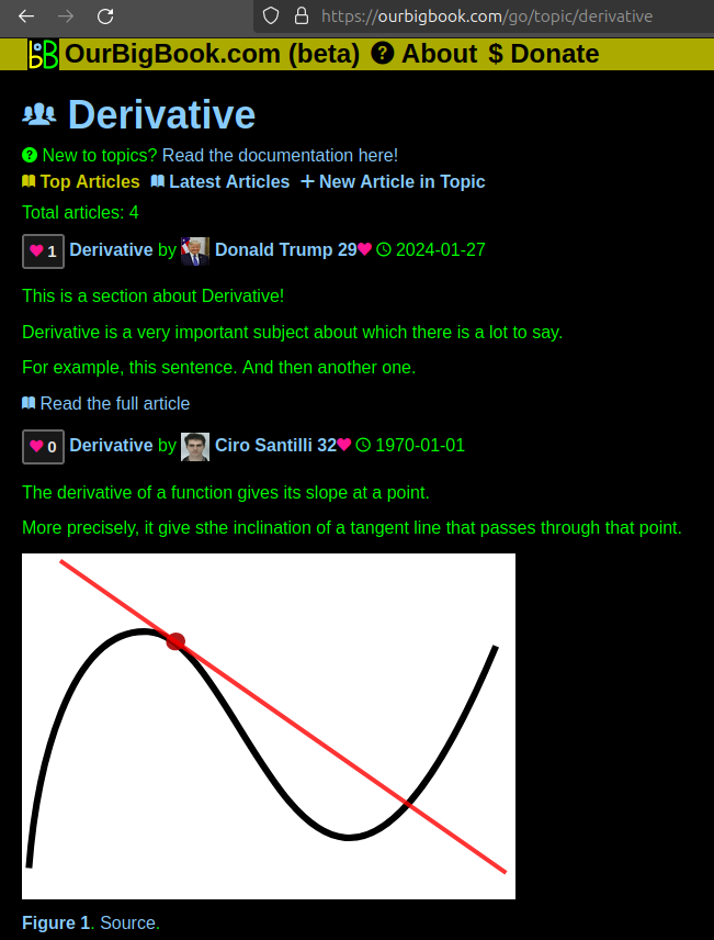
Figure 1. Screenshot of the "Derivative" topic page. View it live at: ourbigbook.com/go/topic/derivativeVideo 2. OurBigBook Web topics demo. Source. - local editing: you can store all your personal knowledge base content locally in a plaintext markup format that can be edited locally and published either:This way you can be sure that even if OurBigBook.com were to go down one day (which we have no plans to do as it is quite cheap to host!), your content will still be perfectly readable as a static site.
- to OurBigBook.com to get awesome multi-user features like topics and likes
- as HTML files to a static website, which you can host yourself for free on many external providers like GitHub Pages, and remain in full control
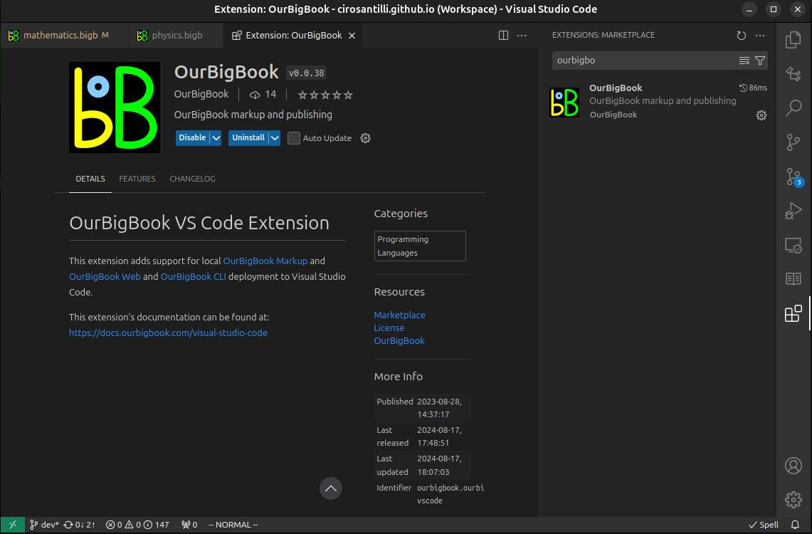
Figure 3. Visual Studio Code extension installation.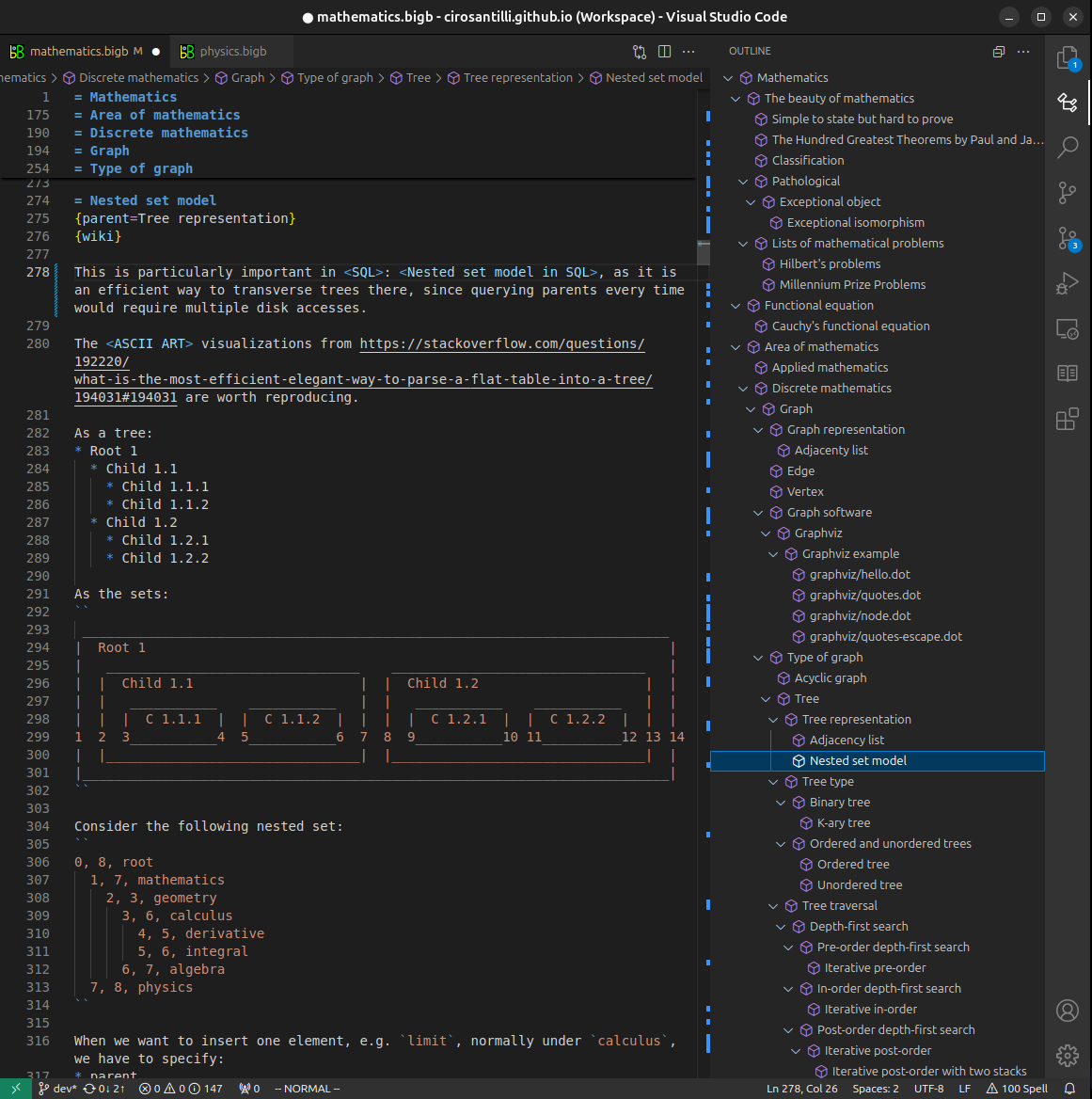
Figure 4. Visual Studio Code extension tree navigation.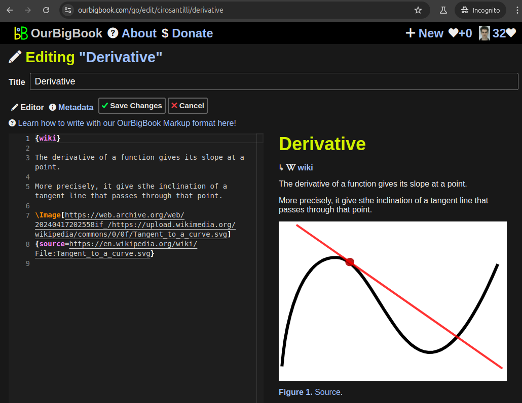
Figure 5. Web editor. You can also edit articles on the Web editor without installing anything locally.Video 3. Edit locally and publish demo. Source. This shows editing OurBigBook Markup and publishing it using the Visual Studio Code extension.Video 4. OurBigBook Visual Studio Code extension editing and navigation demo. Source. 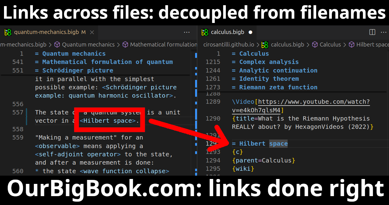
- Infinitely deep tables of contents:


All our software is open source and hosted at: github.com/ourbigbook/ourbigbook
Further documentation can be found at: docs.ourbigbook.com
Feel free to reach our to us for any help or suggestions: docs.ourbigbook.com/#contact