In Respiratory enzymes in oxidative phosphorylation. I. Kinetics of oxygen utilization (1955), Britton Chance and G. R. Williams examined isolated liver mitochondria in isotonic medium with substrate and inorganic phosphate. A one-millilitre optical cuvette held the suspension. A vibrating platinum electrode lay outside the light path; calibrated electric current followed dissolved oxygen, with rapid changes recorded by a galvanometer oscillograph. Air-saturated and oxygen-depleted readings checked sensitivity. Their Figure 2 shows ADP additions accelerating oxygen consumption, followed by return to slow respiration; repeated additions restored acceleration. Figure 4B compares delayed substrate addition after ADP incubation with the reverse order, supporting little unrelated ADP loss in that preparation. These comparisons support coupling to ADP utilization. The authors discuss dilution, sensitivity changes and atmospheric oxygen entry, which especially biases slow respiration. The readout did not directly measure the membrane potential or prove the later chemiosmosis mechanism.[ref]
Respiratory enzymes in oxidative phosphorylation. I. Kinetics of oxygen utilization by Britton Chance and G. R. Williams. pp. 383–386, Preparations and Method, Figures 1–2; pp. 387–389, Endogenous Substrate and Figure 4B; Figure 5: rapid recording
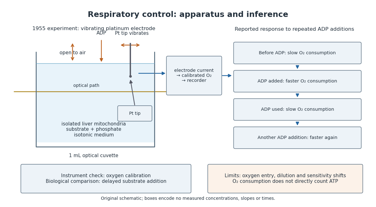

A control can test either the instrument or the biology. Calibrating electric current against oxygen checks the former; comparing responses before and after an ADP addition checks the latter. A valid calibration cannot by itself establish ATP formation, and a repeatable biological response cannot by itself fix an instrument's sensitivity.
Articles by others on the same topic
There are currently no matching articles.