A mitochondrion is an organelle with an outer mitochondrial membrane and an inner mitochondrial membrane. The latter encloses the mitochondrial matrix and folds into cristae; the intermembrane space lies on its opposite side. This architecture separates a chemical workspace from a membrane that couples electron transport to ATP formation.[ref]
4.3 Eukaryotic Cells. Biology 2e by Mary Ann Clark and others; §4.3, Mitochondria and Figure 4.14: two membranes, matrix and cristae
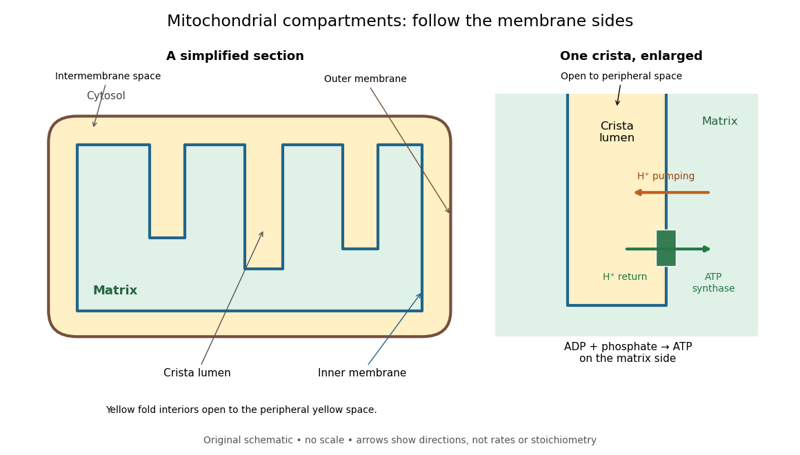

For an original location exercise, place soluble label A in the mitochondrial matrix and soluble label B in the interior of a crista. In the depicted geometry, B can reach the peripheral intermembrane space through the fold's opening without crossing a lipid bilayer. A must cross the inner mitochondrial membrane to reach either location. A fold projecting into the mitochondrial matrix therefore does not make its enclosed fluid part of the mitochondrial matrix. Following a continuous path through the colored regions proves the distinction; merely asking whether a label is “inside the mitochondrion” loses it.
The functional connection is equally spatial. The mitochondrial respiratory chain pumps protons from the mitochondrial matrix side across the inner mitochondrial membrane. Their return through ATP synthase can drive ATP formation. Oxidative phosphorylation develops the mechanism and chemiosmosis gives its free energy bookkeeping. Compartmentalization is useful because it lets a shared electrochemical gradient couple different proteins without direct transfer of a chemical intermediate between each pair.
Under aerobic conditions, mitochondria supply much of the ATP in many eukaryotic biological cells; the contribution varies with biological tissue, demand and oxygen supply. Glycolysis also makes ATP in the cytosol. Mature mammalian red blood cells lack mitochondria, and bacteria have no mitochondria at all. Neither all ATP production nor all biological cells fit the familiar mitochondrial picture.[ref]
Biology 2e by Mary Ann Clark and others. §4.3, Mitochondria; §7.2, glycolysis; §40.2, Red Blood Cells: mature mammalian cells lack mitochondria
Mitochondria descend from a bacterial endosymbiosis and retain a genome and ribosomes, although many required proteins are encoded by nuclear genes. Endosymbiotic theory develops the ancestry argument; organelle protein import explains delivery of nuclear-encoded proteins. A functioning isolated mitochondrion demonstrates biochemical autonomy under supplied conditions, rather than independently establishing that ancestry.
Dynamic remodeling of a mitochondrial population through division and merger. Fusion permits content mixing, while fission supports distribution and can segregate portions for mitophagy.
The inner mitochondrial membrane separates mitochondrial matrix from intermembrane space. Its selective permeability permits distinct chemical conditions, while embedded mitochondrial respiratory chain proteins and ATP synthase support oxidative phosphorylation. Cristae increase its surface area.[ref]
Molecular Biology of the Cell, 4th edition by Bruce Alberts and others. Chapter 14, The Mitochondrion Contains an Outer Membrane, an Inner Membrane, and Two Internal Compartments; Figure 14-8
A selective boundary and a productive enzyme have different jobs. Even active respiratory enzymes cannot sustain a useful electrochemical gradient if protons return freely across the boundary. Conversely, an intact boundary without an electron donor does not supply the energy for continued pumping. The isolated mitochondria respiration experiment separates some of these dependencies by additions to the same preparation.
The electric potential difference across the inner mitochondrial membrane, contributing to the proton-motive force. A change in potential can alter both useful phosphorylation and leak behavior.
A transport protein supplying phosphate across the inner mitochondrial membrane for processes including ATP synthesis.
A carrier exchanging adenine nucleotides across the inner mitochondrial membrane, supplying matrix ADP and exporting synthesized ATP during ordinary oxidative phosphorylation.
A transport protein exchanging suitable dicarboxylates, including succinate and malate, with counter-substrates such as phosphate. Its relevance to respiration depends on the fuel and preparation.
A crista is a fold of the inner mitochondrial membrane into the mitochondrial matrix. Its membrane remains continuous with the surrounding inner mitochondrial membrane; it is not a separately inserted sac like a thylakoid.[ref]
Molecular Biology of the Cell, 4th edition by Bruce Alberts and others. Chapter 12, The Transport of Proteins into Mitochondria and Chloroplasts, Figure 12-22: continuity of cristae and contrast with thylakoids
The diagram idealizes access through the fold's opening. Connectivity alone does not imply instant mixing or identical concentrations throughout the intermembrane space. Narrow geometry and molecular interactions can matter to transport. Increasing surface area also does not, by itself, prove a proportional increase in ATP production: the supply of substrates, available oxygen and the state of the coupling machinery must also support the flux.
The mitochondrial matrix is the compartment enclosed by the inner mitochondrial membrane, containing mitochondrial DNA, ribosomes and many enzymes. Most citric acid cycle steps occur here; succinate dehydrogenase is membrane-associated. The citric acid cycle supplies NADH for the mitochondrial respiratory chain.[ref]
Molecular Biology of the Cell, 4th edition by Bruce Alberts and others. Chapter 14, The Mitochondrion Contains an Outer Membrane, an Inner Membrane, and Two Internal Compartments; Figure 14-8, matrix enzymes and inner-membrane succinate dehydrogenase
Distinguish the location of a chemical reaction from the location of its detection. An external oxygen electrode can report consumption by an intact preparation without entering the mitochondrial matrix. That signal does not locate every intermediate, establish protein targeting or directly count newly formed ATP.
In a mitochondrion, the intermembrane space lies between the outer mitochondrial membrane and inner mitochondrial membrane, extending into the interiors of cristae. It includes the side of the inner mitochondrial membrane from which protons return through ATP synthase to the mitochondrial matrix.
A useful sidedness check is to follow a single arrow: outward pumping goes from green to yellow in the diagram, while coupled return goes from yellow to green. These are opposing movements of protons, not a circuit in which electrons return through ATP synthase. The mitochondrial respiratory chain and chemiosmosis articles distinguish the two ledgers.
The outer mitochondrial membrane separates the surrounding cytosol from the intermembrane space. Channels permit passage of many small molecules, while the inner mitochondrial membrane supplies the main selective barrier between them and the mitochondrial matrix.[ref]
Molecular Biology of the Cell, 4th edition by Bruce Alberts and others. Chapter 14, The Mitochondrion Contains an Outer Membrane, an Inner Membrane, and Two Internal Compartments: outer-membrane channels and inner-membrane selectivity
Small-solute access does not imply unrestricted access for proteins. The cytochrome c addition in the isolated mitochondria respiration experiment tests a consequence of damage to this boundary. Organelle protein import describes purposeful delivery across mitochondrial boundaries.
Respiratory control can be investigated outside an intact biological cell by supplying isolated mitochondria with substrates and ADP. This makes the boundary between oxygen consumption and ATP formation experimentally accessible. The historical mitochondrial oxygen electrode assay describes the 1955 experiment; the following authored protocol adds modern preparation and inhibitor controls.
In Functional Assessment of Isolated Mitochondria In Vitro (2009), Ian R. Lanza and K. Sreekumaran Nair describe human skeletal-muscle biopsy mincing, cold protease treatment, homogenization and differential centrifugation. A low-speed spin removes coarse material; subsequent spins collect and wash mitochondria. A stirred, closed Oxygraph-2k chamber at uses a calibrated polarographic oxygen electrode. Its signal gives dissolved oxygen concentration; the negative time derivative gives consumption. Instrument corrections include electrode consumption and oxygen leakage.[ref]
Functional Assessment of Isolated Mitochondria In Vitro. Methods in Enzymology, volume 457: Mitochondrial Function, Part B; Tissue homogenization; Separation of mitochondria by differential centrifugation; Mitochondrial respiration, Overview; An oxygraph protocol for mitochondrial assessment, steps 1–2
Their additions include glutamate plus malate, an ADP pulse, then saturating ADP. Added cytochrome c tests outer-membrane damage: stimulation suggests loss of endogenous cytochrome c. Later oligomycin inhibits ATP synthase; FCCP produces respiratory uncoupling; antimycin A inhibits respiratory complex III to estimate residual background. Figure 6 includes intervening succinate dehydrogenase and respiratory complex I interventions; the modeled traces below simplify that sequence. Isolation can damage or select mitochondria, and saturating assay conditions differ from those in living biological tissue.[ref]
The control logic matters more than a single steep slope. If ADP accelerates consumption and a finite pulse is followed by slowing, demand is affecting respiratory flux. If oligomycin slows consumption, but subsequent FCCP accelerates it, the electron transport chain can still turn over while ATP synthase remains inhibited. That contrast separates restricted coupled turnover from a completely inactive chain. Failure of FCCP to restore a rate is less specific: substrate limitation, damage, low oxygen or excessive uncoupler can also restrict respiration.
Neither the cytochrome c test alone nor a large respiratory control ratio establishes every aspect of mitochondrial integrity. Likewise, oxygen consumption is not a direct ATP assay: respiratory uncoupling provides a concrete counterexample. The preparation, additions and normalization denominator determine what comparison the experiment can support.
The following original model chooses piecewise constant oxygen consumption rates in a closed, well-mixed suspension. It assumes no gas exchange, negligible addition volume, a corrected sensor and sufficient oxygen throughout. The curves are invented teaching examples, not measured traces, fitted data or a reconstruction of Figure 6.
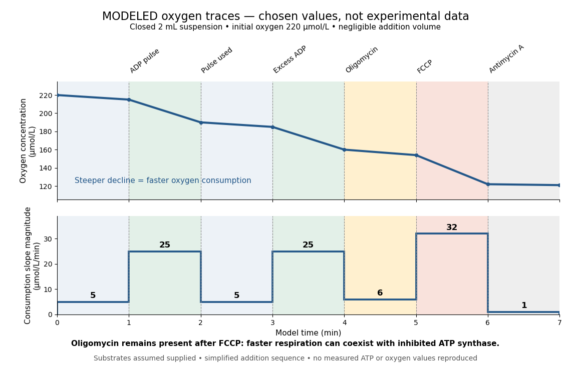

Start at . On successive one-minute intervals, choose positive slope magnitudes . Integration gives the endpoint concentrations . This is just the recurrence , with ; the step plot shows . The trace remains above zero by construction.
During the second ADP interval, the total consumption rate isWith a stipulated mitochondrial protein mass of , that is . Using a denominator twice as large would halve this reported rate without changing the raw oxygen trace. Comparisons therefore need both loading and the chosen denominator.
For a second bookkeeping check, the modeled post-antimycin A slope of corresponds to background. If that background is additive and unchanged in earlier windows, subtraction gives , and for excess ADP, oligomycin and FCCP, respectively. The oligomycin-sensitive difference is . It is a difference in oxygen consumption under the chosen interventions, not an ATP production rate. Converting it into one requires additional coupling assumptions or an independent ATP measurement; the FCCP interval shows why slope alone cannot supply that conversion.
Across the whole seven-minute example, consumption is , also the sum of the seven interval consumptions. This equality checks the rate-to-amount accounting independently of how the intervals are named. Actual records require attention to calibration, mixing transients, background stability and decreasing oxygen availability; a line that steepens or flattens alone does not identify the cause.
Articles by others on the same topic
There are currently no matching articles.