Biochemistry studies the chemistry of living systems: what their molecules are, how they recognize one another, and how their chemical reactions support the organization of a biological cell. It connects biology to chemical kinetics, stoichiometry and thermodynamics. A useful biochemical explanation follows both material and energy: which substrate becomes which reaction product, what sets the reaction rate, and how the transformation is driven.
Consider three connected examples. The stereochemistry of a sugar helps determine how it interacts with an enzyme's active site; the molecular formula alone cannot describe that recognition. Selective enzymes then direct material through metabolism, while ATP can participate in energy coupling that drives otherwise unfavourable transformations. These are different questions about the same working system, rather than three names for its energy supply.[ref]
Chemistry 2e by Paul Flowers and others. §12.7, Enzyme Structure and Function, Figure 12.25: substrate recognition and induced fit
This viewpoint is useful because it separates possible explanations for a stopped process. Missing substrate, inactive enzyme, depleted ATP and accumulated reaction product call for different interventions. Studying a cell-free extract can isolate some of those causes while leaving a substantial chemical reaction network intact.
Dialysis exchanges small solutes between a sample and an external bath through a semipermeable membrane, while retaining suitably large macromolecules, such as proteins. The transport is passive diffusion: random motion carries permeant molecules both ways, with net transport down their concentration gradient. The bath is the dialysate. At equal permeant solute concentrations, microscopic exchange continues but net exchange stops.[ref]
Dialysis: an overview, Pierce Biotechnology technical note. Dialysis: an overview, Tech Tip 20, p. 1, Dialysis is Diffusion Through a Semi-Permeable Membrane
For example, a protein sample containing excess sodium chloride can lose the small ions to a low-salt dialysate without deliberately transferring the protein. Conversely, components of a new buffer solution can enter from the bath. This is useful before studying enzyme kinetics: changing the surrounding chemical solution can remove an interfering small solute while keeping the enzyme available for the next experiment.
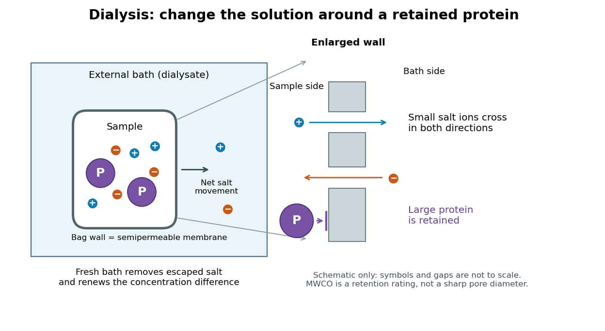
The striking feature is that the retained protein does not have to be captured and released again to change its surroundings. However, retention is conditional on the semipermeable membrane and the particular macromolecule, and dialysis is poorly suited to separating proteins of nearly the same size. The molecular weight cut-off is a nominal retention rating, not a sharp geometrical boundary.
The external chemical solution on the other side of a dialysis semipermeable membrane. Its composition supplies the boundary condition for exchange: it can receive unwanted small solutes or supply desired small solutes. A bath replacement discards the old dialysate and introduces a new reservoir with a specified composition.
The molecular weight cut-off rates retention of reference molecules. One manufacturer's definition uses the mass at which approximately remains after seventeen-hour dialysis, tested with globular proteins. A rating uses dalton units: a mass, not a length or an absolute passage threshold.[ref]
Dialysis: an overview, Pierce Biotechnology technical note. Tech Tip 20, p. 1, Factors Affecting Dialysis Rate: MWCO definition and globular reference molecules
A semipermeable membrane has a distribution of passage sizes, and a molecule's shape affects passage as well as its mass. Near the nominal MWCO, slow passage can make substantial retention compatible with eventual leakage. The protein-retention experiment below makes retention an observable quantity rather than a universal yes-or-no property inferred from the label.
Buffer exchange changes the small components surrounding a retained macromolecule toward those of a desired buffer solution. Components absent from fresh dialysate tend to leave; supplied components can enter. Each permeant solute has its own exchange problem.[ref]
Dialysis: an overview, Pierce Biotechnology technical note. Tech Tip 20, pp. 1-2, renewed gradients and Other Factors in Dialysis
For a dilute, nonbinding permeant solute, an ideal equilibrium calculation approaches the bath's specified concentration. That approximation need not describe a solute bound to a protein or a charged mixture with additional electrical constraints. Retaining a protein also does not by itself establish that its protein structure or catalytic function survives the change of buffer solution.
Consider an original ideal model with sample volume , bath volume , and initial sample solute concentration . Assume each volume is fixed, a freely permeant, nonbinding solute, no chemical reactions, complete mixing on each side, and equal concentrations at equilibrium. Each fresh bath initially contains none of this solute. The protein is perfectly retained in this model.
The first equilibration redistributes the initial amount of substance through both compartments. Conservation givesThis denominator includes the sample itself. For the chosen and , , rather than . Nine-tenths of the initial solute is in the bath and one-tenth remains in the sample; nothing has vanished during equilibration.
Discarding the equilibrated bath removes its solute. A second fresh bath starts with only of solute in the entire system, so the same argument applies again. Induction gives the geometric progressionHere counts completed equilibrations with fresh baths: is the first bath, follows one replacement, and follows two replacements. The normalized sample concentrations are respectively , and . Leaving the first bath in place for longer cannot lower the ideal equilibrium value below .
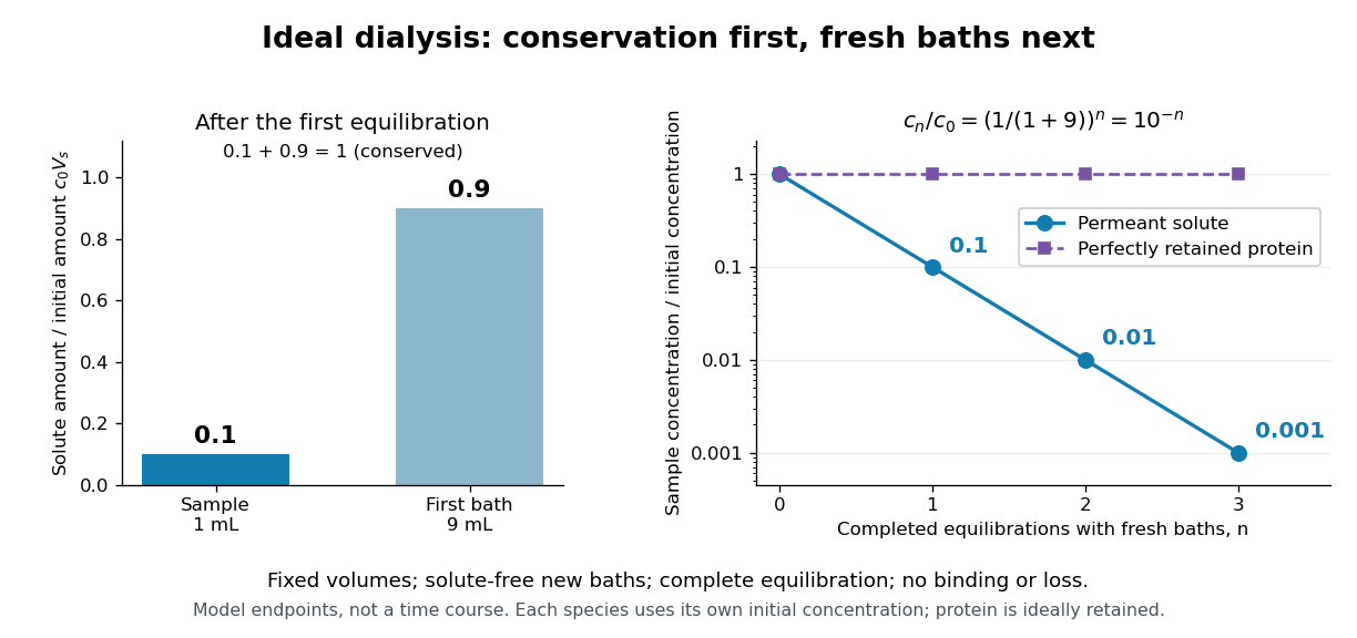
Repeated fresh baths reduce normalized solute concentration geometrically
. Original fixed-volume model, not experimental data. The left panel normalizes the first bath's conserved amount of substance by , distinguishing the sample's share from the bath's share. The right panel plots sample solute concentration divided by after each completed fresh-bath equilibration on a logarithmic vertical axis. The protein curve is normalized independently by its own initial concentration. The perfectly retained protein stays at normalized concentration one only because its amount of substance and the sample volume are fixed. Details.The same conservation argument handles a fresh bath already containing a permeant solute at fixed concentration :Thus the sample approaches , not necessarily zero. For example, if a hypothetical new buffer component starts at zero in the sample and at in every bath, its sample concentration reaches , and in the three equilibrations. Outward removal and inward replacement follow the same mathematics.
Equilibrium fractions do not specify elapsed time. An illustrative well-mixed linear ordinary differential equation model within one bath isHere is an effective transfer coefficient with units of volume per time. Adding the two equations conserves ; subtracting their normalized forms givesThe difference undergoes exponential decay. A bath replacement resets , renewing the driving difference without making jump instantly. The plotted points are equilibrium endpoints, not a measured time course or a timetable for an arbitrary protein.
Real sample volume can change through osmosis; proteins can be lost through leakage or adsorption to the semipermeable membrane. Consequently, a measured decrease in protein concentration alone cannot distinguish dilution from loss of protein. Measuring recovered volume as well as protein concentration permits an estimate of recovered protein mass.[ref]
Dialysis: an overview, Pierce Biotechnology technical note. Tech Tip 20, p. 2, Other Factors in Dialysis: water transport, volume change and nonspecific membrane binding
The manufacturer-issued Protein Preparation Handbook reports a paired dialysis comparison: of bovine serum albumin, in sodium chloride, was held behind a MWCO membrane against of water in a conical tube, shaken at room temperature. The bath was replaced after two hours. A conventional stirred-beaker treatment against of water supplied the comparison control. Residual salt was followed by sample electrical conductivity; results averaged two samples. This readout tests small-ion removal, not protein retention.[ref]
Protein Preparation Handbook, Thermo Fisher Scientific. Protein Preparation Handbook, printed p. 11, Figure 9A and caption: sample preparation, 10K membrane, baths, conductivity and conventional-dialysis reference
Its separate retention experiment used individual proteins at in saline or carbonate-bicarbonate buffer solution, pH , with membranes rated , , , and . After seventeen hours at , retained protein was estimated by a bicinchoninic acid assay. The size series and membrane series compare retention; they do not isolate shape independently of mass. The figure caption supplies neither a no-membrane recovery control nor a complete uncertainty analysis. These are manufacturer comparisons, not proof of universal recovery or unchanged enzyme function.[ref]
Protein Preparation Handbook, Thermo Fisher Scientific. Protein Preparation Handbook, printed p. 11, Figure 8 and caption: membrane series, preparation, duration, temperature and protein assay; the separately plotted vitamin B12 uses a different optical readout
The two experiments answer complementary questions: did the small solute leave, and did the desired macromolecule remain? In a new experiment, a bath blank and a starting salt standard would help interpret electrical conductivity, while a matched undialyzed protein aliquot and recovered volume would help distinguish handling loss from dilution. Those are proposed additional controls, not controls claimed for the published figures. Other ions also contribute to electrical conductivity, so the readout cannot generally identify sodium chloride alone in an unfamiliar buffer solution.
An enzyme accelerates particular chemical reactions, with catalytic selectivity arising from recognition and from the relative ease of the subsequent chemistry. Strong binding alone does not guarantee fast conversion. Nor does catalysis change the chemical equilibrium for an unchanged net chemical reaction. These distinctions explain how enzymes can direct the flow of material without creating an extra source of free energy.[ref]
Chemistry 2e by Paul Flowers and others. §12.7, opening paragraphs and Enzyme Structure and Function: alternative mechanisms, regeneration and specificity
For an original model of competing routes, let a substrate become either a desired reaction product or another reaction product :The labels represent hypothetical chemical species with the same elemental composition, not molecular structures. Assume fixed volume, irreversible first-order reactions, constant effective rate constants, and initially of with neither reaction product present. The model approximates a limited operating regime; real enzyme kinetics can include saturation and reverse reactions.
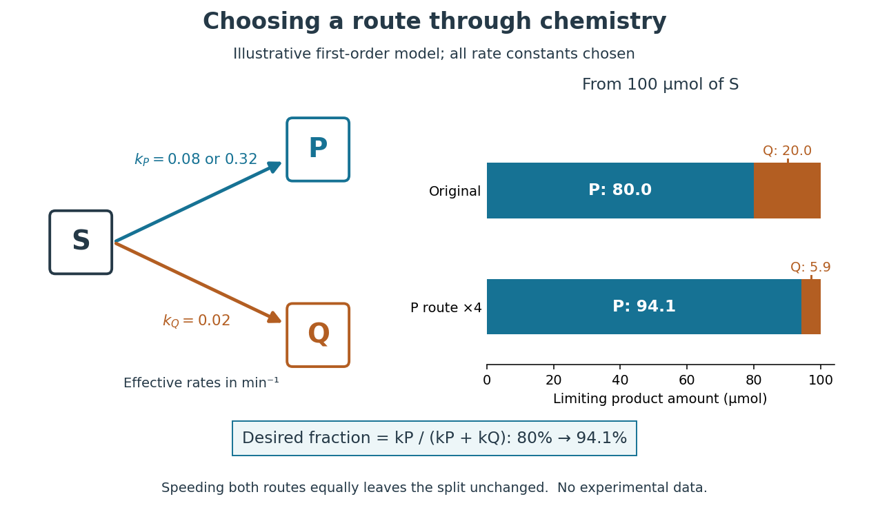
Selective acceleration changes the split between competing biochemical reaction products
. Original illustrative chemical reaction network and limiting product amounts from of hypothetical . Chosen effective rate constants are or and . The boxes label chemical species; they are not molecular structures. The bars are model limits, not experimental data. Details.The reaction-rate equations give , and , where denotes amount of substance. Consequently,With and , the eventual split is of and of . At ten minutes, the calculated amounts are approximately , and for , and . Selectively increasing fourfold changes the eventual desired fraction to . Increasing both rate constants fourfold instead preserves the split and only shortens the conversion time. Speed and selectivity answer different questions.
The bookkeeping matters independently of that model. In ideal alcoholic fermentation, complete conversion of of glucose yields each of ethanol and carbon dioxide. Of the initial of carbon chemical atoms, remain in ethanol and enter carbon dioxide. This is a chosen material-balance example using the net stoichiometry explained under Saccharomyces cerevisiae; it gives neither an observed yield nor the mechanism of the individual steps. Equal amounts of the two reaction products also do not mean equal masses.
Hexokinase provides a concrete connection between sugar chemistry and energy coupling: it transfers a terminal phosphate group from ATP to glucose, forming a phosphorylated sugar and adenosine diphosphate. The enzyme accelerates the transfer, while consuming ATP changes the net chemical reaction being carried out. The donated group reaches the substrate through the catalytic route.[ref]
Biology 2e by Mary Ann Clark and others. §7.2, First Half of Glycolysis, step 1: hexokinase and ATP-dependent glucose phosphorylation
For a symbolic teaching model, write for an unphosphorylated sugar, for its phosphorylated form, and for inorganic phosphate. These are labels for chemical states, not complete elemental formulas. Using one consistent reference convention at fixed pH and temperature, stipulate the following molar changes in Gibbs free energy:All three numbers are chosen illustrative values, not measured properties of glucose or a particular enzyme. The prime marks a biochemical reference convention with pH held fixed; explicit proton and metal-binding bookkeeping is suppressed consistently. The first two lines are a thermodynamic decomposition: cancelling water and inorganic phosphate gives the net transfer. They do not assert that the reaction mechanism first releases free inorganic phosphate and then recaptures it.
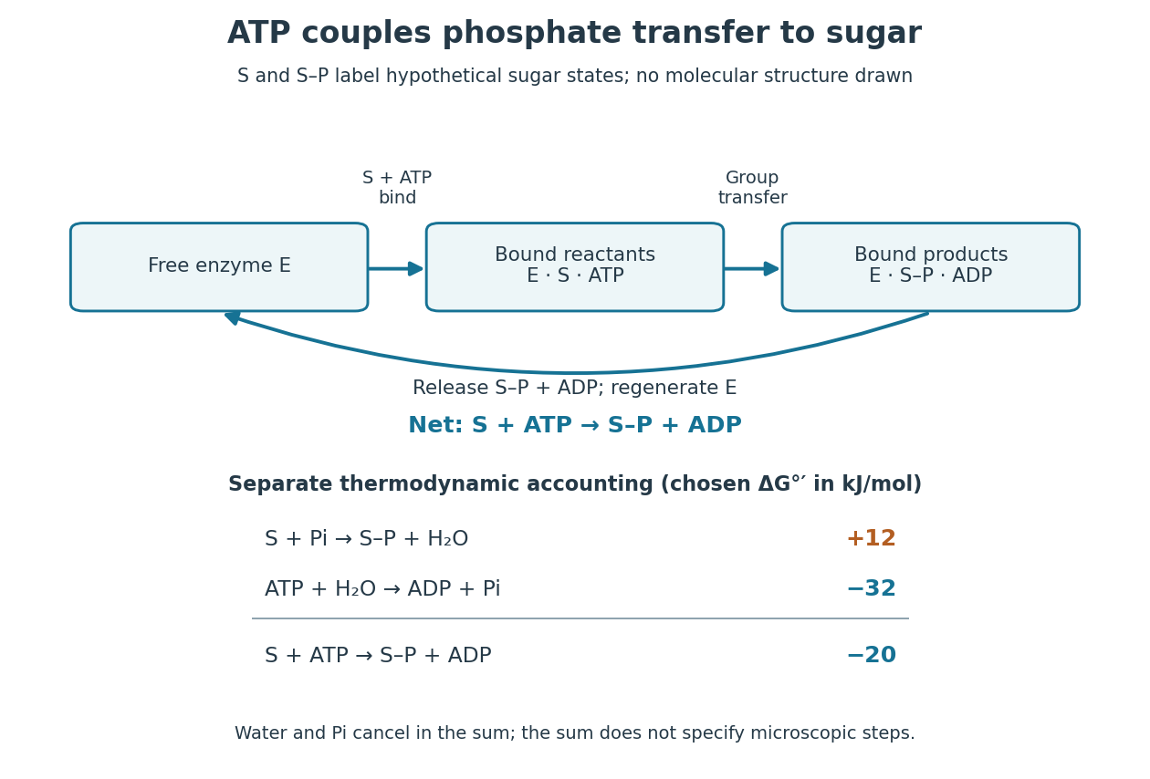
Because Gibbs free energy is a state function, consistently defined reaction changes add. A working reaction mechanism must also connect the desired conversion to ATP consumption; independently allowing ATP hydrolysis beside the sugar does not establish energy coupling. The general distinction is developed under energy coupling. ATP-dependent phosphorylation of a membrane protein can likewise connect a chemical change to active transport.[ref]
Biology 2e by Mary Ann Clark and others. §6.4, Energy Coupling, Figure 6.14: transfer to the sodium-potassium pump and its conformational cycle
Actual Gibbs free energy changes depend on thermodynamic activities. Even a favourable reference sum can be overcome by accumulation of reaction products or depletion of ATP; it supplies no prediction of the reaction rate. A living biological cell also regenerates ATP. Sustained operation therefore requires a connected metabolism, not an inexhaustible stock of a special molecule.
A cell-free extract is material obtained from disrupted biological cells after removing intact biological cells. Its biochemical activity can be tested separately from the whole organism. It can contain many enzymes and substrates, together with cofactor components; cell removal is different from purification of one component.
The cell-free extract experiment under yeast asks whether fermentation requires intact living biological cells. In his 1907 lecture, Eduard Buchner described grinding yeast with quartz sand and diatomite in a large mortar using a heavy pestle, wrapping the dough in canvas, then squeezing it in a hydraulic press. Adding concentrated sugar syrup to the expressed liquid produced carbon dioxide bubbles and foam; separate investigations identified ethanol.[ref]
Cell-free fermentation by Eduard Buchner. Cell-free fermentation, Nobel lecture delivered 11 December 1907, printed pp. 110–112, Figures 2–4 and accompanying preparation/readout text
Activity persisted after filtration through cell-removing porcelain candles. Toluene inhibited live yeast fermentation while the cell-free extract retained activity, addressing a different objection from cell removal. These comparisons support extracted chemical machinery as the cause; bubbles alone would not identify alcoholic fermentation.[ref]
Cell-free fermentation by Eduard Buchner. Printed pp. 112–114: residual-cell objection, biscuit-porcelain filtration, toluene comparison and extract activity
The inference is activity outside intact living biological cells, not that biological cells are unnecessary for making or maintaining the machinery. Nor was this a purified single-enzyme mechanism: the lecture acknowledges unsuccessful purification and reports that two separated inactive fractions recovered fermentation when reunited. Such a reconstitution asks which components must work together, extending the initial separation of organism from chemical activity.[ref]
Cell-free fermentation by Eduard Buchner. Printed p. 115: unsuccessful zymase separation; pp. 117–118: gelatine-filter fractions and restored activity on reunion
Identity source: pmc.ncbi.nlm.nih.gov/articles/PMC275499/
Identity source: pmc.ncbi.nlm.nih.gov/articles/PMC275499/
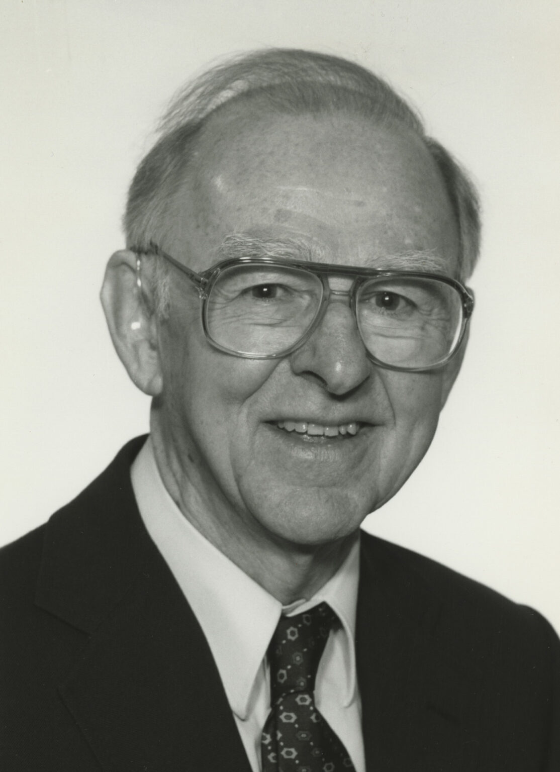
Portrait of Edwin G. Krebs
. Photograph identified on the named portrait page.Homepage: www.phar.cam.ac.uk/research/Irvine

Portrait of Eduard Buchner
. Photograph reproduced in Les Prix Nobel (1907), credited to the Nobel Foundation on Wikimedia Commons, which identifies it as public domain. Source.
Portrait of Kary Mullis
. Photograph by Dona Mapston, shown in the crop provided by Wikimedia Commons, under CC BY-SA 3.0. Source. Articles by others on the same topic

Biochemistry is the branch of science that explores the chemical processes and substances that occur within living organisms. It combines principles from both biology and chemistry to understand the molecular mechanisms of life. Biochemists study the structure, function, and interactions of biological molecules such as proteins, nucleic acids (DNA and RNA), carbohydrates, and lipids. Key areas of study within biochemistry include: 1. **Metabolism**: The biochemical pathways that convert nutrients into energy and the building blocks for cellular structures.



iubmb.onlinelibrary.wiley.com/doi/full/10.1002/bmb.2002.494030030067 Surprises and revelations in biochemistry: 1950-2000 by Perry A. Frey (2006). This should be worth a read.