1 = AB, 2 = AC, 3 = BC. A and B are within the N-terminal portion of the chain and correspond to the darker, lower-error block. C lies in the later portion; the off-diagonal comparisons to it are paler and less confidently positioned.
The matrix reports predicted aligned error, not physical separation: a large matrix value does not mean that the two residues are far apart. It indicates uncertainty in their relative placement after alignment on one residue.[ref]
PAE: A measure of global confidence in AlphaFold2 predictions by EMBL-EBI. Definition of PAE and interpretation of dark versus pale inter-domain blocks
The main diagonal contains relatively dark blocks separated by much paler off-diagonal regions. This supports a modular protein with internally more confidently predicted regions but uncertain relative packing. In particular, the broad early region ending near residue 650 and the later region are poorly positioned relative to one another. Smaller blocks within these regions suggest further domain subdivision.
Thus the compact-looking arrangement in the ribbon drawing should not be treated as a uniquely established structure of the whole enzyme. The matrix can suggest protein domain boundaries and flexible inter-domain relationships; it neither proves domain independence experimentally nor establishes that uncertainty is caused by a particular motion.
Using the supplied cluster counts,These are structural clusters represented across the three domains of life under the stated classification. They are not necessarily folds present in every individual species, nor must every cluster be a distinct topological fold. The distinction between a cluster and a fold depends on the structural-similarity criterion. The underlying large-scale analysis explicitly used structural clustering of predicted models.[ref]
Clustering predicted structures at the scale of the known protein universe by Inigo Barrio-Hernandez and others. Structural cluster construction and taxonomic distributions
All three groups retain broadly shared structural solutions, while also containing more restricted clusters. The displayed domain-restricted counts are much larger for bacteria and eukaryotes, approximately and , than for archaea, approximately . Restricted clusters also occur at lower taxonomic levels, consistent with continuing diversification rather than a single early episode of structural innovation.
These counts are not direct rates of new-fold generation. The groups differ in sampling, numbers of represented sequences and species, evolutionary depth, and detectability of relationships. Restriction to a lineage can reflect divergence, differential loss or incomplete sampling as well as origin in that lineage. The figure therefore supports unequal observed structural diversity, but does not by itself identify how rapidly each lineage invents new folds.
There arepossible sequences of length 300 over the standard twenty-amino-acid alphabet. Sampling sequences would cover only about of that space, even before allowing repeated sequences. We cannot conclude that all possible protein folds have been explored.
However, this sequence count is not a count of folds: many sequences can share a fold, and many arbitrary sequences may not form a stable folded protein. Evolution also samples sequences non-uniformly. The calculation establishes vastly incomplete sequence-space exploration; proving either exhaustive or non-exhaustive coverage of physically realizable folds needs an additional model relating sequences to structures. The observed structural-cluster count alone supplies no such proof.
Protein structure can remain similar after extensive protein sequence divergence has obscured a sequence-only relationship. A structural comparison may therefore reveal a conserved protein fold, plausible binding pocket or spatial arrangement of candidate catalytic residues that a direct sequence search misses. Such evidence can suggest a functional family and useful hypotheses.
A similar fold does not uniquely determine function. The same architecture can support different activities, and a predicted pocket does not demonstrate binding or catalysis. Confidence in the model, residue-level chemistry and independent biochemical evidence must be considered together. Structural classification complements rather than replaces sequence and experimental analysis.
Reusable protein domains make evolution combinatorial: a new domain combination can join an existing catalytic activity to a binding, localization or regulatory module without inventing every component anew. Gene duplication allows one copy to retain an established role while another diverges; domain shuffling and fusion can create new combinations. Connecting domains can also increase effective local concentration of interacting sites or enable allostery.
These are potential advantages, not guarantees that every fusion is beneficial: domains must still fold and work together. The domain-level analysis behind this example found extensive reuse and new combinations of recognized structural units.[ref]
Exploring structural diversity across the protein universe with The Encyclopedia of Domains by Andy M. Lau and others. Domain classification and inter-domain packing relationships
In surface plasmon resonance, target protein in the mobile phase passes over the immobilized antibody domain. Binding changes the optical response near the sensor surface; response increases during association and falls when unbound target is washed away. The successive injections give larger responses as the supplied target concentration increases.
For a simple 1:1 model,The reported fit gives . This is a binding estimate for the tested preparation and model. Surface heterogeneity, transport limitations or rebinding could cause deviations; the visible mismatch between parts of the trace and fit discourages treating the binary model as exact.
Antibody specificity requires comparison with alternatives. Measure binding to unrelated proteins and to appropriate related antigens under comparable conditions, with a reference surface controlling nonspecific sensor binding. Preferential binding to the intended target would support specificity within that tested panel.
Competition with a soluble target, or an independent interaction assay, can help establish that the signal reflects the intended interaction. Specificity for a particular epitope is a stronger claim than specificity for the target protein and needs evidence localizing the contact. A single good-looking binding curve establishes neither universal absence of cross-reactivity nor epitope identity.
A complete bivalent antibody could show greater apparent avidity, provided both protein binding sites can engage suitably spaced antigens on the particle. Dissociation of one arm then need not release the antibody because the other arm retains it nearby. The observed release can consequently be slower than for an isolated monovalent domain.
This does not imply that the intrinsic single-site antibody affinity has improved. Geometry, accessibility and the antibody context could prevent bivalent engagement or even impair the isolated domain's interaction. Thus an altered apparent binding strength is plausible, but its direction and size cannot be inferred solely from conversion to a full antibody.
The unliganded and peptide-bound preparations elute at almost the same volume, near 14 ml. They therefore have similar effective hydrodynamic sizes in this separation. A conformational change can occur without a large change in that measurement. The GFP–peptide-containing complex elutes earlier, near 10.5–11 ml, consistent with a larger particle; the later peak is consistent with excess unbound GFP–peptide.
The experiment supports ligand association but does not alone determine subunit number. Size-exclusion chromatography separates by access to pores, which depends on size and shape rather than mass alone. The similar peptide-free and peptide-bound elution volumes therefore do not establish identical conformations or exact molecular masses.
The main particles are approximately 120 kDa in both conditions. With 30 kDa per protein subunit, this supports a tetramer:The small peptide contribution is not clearly resolved as a separate mass increase at this scale. Together with the microscopy, the results support a conformational change of an existing tetramer, rather than a large change in oligomer number. Mass photometry reports the scattering-derived mass distribution of detected particles; it does not by itself identify which conformation a particle adopts or count small bound ligands precisely.
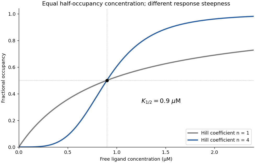
Yes: the binding data support strong positive cooperativity. On the linear concentration axis, occupancy rises sigmoidally, with reported Hill coefficient , far above the noncooperative value one. The Hill equation gives the useful empirical descriptionThe steep response is consistent with the independent evidence for a four-subunit assembly. The fitted half-occupancy concentration is not automatically the microscopic dissociation constant of one isolated site.
Binding alone does not demonstrate which conformational change is coupled to occupancy. Here the microscopy supplies the complementary structural evidence, so together the observations support a cooperative ligand-dependent switch. A Hill coefficient near four is consistent with strongly coupled binding in a tetramer, but is not an independent exact count of ligands.

A: downward when ligand is present; B: downward; C: upward, over the intended protein and ligand concentration range. Ligand should favour its bound Y state, and the ligand-bound subunits should favour tetramer assembly. Without ligand, the unassembled state should predominate, rather than forming a stable ligand-free tetramer.
These preferences are thermodynamically coupled. The ligand must bind preferentially to states participating in the assembled structure strongly enough to compensate for the cost of shifting the unliganded conformational and assembly equilibria. At extremely low protein concentration, association entropy can still favour monomers; “ligand-dependent” does not mean concentration-independent assembly. This is a thermodynamic linkage argument, rather than three independently adjustable arrows.
A single predicted monomer structure is insufficient. It may suggest a possible binding pocket, domain hinge or subunit interface, and a confident multimer model may support a particular assembly hypothesis. Those features do not establish that alternative conformations are populated, that ligand binding shifts their populations, or that occupancy at one site changes another site's binding strength.
Cooperative allostery concerns free-energy relationships among states, not only one static geometry. Structural comparisons across conditions, independent oligomeric-mass measurements and ligand-dependent binding or activity curves are needed to test the proposal. A low-confidence inter-domain orientation is likewise uncertainty about a model, not a prediction that the protein is necessarily an allosteric oligomer.
Articles by others on the same topic
There are currently no matching articles.