Protein structure describes both the chain's chemical organization and its spatial arrangement. Four levels answer different questions: protein primary structure gives the order; protein secondary structure gives recurring local patterns; protein tertiary structure gives one chain's overall arrangement; protein quaternary structure gives the assembly of multiple chains. These are levels of description, not four mandatory stages that every protein must pass through. See Clark, Douglas and Choi: four levels of protein structure.
A Ramachandran plot displays backbone angles. glycine has a broader sterically allowed region because its side chain is hydrogen, whereas proline ring closure strongly restricts .
A segment of a protein that does not maintain one stable folded structure under the specified conditions. An otherwise folded protein can contain such a region, so a region should not be equated with an entire intrinsically disordered protein.
A recurring organization of protein secondary structure elements in three dimensions. Structural-cluster counts depend on comparison criteria and should not automatically be equated to counts of distinct topological folds.
A beta sheet closed into a barrel-like protein fold by joining its edge strands through backbone hydrogen bonds. A membrane-spanning barrel can present nonpolar side chains to lipid while lining a pore with polar groups; soluble barrels instead have a different solvent-facing pattern. The topology alone does not imply a membrane location.
The predominantly alpha-helical structural framework characteristic of globins. Conserved haem-pocket architecture in myoglobin and haemoglobin supplies evidence for divergence from a shared ancestor.
A conformational ensemble is a distribution over molecular conformations under specified conditions. Different measurements average different functions of that distribution; a single mean structure can conceal rare states important for binding or activity.
protein dynamics describes structural fluctuations and transitions between conformations of a protein. Functional interpretation needs the populations and timescales of these transitions, not merely a list of static structures.
The distribution of molecular conformations and their interconversion in an enzyme under specified chemical conditions. A minor population can be functionally important even when a mean structure changes little.
A model-based measure of the amplitude of internal bond-vector motion inferred from suitable relaxation measurements. It requires assumptions about overall tumbling and timescale separation.
AlphaFold 3 is a learned model for predicting structures of biomolecular assemblies. A predicted interface is a structural hypothesis; its occurrence, occupancy and function in a particular biological condition need experimental support.[ref]
A protein domain is a distinguishable structural and functional region of a protein. A catalytic protein domain and a binding protein domain can carry out different tasks within one chain.
A protein structural motif stabilized by zinc coordination. Different zinc-finger families can bind DNA, RNA or proteins; the motif alone does not establish a transcriptional role.
A carbohydrate-binding module targets an associated protein to a carbohydrate substrate. Surface enrichment can help insoluble-substrate catalysis, but binding alone does not establish productive access.
A helix-turn-helix is a structural DNA-binding motif in which an intervening turn relates two helices, one often contributing major-groove recognition contacts. The motif alone does not specify which DNA sequence is recognized.
An activation domain is a protein region that promotes transcription when appropriately recruited, often through cofactor interactions. Tethering experiments can distinguish activation capacity from the sequence specificity supplied by a DNA-binding domain.
A DNA-binding domain is a region of a protein contributing to DNA recognition or binding. Its binding specificity can be separable from an activation domain, allowing experimental tests of recognition and transcriptional stimulation.
A protein domain that can recognize particular methylated histone states, with specificity depending on the particular protein.
An SH2 domain is a protein domain that recognizes a phosphotyrosine residue together with surrounding sequence features. This selective binding recruits a protein to a modified receptor or docking protein; it does not itself imply catalysis.[ref]
Signaling through Enzyme-Linked Cell-Surface Receptors. “Phosphorylated Tyrosines Serve as Docking Sites For Proteins With SH2 Domains”, Figure 15-53
The protein primary structure specifies the ordered amino acid residues of each polypeptide chain, joined through peptide bonds. A chain is conventionally written from its N-terminus to its C-terminus. In the broader chemical sense, a complete protein primary structure also specifies covalent bond crosslinks between chain positions, including protein disulfide bonds; a bare sequence string leaves that information unstated. This describes chemical connectivity rather than the spatial arrangement of protein tertiary structure or protein quaternary structure. See IUPAC Gold Book: primary structure, sequence and chain crosslinking and IUPAC-IUB, 3AA-16: writing residue sequences from the amino end. Some conventions reserve protein primary structure for sequence alone and call the complete chemical connectivity the covalent structure; the distinction should be stated when comparing descriptions. See IUPAC-IUB polypeptide-conformation recommendations, section 6.1: the narrower sequence-only convention.
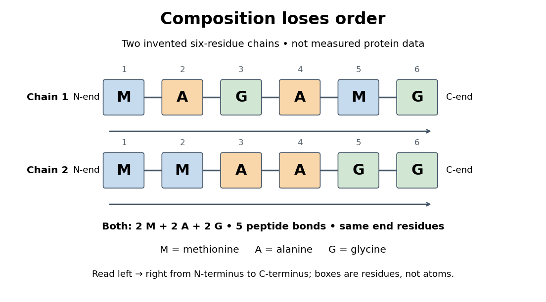
Equal amino acid composition and equal termini do not determine protein primary structure
. Original toy polypeptide chains MAGAMG and MMAAGG, read from the N-terminus to the C-terminus. Each contains two methionine, two alanine and two glycine amino acid residues, but their internal order differs. Letters denote chain positions rather than free amino acids; lines denote peptide bonds. Neither string is identified as a natural protein or a measured sequence. Details.
In this toy example M denotes methionine, A alanine and G glycine. Both polypeptide chains have six amino acid residues, five peptide bonds, an M at the N-terminus and a G at the C-terminus. Their amino acid composition is identical, yet position 2 is A in MAGAMG and M in MMAAGG. There are distinct orders with these counts, a multinomial coefficient. Even fixing those two end residues leaves orders. The counts constrain the answer without determining it.
For multiple polypeptide chains, record each sequence separately and specify any protein disulfide bonds between cysteine positions. Mature insulin, for example, has an A chain of 21 amino acid residues and a B chain of 30 amino acid residues. Their combined 51 residues give peptide bonds, rather than the 50 of one unbroken 51-residue chain. See Cooper, The Cell: insulin's two chains and cysteine crosslinks, figure 2.16. A protein disulfide bond joins side groups; it does not concatenate the C-terminus of one chain with the N-terminus of another. The three insulin protein disulfide bonds connect A6 to A11 within the A chain, and A7 to B7 and A20 to B19 between chains. Positions are numbered from each chain's N-terminus. See Replacement of the interchain disulfide bridge-forming amino acids A7 and B7 by glutamate impairs the structure and activity of insulin: native disulfide positions.
Why keep this information? Protein primary structure lets us distinguish changes of chemical identity from changes of shape, and compare corresponding positions between proteins. Protein folding can bring distant amino acid residues together at a protein binding site while preserving their sequence. The attractive idea is that a readable linear record can constrain a complicated three-dimensional object; it is not, by itself, a guarantee of a unique fold in every environment. See protein folding and the ribonuclease A refolding experiment. Merely bending an intact chain preserves its protein primary structure; hydrolysis of a peptide bond, or changing which positions a protein disulfide bond connects, changes its chemical connectivity.
A protein sequence records the order of amino acid residues in a polypeptide chain. The sequence alone does not specify every modification, covalent crosslink, assembly state or conformation present in a purified sample.
For fixed length and the standard twenty-amino-acid alphabet, there are sequences: each position has twenty choices. This counts possible sequences, not stable protein folds; the sequence-to-structure relationship is many-to-one and some sequences do not form a stable folded protein.
The amino acid composition lists amounts or proportions of the different amino acid residues in a protein or specified polypeptide chain, without giving their order. The toy MAGAMG and MMAAGG each have counts M:2, A:2, G:2, or a proportion of each. Knowing composition therefore does not supply protein primary structure. Sanger's account begins by distinguishing the already available composition measurements from the largely unknown residue order. See Sanger's authored 1985 account of the move from composition to terminal-residue analysis.
Glycine is an amino acid, represented by Gly or G for an incorporated amino acid residue. Its amino acid side chain is a hydrogen chemical atom. It was identified at one insulin N-terminus in Sanger's insulin sequencing experiment. See IUPAC Blue Book, P-103.1: amino-acid names, symbols and structures.
Phenylalanine is an amino acid, represented by Phe or F for an incorporated amino acid residue. It was identified at the other insulin N-terminus in Sanger's insulin sequencing experiment. See IUPAC Blue Book, P-103.1: amino-acid names and symbols.
Cysteine is an amino acid whose amino acid side chain contains sulfur, represented by Cys or C for an incorporated amino acid residue. Two cysteine positions can be joined through a protein disulfide bond. See IUPAC Blue Book, P-103.1: cysteine's structure and symbols and Cooper: cysteine crosslinks in proteins. The C in a sequence means cysteine, whereas the C-end label in the toy diagram means C-terminus.
The motivating question was experimental: how could one recover the order in a protein when breaking all its peptide bonds destroys that order? Frederick Sanger and colleagues used complementary tests on portions of insulin. The following is an explanation of their evidence, not a laboratory recipe.
First, mark ends before destroying the chains. Sanger used the colored reagent fluorodinitrobenzene, abbreviated FDNB, to label free amino groups. After acid hydrolysis, the attached label remained on the end-residue products. Separating these derivatives by their different movement through solvents and comparing them with reference derivatives made the labels readable. The N-terminus products identified glycine and phenylalanine. Sanger emphasized that finding a stable label and a way to separate its products were both essential. See Sanger, Citation Classic (1985), discussing The free amino groups of insulin (1945). FDNB also labels suitable free amino groups on amino acid side chains, so a labeled product is not automatically an N-terminus product. See Sanger, The Chemistry of Insulin, Nobel lecture (1958), pp. 546–547.
Next, separate chains and preserve local order. Oxidation with performic acid broke the protein disulfide bonds, allowing A and B chains to be isolated. Partial hydrolysis and enzyme treatments produced fragments; these were separated and their composition and local sequences characterized. Overlaps between different fragment sets linked smaller sequences into longer ones. Many more fragments than minimally necessary were checked for consistency. See Sanger's authored Nobel lecture, pp. 547–552: chain separation, fragment analysis and sequence reconstruction.
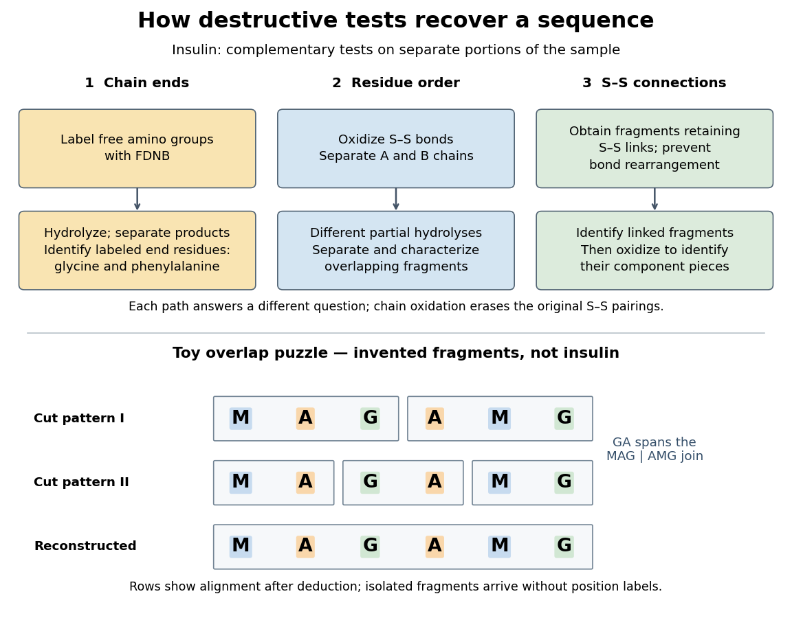

The toy fragments make the logic visible. Suppose one cut pattern yields MAG and AMG, and another yields MA, GA and MG. The first set alone permits MAGAMG or AMGMAG. The fragment GA in the second set crosses the MAG–AMG boundary in MAGAMG; it does not occur in AMGMAG. Among concatenations of the first set, therefore, the second set selects MAGAMG. This example assumes intact, correctly identified fragments from one homogeneous chain and known fragment membership in each cut pattern. Repeated stretches, missing fragments or sample mixtures can leave ambiguities; the picture illustrates inference from overlaps, not a universal guarantee of reconstruction.
Finally, determine the protein disulfide bonds separately. Oxidation used to separate whole chains erases their original connections. Sanger and colleagues instead studied fragments still joined by protein disulfide bonds, then oxidized the isolated joined fragments to identify their component pieces and locate the connected chain regions. An important trap was rearrangement of disulfide bonds during treatment: products can acquire connections absent from the starting protein. Their work therefore had to establish conditions that prevented rearrangement before interpreting a linked fragment as evidence. See Ryle and Sanger, A Disulphide Interchange Reaction, Nature 171, 1025–1026 (1953) and Sanger's Nobel lecture, pp. 553–554: preserving and identifying disulfide connections. The result was an experimentally constrained chemical structure rather than a composition list or a photograph of a fold.
Insulin is a protein hormone. Its mature form has two polypeptide chains; identifying their sequences and protein disulfide bonds provided the motivating example in Sanger's insulin sequencing experiment. See Cooper: insulin as a two-chain hormone, figure 2.16.
A meal illustrates the insulin side of blood glucose homeostasis: rising glucose stimulates beta cells in the pancreatic islets, and released insulin favors uptake and storage of fuel. Alpha cells produce glucagon, while delta cells produce somatostatin, which inhibits release of both insulin and glucagon. The pancreas therefore contains interacting sensor and secretory biological cells, rather than one undifferentiated source of hormones. [ref]
Insulin and glucagon are peptide hormones, built from amino acid residues joined by peptide bonds; glucose is a small carbohydrate molecule. A hormone changes signal transduction, not the identity of the fuel it regulates. Human mature insulin has 51 amino acid residues in two polypeptide chains. Beta cells process proinsulin to insulin and C-peptide, storing the products in secretory granules for exocytosis. Increased glucose metabolism raises the ATP-to-ADP ratio, tending to close ATP-sensitive potassium channels; depolarization and entry of calcium ions then trigger exocytosis. This is an important triggering pathway, with additional amplification and modulatory inputs.[ref]
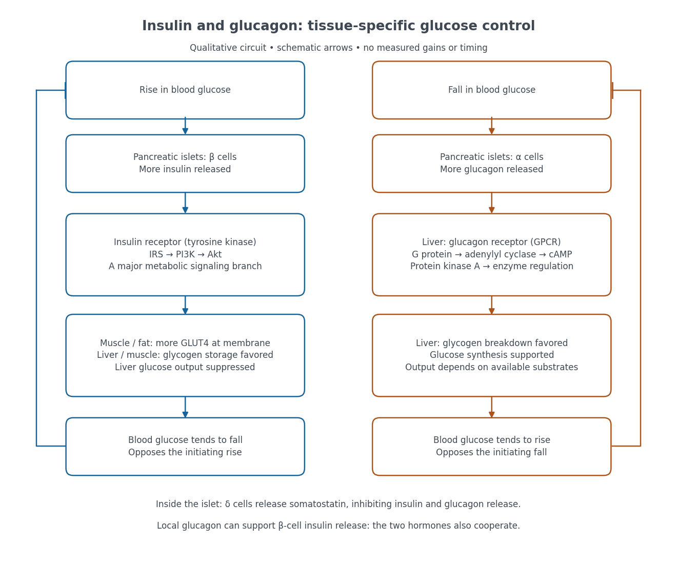

Binding to the insulin receptor, a receptor tyrosine kinase, starts phosphorylation of the receptor and insulin receptor substrates. Recruitment of phosphoinositide 3-kinase and activation of Akt transmit a major metabolic branch. In skeletal muscle and adipose tissue, that branch helps move GLUT4 to the cell membrane, increasing glucose uptake by facilitated diffusion. GLUT4 is a membrane transport protein, not an insulin receptor. Insulin also favors glycogenesis in skeletal muscle and the liver, and inhibits lipolysis in adipose tissue. In the liver, suppression of glucose output involves enzyme regulation, gene expression, and indirect changes in substrate supply. Its relative direct and indirect contributions are more complex than a single Akt arrow. [ref]
Insulin is not a universal permission slip for glucose entry: red blood cells and much of the nervous system use uptake mechanisms that are not acutely dependent on insulin. Nor does the liver use the same GLUT4 switch as skeletal muscle. This is why reduced insulin action can change blood glucose dramatically without stopping all glucose uptake. [ref]
Diabetes can disrupt production, response, and counterregulation. Type 1 diabetes usually involves destruction of beta cells and severe loss of endogenous insulin; type 2 diabetes combines insulin resistance with insufficient beta cell compensation. High circulating insulin can therefore coexist with impaired insulin action. Inappropriate glucagon release can add to excessive liver glucose output, and the response to low blood glucose can also be impaired: "more glucagon" is not a complete description of diabetes. These distinctions explain mechanisms, rather than prescribe treatment. [ref] [ref]
A single polypeptide precursor whose processing yields mature insulin and C-peptide; it is not the two-chain mature hormone. The biosynthetic distinction is described under insulin.
Glucose clamp technique: a method for quantifying insulin secretion and resistance. by R. A. DeFronzo and others separates two questions. In a hyperglycemic clamp, variable glucose infusion holds blood glucose above baseline while sampled insulin reports the response. The 1979 article's abstract reports an early release burst followed by a slower rise. In a euglycemic insulin clamp, infused insulin is held elevated and variable glucose infusion holds blood glucose at baseline. Frequent blood sampling, a glucose analyzer, and controlled infusion apparatus make the imposed concentration a maintained condition rather than an uncontrolled consequence. [ref][ref]
The maintained glucose level controls an important confounder: a change in blood glucose would itself change uptake and hormone release. At a steady state, conservation of mass gives uptake equal to infused supply plus endogenous production, after accounting for losses. Consequently, infusion alone estimates uptake only if endogenous production is negligible and the other balance assumptions hold. Labelled glucose measurements can estimate residual endogenous production; matching infused insulin across comparisons matters. Whole-body uptake cannot by itself identify the responding tissue, and plasma insulin reflects both release and removal. This interpretation uses the accessible 1979 abstract and the later authored methods chapter; the original article's full methods were not available for inspection.[ref]
Protein secondary structure consists of recurring local arrangements of the protein backbone, notably the alpha helix and beta sheet. Backbone hydrogen bonds help stabilize these patterns. A beta sheet can bring together stretches far apart in the protein primary structure; local refers to the pattern of backbone geometry, not necessarily neighboring sequence positions. See Alberts et al.: alpha helices, beta sheets and backbone interactions.
A pi helix contains carbonyl-i to NH-(i+5) hydrogen bonds, sixteen-membered hydrogen-bonded rings, and approximately 4.4 residues per turn.
A 3-10 helix repeats carbonyl-i to NH-(i+3) hydrogen bonds and has about three residues per turn. The “10” counts atoms in its hydrogen-bonded ring.
A gamma turn commonly contains a seven-membered backbone hydrogen-bonded ring, connecting a residue-i carbonyl to the NH of residue i+2.
An amphipathic helix presents chemically different faces, often a relatively polar face and a hydrophobic face. Shallow membrane insertion can change leaflet packing without making a full transmembrane segment.
An alpha helix is a coiled protein secondary structure with a repeated pattern of backbone hydrogen bonds. In the standard pattern, the backbone group of amino acid residue bonds to that of residue : for example, positions 2 and 6. This is an additional spatial interaction, not a replacement peptide bond between those positions. Pauling, Corey and Branson proposed the helical arrangement in their 1951 paper by reasoning from the geometry of small molecules. See Pauling, Corey and Branson, The Structure of Proteins: Two Hydrogen-Bonded Helical Configurations of the Polypeptide Chain, PNAS 37, 205–211 (1951).
A beta sheet consists of extended stretches of protein backbone lying alongside one another, joined by backbone hydrogen bonds. The stretches can belong to different portions of the same polypeptide chain or to different chains.
Each extended stretch is a beta strand. Neighboring beta strands pack side by side, with repeated backbone hydrogen bonds between their backbone and groups: the oxygen is a hydrogen bond acceptor, and the backbone nitrogen with its attached hydrogen provides a hydrogen bond donor. These are interactions of the shared protein backbone, not merely contacts between amino acid side chains. Unlike an alpha helix, a beta sheet joins adjacent extended stretches rather than coiling one stretch around an axis. See Clark, Douglas and Choi, Biology 2e, 3.4: backbone groups in secondary structure.
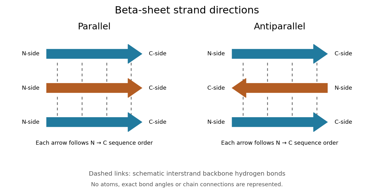
Read each beta strand in the sequence direction from the N-terminus toward the C-terminus, even when it is an internal part of a longer polypeptide chain. In a parallel beta sheet, neighboring beta strands run in the same N→C direction; in an antiparallel beta sheet, they run in opposite N→C directions. A three-strand example can therefore have arrows right–right–right or right–left–right. A beta sheet can also mix the two arrangements. Direction does not identify chain membership: a single polypeptide chain can fold back to supply several beta strands, and different polypeptide chains can contribute to one beta sheet. See Alberts et al., Molecular Biology of the Cell: beta-sheet strand arrangements.

The pleated appearance comes from the geometry of the protein backbone. Successive amino acid residues carry amino acid side chains on alternating faces of an ideal beta strand, conventionally drawn above and below the sheet. The attachment positions are the amino acid alpha carbons. Thus a six-position cartoon has groups 1, 3 and 5 on one face and 2, 4 and 6 on the other. This alternation is a spatial pattern, not a rule that alternating positions must contain identical amino acids. See Clark, Douglas and Choi: side chains above and below the pleats.
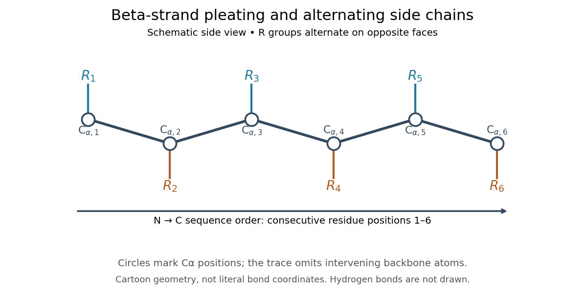

Real beta sheets commonly twist and curve; pleating does not make them perfectly flat plates. Which beta strands are neighbors, how connecting regions join them, and how their faces pack are features of the three-dimensional protein structure. An alternating pattern in the protein primary structure alone neither establishes a beta sheet nor specifies a unique fold independently of surroundings and interacting molecules. See Breda et al., Protein Structure, Modelling and Applications: extended strands, sheet twist and three-dimensional organization and protein folding.
Why this arrangement is useful: repeated backbone hydrogen bonds organize extended chain regions into a framework that can contribute to the interior of a folded protein. Amino acid side chains and connecting loops give that framework chemical variety and can shape a protein binding site for a ligand. Its attractive feature is the combination of a reusable backbone pattern with variable exposed groups: similar beta sheets can support different recognition surfaces. See Alberts et al.: beta-sheet cores and loops that form binding sites.
Historically, Linus Pauling and Robert B. Corey published The Pleated Sheet, a New Layer Configuration of Polypeptide Chains in PNAS 37, 251–256 (1951). They reasoned from peptide bond geometry and possible backbone hydrogen bonds, proposing an idealized layer rather than measuring every modern beta sheet. Their initial model should not be identified with all present-day beta sheet geometries; they published further pleated-sheet configurations later in 1951. See Pauling and Corey: original 1951 paper and scan, [ref]
https://williams.chemistry.gatech.edu/course_Information/6572/papers/Pauling_PNAS-1951_sheet.pdf. accessible scan of the original paper
, and Pauling and Corey, Configurations of Polypeptide Chains With Favored Orientations Around Single Bonds: Two New Pleated Sheets, PNAS 37, 729–740 (1951).A beta strand is an extended segment of protein backbone in the geometry characteristic of a beta sheet. Several beta strands can associate through backbone hydrogen bonds. A beta strand need not be an entire polypeptide chain.
A parallel beta sheet has neighboring beta strands running in the same direction along their amino acid residue sequences, from the N-terminus toward the C-terminus.
An antiparallel beta sheet has neighboring beta strands running in opposite N→C sequence directions. This specifies strand orientation rather than whether the beta strands belong to one polypeptide chain or several.
A backbone hydrogen bond is a hydrogen bond involving groups of the protein backbone, typically a backbone oxygen chemical atom and a hydrogen chemical atom already joined by a covalent bond to backbone nitrogen. It can connect positions that are not adjacent along the polypeptide chain. The peptide bonds still determine which amino acid residues are consecutive. See the original structural argument cited under alpha helix.
The protein tertiary structure is the overall spatial arrangement of one polypeptide chain. It includes how its protein secondary structures and intervening regions fit together. For example, the schematic under protein places positions 4 and 9 near each other despite their separation in protein primary structure. Such cooperation can help construct an active site; sequence distance and spatial separation answer different questions. Hydrophobicity, backbone hydrogen bonds and other interactions help stabilize folds, and some proteins additionally contain protein disulfide bonds. See Cooper: tertiary structure and interactions between chain regions.
A protein binding site is a region of a protein structure that interacts with a ligand. Its arrangement of amino acid residues helps determine recognition; connecting loops supported by a beta sheet can contribute to it. An enzyme's active site binds a substrate and accelerates a chemical reaction. See Alberts et al.: protein binding sites and active sites.
A protein disulfide bond is a disulfide bond linking two sulfur chemical atoms carried by suitable amino acid residues. It can connect distant portions of a polypeptide chain or different chains. See Cooper: sulfur-containing residues and disulfide crosslinks. The four internal protein disulfide bonds of ribonuclease A are central to the ribonuclease A refolding experiment; breaking these crosslinks need not break the peptide bonds along its protein backbone.
The protein quaternary structure is the arrangement of multiple polypeptide chains in a functional assembly. A single-chain protein has protein tertiary structure without requiring protein quaternary structure. For an exact bookkeeping example, two intact model chains of twelve amino acid residues each contain residues and peptide bonds. Assembly alone adds no peptide bond joining their ends. Stable functional assembly should also be distinguished from protein aggregation. See Alberts et al.: proteins as assemblies of polypeptide subunits.
A protein complex is an assembly of associated proteins. Its composition specifies component identities and their stoichiometry; variable accessory occupancy and transient partners can make the assembly a population of related forms rather than one fixed object.
A vesicle-tethering complex connecting membrane traffic to a selected cell surface. Recruitment to an apical initiation site helps coordinate delivery during epithelial lumen formation.
A cellulosome organizes multiple carbohydrate-degrading enzymes on a scaffold. Local cooperation, substrate attachment and diffusion constraints jointly influence its performance.
ERMES is the yeast ER–mitochondria encounter structure, a contact-associated complex. Synthetic-tether rescue and lipid-conversion assays help distinguish its architectural and functional contributions.
Articles by others on the same topic
There are currently no matching articles.