For a megavoltage X-ray beam, the surface absorbed dose is below the subsurface maximum. Photons generate energetic secondary electrons, mainly through Compton scattering; the downstream electron fluence initially builds up until approximate charged-particle equilibrium is established. Beyond this buildup region, decreasing primary photon fluence, scattering and geometric spreading make the depth-dose curve fall gradually. A penetrating photon beam has a substantial exit dose.
A nearly monoenergetic proton beam deposits a moderate entrance dose, then a sharp Bragg peak near the end of its range. As nonrelativistic protons slow, their electronic stopping power rises approximately as 1/v2 over the relevant part of the track. Once the protons stop, the dose falls steeply. Range straggling and energy spread broaden the peak; nuclear interactions add a smaller nonideal tail, so an exact hard cutoff is an idealization. Superposing appropriate incident energies produces a spread-out Bragg peak covering an extended target.[ref]
Proton depth-dose proposal (1946). pp. 487–489, increasing specific ionization near the end of range
The label MV for an X-ray treatment beam specifies the nominal accelerating potential of the electron accelerator. Its bremsstrahlung beam is a broad photon energy spectrum with an endpoint of order eV, not a monoenergetic beam whose individual photons all carry the quoted numerical energy. Proton therapy beam energies can instead be specified directly in MeV per incident proton.
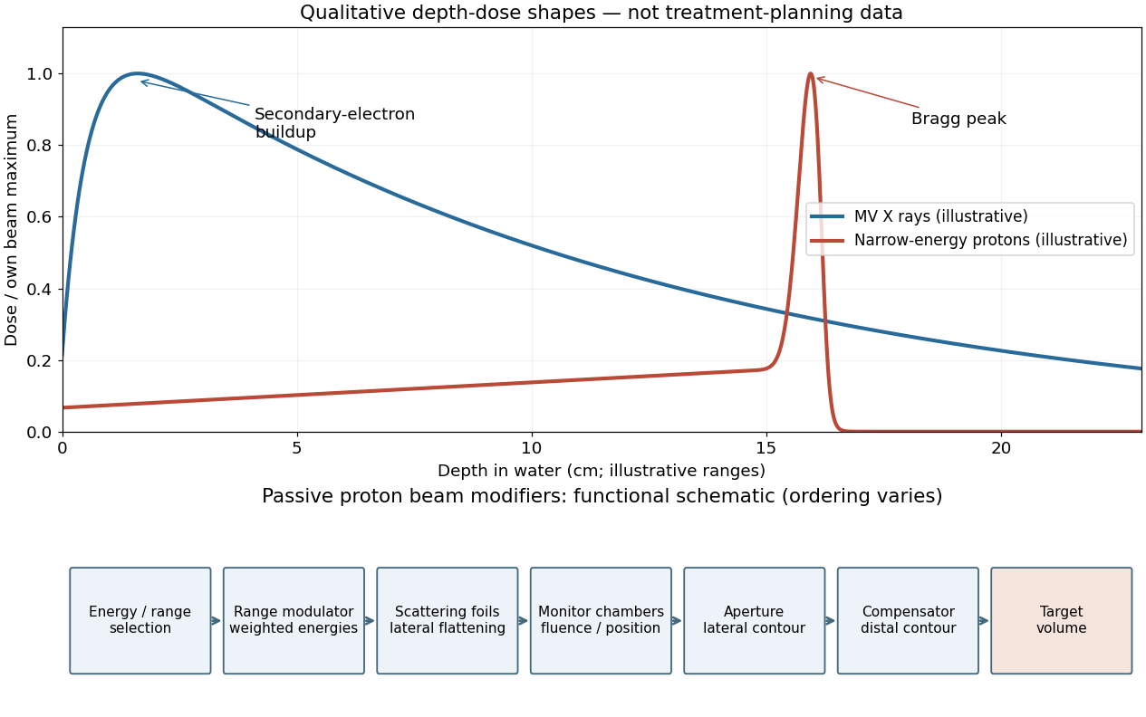

A passively scattered proton therapy beam uses distinct devices to control lateral spread, range and distal shape. The lower panel of the figure gives a functional schematic; the exact ordering varies among beamlines. An energy selector or range shifter sets the maximum range. A stepped range-modulation wheel or ridge filter gives a weighted collection of proton energies, forming the spread-out Bragg peak. One or more scattering foils broaden the narrow beam; a double-scattering system can improve lateral uniformity. Ionization chambers monitor delivered fluence and position. A patient-specific aperture or collimator blocks unwanted lateral regions. A shaped range compensator makes the remaining range vary across the field, matching the distal target boundary. Their effects must be combined: lateral collimation alone cannot flatten the depth distribution, and an energy modulator alone cannot set the lateral contour.[ref]
Passive proton double-scattering study (1977). original experiments and design: blocking the central peak followed by rescattering to flatten a large field
Use the linear-quadratic radiobiology model, independent fractions with complete inter-fraction repair, and no tumour repopulation. For N fractions of dose d, the logarithmic cell kill is N(αd+βd2). Dividing by α defines the biologically effective dose,For the tumour, the original schedule gives 26(2)(1+2/10)=62.4Gy10. A 3Gy fraction contributes 3(1+3/10)=3.9Gy10, so precisely 62.4/3.9=16 such fractions give the same tumour biologically effective dose. The physical total dose changes from 52 to 48Gy.
BED=Nd(1+α/βd).
For the specified normal-tissue α/β=3Gy, the original and altered schedules giveThus the normal-tissue model predicts an increase of 9.33Gy3, or about 10.8%, despite the smaller total physical dose. The smaller normal-tissue α/β makes increasing fraction size relatively more costly. These are comparisons within the source's hypothetical radiobiological model and equal-dose geometry, not individualized treatment prescriptions.[ref]
BEDold=52(1+32)=86.7Gy3,BEDnew=48(1+33)=96.0Gy3.
Linear-quadratic fractionation study (1989). BED formulation and dependence on fraction size and alpha/beta
Ancestors (10)
- S10
- 2015
- Past exam of the short options paper shared by Prelims and Parts A and B of the physics course of the University of Oxford
- Short options paper shared by Prelims and Parts A and B of the physics course of the University of Oxford
- Physics course of the University of Oxford
- Undergraduate course of the University of Oxford
- Course of the University of Oxford
- University of Oxford
- List of universities
- Home
Discussion (0)
New discussionThere are no discussions about this article yet.
Articles by others on the same topic (0)
There are currently no matching articles.