A structural biologist studies the three-dimensional organization of biological macromolecules and how it relates to their function. Structural biology combines measurement with a macromolecular structural model: a representation supported by experimental observations, rather than a direct photograph of every atom. Common approaches include X-ray crystallography, cryo-electron microscopy and NMR spectroscopy.
In X-ray crystallography, a crystal produces a diffraction pattern from which researchers infer an electron density map and refine a molecular model. A concrete dataset is calmodulin structure1CLL in the Protein Data Bank. Its primary record reports X-ray diffraction at resolution and a refined model containing four calcium ions.[ref]
Calmodulin structure 1CLL in the Protein Data Bank. Experimental Data Snapshot and Primary Citation abstract
An original unit conversion gives the scale:That resolution value is not a universal error bar of on every modeled atom. Some regions can be well constrained while flexible or disordered regions are poorly resolved. In1CLL, the primary paper's abstract explicitly identifies poorly defined terminal residues.[ref]
Calmodulin structure 1CLL in the Protein Data Bank. Primary Citation abstract, poorly defined residues and refinement
A structural biologist can then test a mechanistic proposal with complementary evidence. A model may suggest a calcium ion binding environment; comparing binding or activity measurements can test its functional consequences. A static arrangement alone does not establish every reaction step or rate. The scientific work lies in connecting the model, its uncertainty and the behavior of the molecular system.
John Cowdery Kendrew helped establish how X-ray crystallography can reveal the three-dimensional arrangement of a protein. Nobel's primary nomination archive names John Cowdery Kendrew, also identifying J. C. Kendrew in its award-sharing comment.[ref]
John Cowdery Kendrew in the Nobel nomination archive. 1962 nomination64-0,nominee2 and Comments
His team's1960 myoglobin paper presented a Fourier analysis at2-angstrom resolution. Its opening recalls how earlier phase determination compared native crystals with heavy-atom derivatives containing localized mercury or gold sites. Such comparisons supply information missing from a simple measurement of diffraction intensity.[ref]
Structure of Myoglobin: A Three-Dimensional Fourier Synthesis at 2 Å. Resolution by J. C. Kendrew and others. Page422,opening discussion of earlier phasing and the2Å analysis
An original one-dimensional example explains that phase problem. Write a periodic electron density using Fourier series coefficients . The two positive densitieshave , and , with the final sign distinguishing them. Their squared coefficient magnitudes are identical:9 for mode zero,1/4 for modes and1/16 for modes . The relative phase of the second harmonic has changed while the first harmonic is fixed. Thus equal intensities need not identify one density.
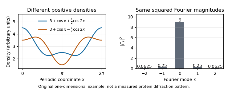
The myoglobin experiment was more demanding than this toy calculation, but the distinction motivates its chemical strategy: modifying crystals at known heavy-atom sites changes their scattering in a way that constrains phase information. Reconstructing an electron density map therefore requires both measured diffraction and a justified phase-determination method, rather than treating the recorded spots as a direct photograph of the molecule.


Chris Oubridge investigated the structural basis of RNA splicing and RNA recognition by proteins. The original 1994 publisher record names him as first author of the U1A protein–RNA hairpin structure previously cited under C. Oubridge.[ref]
Crystal structure at 1.92 A resolution of the RNA-binding domain of the U1A spliceosomal protein complexed with an RNA hairpin by C. Oubridge and others. Publisher byline and Abstract, DOI10.1038/372432a0
His institutional obituary records 1966–2020 and describes his contributions to spliceosome structural studies.[ref]Chris Oubridge obituary at the MRC Laboratory of Molecular Biology. Named heading and research account
The 1994 X-ray crystallography study resolved an RNA-binding domain with a 21-nucleotide RNA hairpin with a resolution of 1.92 angstroms. Its ten-nucleotide loop lies on the domain's beta sheet; contacts involving the loop sequence include hydrogen bonds. This provides a physical explanation of sequence recognition, rather than treating a matching letter sequence as sufficient without a compatible RNA structure.[ref]
For an original affinity example, suppose changing a loop raises a measured dissociation constant from to . At 298 kelvin, the corresponding change in standard binding Gibbs free energy iswhere is the molar gas constant. These are stipulated values, not measurements from the 1994 paper. A structure can motivate which contacts to test, while an independent binding affinity measurement establishes the net energetic effect; counting visible contacts alone does not supply that effect.
Oubridge also shared first authorship of the 2009 U1 snRNP functional-core structure, connecting the study of an individual recognition domain with the arrangement of RNA and proteins in a larger assembly.[ref]
Crystal structure of human spliceosomal U1 snRNP at 5.5 Å resolution by Daniel A. Pomeranz Krummel and others. Byline, equal-contribution note and Abstract

Chris Oubridge in the laboratory, named photograph in the MRC Laboratory of Molecular Biology obituary
. Articles by others on the same topic
There are currently no matching articles.