Overspeed generally refers to a situation in which a vehicle or machinery exceeds its safe or intended speed limit. This term can be applied in various contexts, including: 1. **Automotive and Aviation:** In vehicles like cars, trucks, and airplanes, overspeed can refer to exceeding the maximum operational speed. This can lead to mechanical failure, loss of control, or accidents. 2. **Machinery and Equipment:** In industrial settings, certain machines have specific speed ratings to ensure safe operation.
A turbine map is a graphical representation that displays information about the performance and characteristics of wind turbines. It typically illustrates various parameters such as power output, wind speed, and efficiency across different operational conditions. Turbine maps can include: 1. **Power Curve**: A chart showing the relationship between wind speed and generated power, indicating the turbine's efficiency at various wind speeds.
The term "sex machine" can refer to a few different things, depending on the context: 1. **Mechanical Device**: In the most literal sense, a sex machine is a device designed for sexual stimulation. These machines typically use mechanical means to simulate sexual activity and can be operated in various ways (manually or automatically). They are often used in adult entertainment settings or by individuals for personal use.
A barrel organ, also known as a street organ or hand-cranked organ, is a mechanical musical instrument that produces music using pipes and a rotating cylinder or barrel that is fitted with pins or pegs. When the barrel is turned, the pins or pegs activate the mechanism that opens vents, allowing air to flow through the pipes and produce sound. They typically feature a variety of tunes and are often used for entertainment at fairs, parades, and public spaces.
A lifting bag, also known as an air lift bag or inflatable lifting bag, is a device designed to lift heavy objects, typically by utilizing compressed air or another gas to create buoyancy. These bags are commonly made from durable materials such as reinforced rubber or some synthetic fabrics that can withstand the stress of lifting heavy loads.
The chemtrail conspiracy theory posits that the condensation trails (contrails) left by aircraft are actually chemical or biological agents deliberately sprayed for nefarious purposes, such as weather modification, population control, or other undisclosed government agendas. Proponents of this theory believe that the trails are not merely the result of water vapor condensing at high altitudes and that they contain harmful substances. The theory gained popularity in the late 1990s and has persisted despite scientific evidence to the contrary.
Astolat Dollhouse Castle is a highly detailed and intricately designed dollhouse that is known for its elaborate architecture and craftsmanship. It was created by artist and designer Brenda Walton and is inspired by the Arthurian legend of Avalon. The dollhouse is a miniature representation of a castle, complete with multiple rooms, exquisite furnishings, and decorations that reflect a variety of historical and fantastical styles. Astolat Dollhouse Castle is particularly famous for its impressive scale and the artisanal quality of its construction.
The Essex Model House is a notable project in architectural design and urban planning, often associated with the ongoing discussions around sustainable building practices and modern living. While specific details about "The Essex Model House" may vary depending on the context, it generally serves as an example of innovative and efficient residential design.
Danger UXD (User Experience Design) is a design and consultancy studio that focuses on creating innovative user experiences for digital products and services. Founded by a group of designers, developers, and strategists, Danger UXD emphasizes user-centered design principles to ensure that the end products are not only functional but also enjoyable to use. Their approach often includes conducting user research, usability testing, and iterative design processes to refine and enhance the user experience.
Huishan clay figurines refer to a traditional form of Chinese folk art that originates from the Huishan area in Wuxi, Jiangsu Province. These figurines are made from a special type of clay known for its fine texture and plasticity, making it suitable for detailed craftsmanship. The creation of Huishan clay figurines dates back to the Ming and Qing dynasties and has continued to be a significant cultural practice.
Yokomo is a Japanese company known for its manufacturing of radio-controlled (RC) cars, parts, and accessories. Founded in 1972, Yokomo initially focused on the production of high-quality R/C car kits and components, gaining a reputation for innovation and performance in the RC racing community. The company is particularly well-regarded in the realms of on-road and off-road RC racing, including drift cars, touring cars, and buggies.
Lilliput Lane is a brand known for its collection of handcrafted miniature ceramic houses and figures that are designed to capture the charm of traditional British architecture. Founded in 1973 by artist David Tate, Lilliput Lane began as a small business creating detailed models of iconic buildings and cottages, often inspired by real-life structures. The items produced by Lilliput Lane are typically characterized by their intricate detailing and craftsmanship, appealing to collectors and enthusiasts of model villages and architectural miniatures.
Varney Scale Models is a company known for producing model trains and related products, particularly in the hobby of model railroading. They specialize in various scales of model railroad supplies, which may include locomotives, rolling stock, track systems, and scenery materials. Varney has a historical significance in the model railroading community, having been one of the earlier manufacturers in the industry.
Rail transport modelling scales refer to the various size ratios used in the modeling of railway systems, including rolling stock (trains), infrastructure (tracks, stations, signals), and landscapes. These scales are essential for hobbyists, engineers, and planners as they help to standardize the sizes of the components used in model train setups, allowing for realistic portrayals of rail systems at different sizes.
The TT scale, or the Tertiary-Tertiary scale, is primarily used in the context of the classification of geological time and rock layers. However, it seems that your question might be referencing a specific context that could vary, as "TT scale" could also refer to other domains, such as educational assessments, musical scales, or technical specifications.
The Great American Train Show is a popular event in the United States that focuses on model trains and railroading in general. Typically held in various locations across the country, the show features a wide range of exhibits, which may include: - Model train displays and layouts created by hobbyists and clubs. - Vendors selling model trains, accessories, books, and other related products. - Workshops and seminars to educate attendees on various aspects of modeling, building, and operating trains.
KCNG1 (potassium voltage-gated channel subfamily G member 1) is a gene that encodes a protein involved in the formation of voltage-gated potassium channels. These channels are essential for the proper functioning of electrical signaling in neurons and muscle cells. The KCNG1 protein specifically contributes to the regulation of membrane potential and the excitability of cells.
Mitsutaka Fujita is a Japanese artist known for his unique art installations and works that often blend elements of traditional Japanese culture with contemporary themes. His pieces may explore concepts such as nature, technology, and the human experience, using various materials and techniques.
Indian nuclear physicists are scientists from India who specialize in the study of nuclear physics, which is the branch of physics that deals with the constituents and interactions of atomic nuclei. This field encompasses various aspects such as nuclear structure, nuclear reactions, and the properties of nuclear matter. Indian nuclear physicists have made significant contributions to both basic research and practical applications, including nuclear energy, medical imaging, and nuclear safety. India has a robust community of nuclear physicists working in various research institutions and universities.
Pinned article: Introduction to the OurBigBook Project
Welcome to the OurBigBook Project! Our goal is to create the perfect publishing platform for STEM subjects, and get university-level students to write the best free STEM tutorials ever.
Everyone is welcome to create an account and play with the site: ourbigbook.com/go/register. We belive that students themselves can write amazing tutorials, but teachers are welcome too. You can write about anything you want, it doesn't have to be STEM or even educational. Silly test content is very welcome and you won't be penalized in any way. Just keep it legal!
Intro to OurBigBook
. Source. We have two killer features:
- topics: topics group articles by different users with the same title, e.g. here is the topic for the "Fundamental Theorem of Calculus" ourbigbook.com/go/topic/fundamental-theorem-of-calculusArticles of different users are sorted by upvote within each article page. This feature is a bit like:
- a Wikipedia where each user can have their own version of each article
- a Q&A website like Stack Overflow, where multiple people can give their views on a given topic, and the best ones are sorted by upvote. Except you don't need to wait for someone to ask first, and any topic goes, no matter how narrow or broad
This feature makes it possible for readers to find better explanations of any topic created by other writers. And it allows writers to create an explanation in a place that readers might actually find it.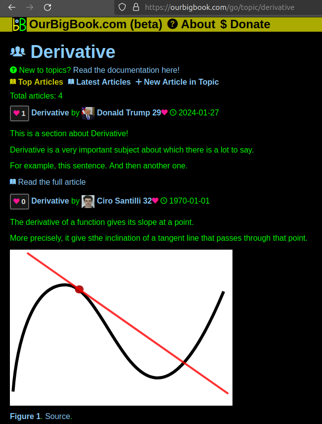
Figure 1. Screenshot of the "Derivative" topic page. View it live at: ourbigbook.com/go/topic/derivativeVideo 2. OurBigBook Web topics demo. Source. - local editing: you can store all your personal knowledge base content locally in a plaintext markup format that can be edited locally and published either:This way you can be sure that even if OurBigBook.com were to go down one day (which we have no plans to do as it is quite cheap to host!), your content will still be perfectly readable as a static site.
- to OurBigBook.com to get awesome multi-user features like topics and likes
- as HTML files to a static website, which you can host yourself for free on many external providers like GitHub Pages, and remain in full control
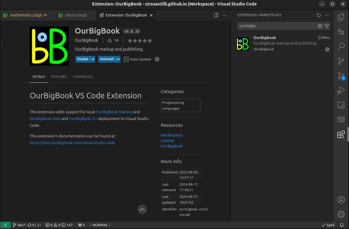
Figure 3. Visual Studio Code extension installation.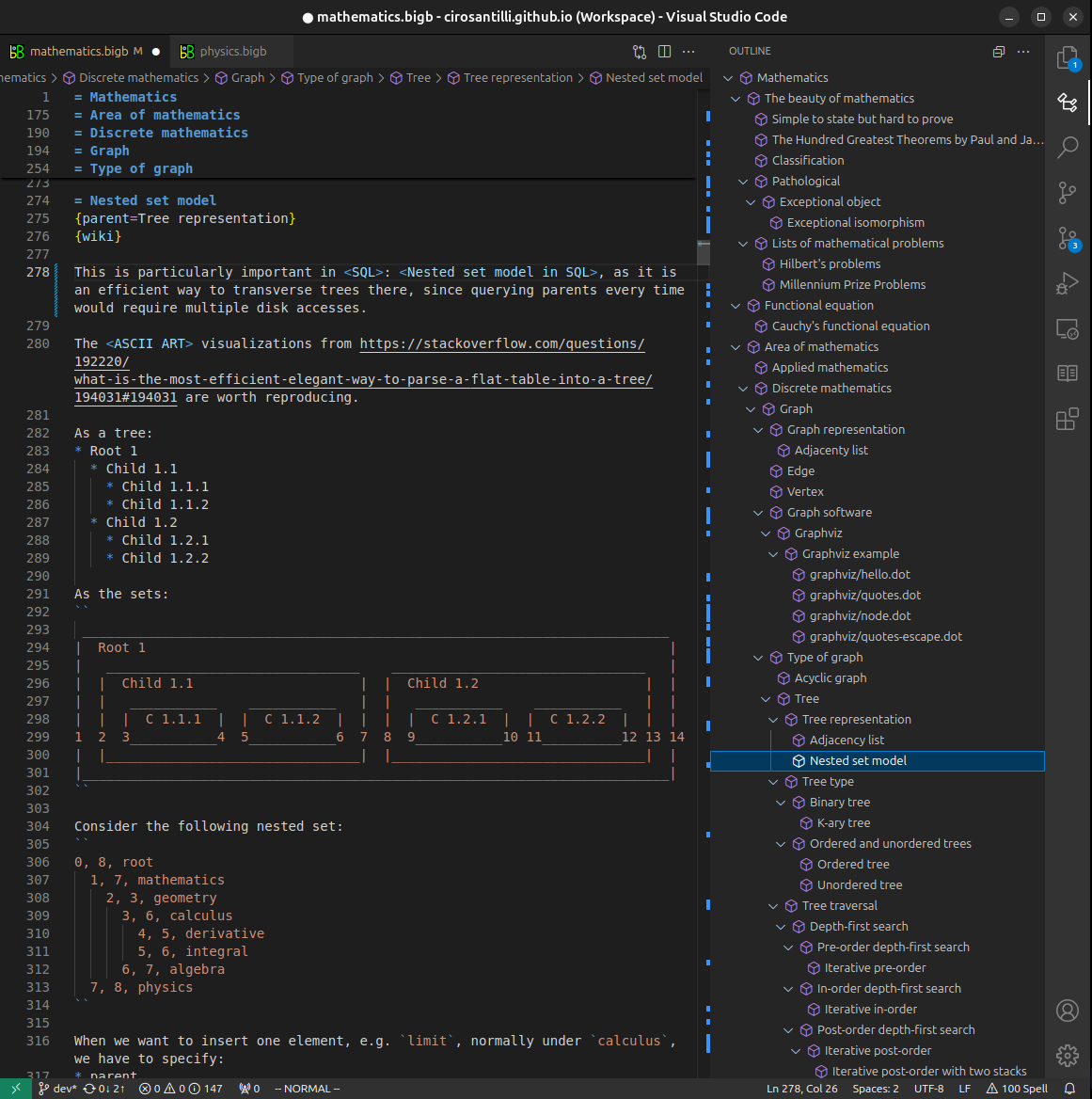
Figure 4. Visual Studio Code extension tree navigation.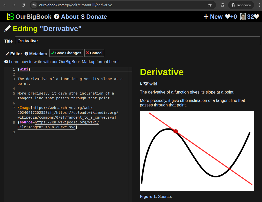
Figure 5. Web editor. You can also edit articles on the Web editor without installing anything locally.Video 3. Edit locally and publish demo. Source. This shows editing OurBigBook Markup and publishing it using the Visual Studio Code extension.Video 4. OurBigBook Visual Studio Code extension editing and navigation demo. Source. 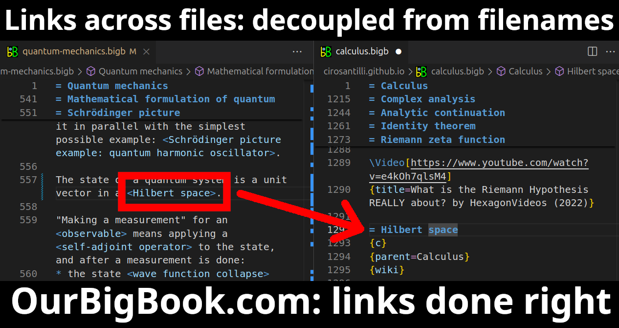
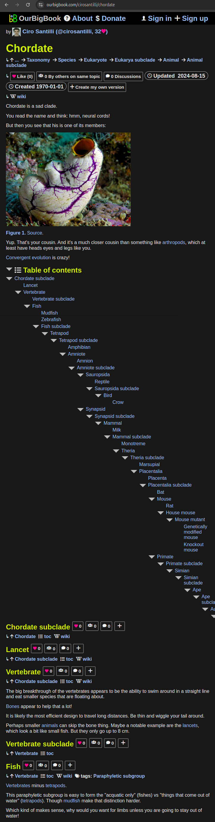
- Infinitely deep tables of contents:

All our software is open source and hosted at: github.com/ourbigbook/ourbigbook
Further documentation can be found at: docs.ourbigbook.com
Feel free to reach our to us for any help or suggestions: docs.ourbigbook.com/#contact