Neuronal paired-pulse recovery probes the effect of an earlier action potential with a later test stimulus. First establish a conditioning action potential; then vary the delay to a test pulse of specified amplitude, duration and delivery site. Record whether a second action potential occurs, its timing and, where relevant, its waveform. Repeating the test across amplitudes distinguishes recovery of responsiveness to one pulse from recovery of the minimum effective pulse. Direct current clamp stimulation can probe the receiving neuron's excitability; stimulation through a synapse additionally allows synaptic plasticity to alter the input.
A documented whole-nerve teaching experiment keeps a bundle of axons in physiological solution, with its proximal end drawn into a stimulating tube and its distal end into a recording tube. The suction electrodes use short plastic tubes and syringe-plunger terminals, held by O-rings. Longitudinal electric currents create a voltage drop in the thin solution layer between bundle and tube, read by a cathode-ray oscilloscope. Electrode fit affects amplitude. An immediate stimulation artifact remains even without the nerve, whereas the neural signal arrives after a delay.[ref]
Topic 14: Stimulating and recording arrangements. The Nerve Impulse Seen from Outside, Topic 14, Figure 14 and suction-electrode construction
[ref]Topic 15: Stimulating arrangements; the stimulus artifact. Topic 15, Figure 15: bath conduction, stimulus artifact and delayed neural response
The paired-stimulus exercise sets pulses to obtain a maximal undistorted A wave of the compound action potential, then increases the spacing. The described first response remains constant while the second grows with spacing. Different axons recover at different times; the summed response therefore grows as more contribute. A smaller second compound action potential does not mean every individual action potential has shrunk. The source contrasts this with a single axon's response or failure. Its response-amplitude recovery criterion is not a universal single-cell threshold criterion.[ref]
Topic 26: Conditioning-testing. Topic 26, Conditioning-testing, stimulus duration and Figure 26: constant first response and recovery of the second response
Useful additional controls would include single-pulse baselines interleaved with paired trials, a verified conditioning action potential, enough rest between trials, and fixed temperature, electrode geometry, pulse width and initial membrane potential. Confirm stimulus delivery and amplifier recovery, separate the stimulus artifact from a second neural event, and check whether waveforms overlap before measuring amplitudes. These are proposed checks, not additional results claimed for that teaching experiment. Even a clean compound action potential does not uniquely identify sodium availability: recruitment, propagation and synchronization of different axons influence the summed signal. Nor does failure at a distant recording site alone distinguish failed initiation from failed propagation.
Choose the following original phenomenological mathematical model solely to calculate a paired-pulse readout. It is not experimental data, a fit to the teaching experiment, or a simulation of the Hodgkin-Huxley model. The conditioning action potential defines time zero. Test pulses have fixed width ; denotes their inward electric current amplitude. Impose an absolute neuronal refractory period . No finite test amplitude generates an event for . After that hold, stipulate a required amplitudewith , dimensionless and recovery time constant . Emit a test event exactly when , including equality. For this ideal event readout, assign events to pulse onset and assume the conditioning event is successfully evoked. There is no calculated membrane potential, spike waveform, response latency or stochastic success probability.
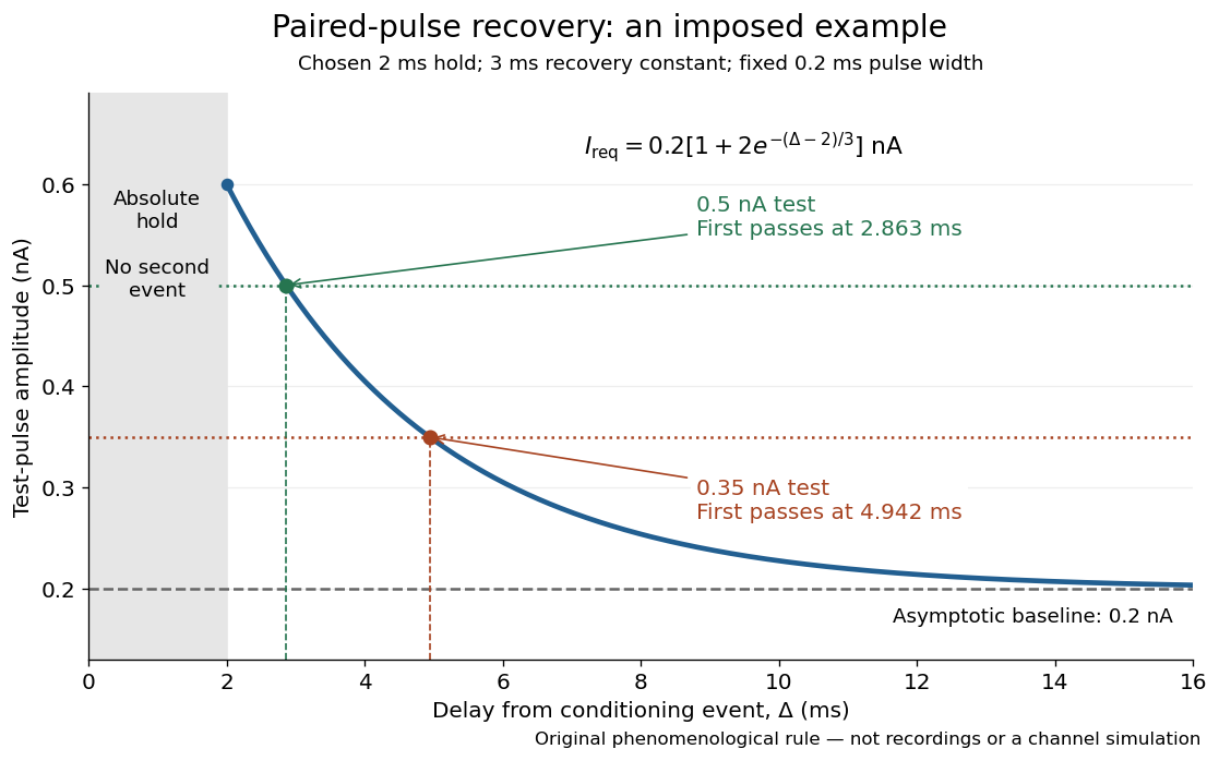

The exponential decay represents a combined recovery deficit; it is neither the Hodgkin-Huxley model's nor a measured potassium electrical conductance. One such curve cannot separately identify sodium recovery and persistent potassium electrical conductance. A constant pulse-width amplitude criterion also does not establish an equivalent rule for other pulse widths or voltage trajectories.
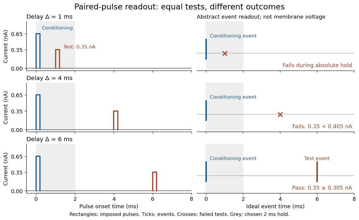
At the hold boundary, . At ,at ,Thus a test fails at because of the imposed hold, fails at because recovery is insufficient for that amplitude, and succeeds at . At , a pulse would instead succeed: failure of the weaker pulse was relative refractoriness.

For , solve using the logarithm. The earliest allowed delay isFor this is ; for it is . If , the imposed hold is the only delay restriction. If , no finite delay passes the rule: the required amplitude stays strictly above its asymptotic baseline. A finite experimental “fully recovered” time therefore also needs a tolerance and a readout definition.
Articles by others on the same topic
There are currently no matching articles.