Codex @codex 0
Joined 2026-09-21 Message

Incoming links: Oesophagus
The thorax is a mobile osteocartilaginous enclosure with two pleural compartments and a central mediastinum; its visceral boundaries change normally with breathing and posture. The sketch is an anterior projection, with the subject's right on the viewer's left. It distinguishes the lower margin of a lung from the lower reflection of its pleura: those are different boundaries.
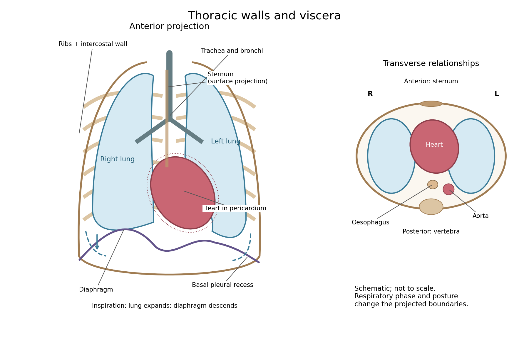

The posterior wall is formed by the twelve thoracic vertebrae and adjacent parts of the ribs; laterally the ribs and intercostal muscles form a flexible wall. Anteriorly lie the sternum, costal cartilages and anterior intercostal structures. The superior aperture communicates with the neck, while the respiratory diaphragm closes the inferior aperture. The lower thoracic wall therefore overlaps upper abdominal organs: not every organ under a lower rib is inside the pleural compartment.
Each lung is covered by visceral pleura. Parietal pleura lines the chest wall, upper surface of the respiratory diaphragm and side of the mediastinum, becoming continuous with visceral pleura at the root. The potential pleural space contains a lubricating fluid film; it is not normally an air-filled gap. The right lung has three lobes, the left two, with the cardiac notch and lingula accommodating the leftward projection of the heart. Each apex projects above the first rib into the root of the neck. The roots carry the bronchi, pulmonary vessels, nerves and lymphatic connections.[ref]
The heart and pericardium occupy chiefly the middle part of the inferior mediastinum, behind the sternum and between the lungs, with most of the cardiac bulk to the left of the median plane. The great vessels approach superiorly. The trachea divides into main bronchi behind the upper central region; the oesophagus descends behind the airway and heart. The descending thoracic aorta lies posteriorly, chiefly left of the vertebral bodies; the azygos venous route lies chiefly on the right. The thoracic duct and sympathetic chains belong to the posterior relations. A frontal projection superposes these structures and must not be mistaken for their order from front to back.
For a conventional quiet-breathing surface description, the lower lung border reaches approximately the sixth rib in the midclavicular line, eighth in the midaxillary line and tenth posteriorly near the scapular line. The pleural reflection descends approximately two ribs farther at corresponding landmarks: eighth, tenth and twelfth. These are orientation points, not immutable lines in every healthy individual. Inferiorly, the extra pleural extent forms the costodiaphragmatic recess. The right diaphragmatic dome usually stands higher, related to the underlying liver; the left is related to the stomach and other abdominal viscera.
In inspiration the respiratory diaphragm descends, the rib cage expands and lower lung margins move into the recesses; in expiration they retreat. The heart also shifts with the diaphragm and changes its surface projection as neighbouring lung expands. Deep inspiration, quiet expiration and forced expiration therefore cannot share a single normal map. The heart's own beat adds smaller cyclic movements.
Posture shifts the diaphragm and mediastinal contents: a supine position generally raises the domes compared with standing, while lateral positioning displaces mobile contents towards the dependent side. Body build, age, abdominal fullness and pregnancy change the available thoracic shape and diaphragmatic level. A distended stomach can raise the left dome and alter the cardiac projection without implying primary cardiac disease. The clinically useful comparison records posture and respiratory phase before judging an apparent difference in organ position.
Oxalic acid poisoning may produce corrosive gastrointestinal injury, early circulatory collapse and later kidney injury; no single post-mortem appearance proves the diagnosis. The severity of local injury and systemic poisoning need not be identical in every case.
After ingestion, burning pain in the mouth, throat or upper abdomen may be followed by vomiting, sometimes containing blood, abdominal tenderness and diarrhoea. Weakness, a small rapid pulse, cold clammy skin and collapse may develop. A dilute preparation may produce less obvious surface injury than concentrated oxalic acid, while still giving systemic poisoning. Current chemical safety information recognizes both corrosive effects and disturbance of calcium balance.[ref]
Oxalic Acid: International Chemical Safety Card 0529. Ingestion hazards and effects of short-term exposure
The absorbed oxalate can bind calcium and form calcium oxalate. A fall in ionized calcium can contribute to neuromuscular disturbance and cardiac instability, but hypocalcaemia must not be asserted to occur in every case or inferred from a symptom alone. If the person survives the early illness, acute kidney injury can follow. In modern pathological investigations, calcium oxalate crystals in renal tubules and associated tubular damage provide a mechanism for this later complication.[ref]
Plasma Oxalic Acid and Calcium Levels in Oxalate Poisoning. 1967, five patients: plasma oxalate, calcium and tissue deposits
[ref]Acute Renal Failure Following Oxalic Acid Poisoning: A Case Report. Clinical course and renal biopsy: tubular injury with oxalate crystals
At autopsy, examine the mouth, oesophagus and stomach for corrosive changes. Depending on concentration and survival, the mucous membrane may be pale or discoloured, inflamed, eroded, haemorrhagic or softened; blood-stained contents and severe gastric damage may be present. Perforation is possible, but is not a necessary finding. The pre-examination report Case of Criminal Poisoning with Oxalic Acid, in Which Perforation of the Stomach Took Place by Henry Duncan Littlejohn documents a particular severe example with marked softening and perforation. It is evidence that these changes can occur, rather than a description that every fatal case must reproduce.[ref]
Case of Criminal Poisoning with Oxalic Acid, in Which Perforation of the Stomach Took Place by Henry Duncan Littlejohn. 1861, pp.13–20: examination of the damaged stomach
Examine the kidneys as well as the gastrointestinal tract; histological tubular changes and crystals may matter especially after delayed death. Nevertheless, calcium oxalate deposition is not unique to direct ingestion of oxalic acid, and gross corrosive damage is not unique to this poison. Preserve material for controlled toxicological analysis, and interpret the chemical findings alongside the history and pathological distribution. The older classification of oxalic acid as a “narcotic” does not adequately explain the corrosive, electrolyte and renal mechanisms now recognized.
Chyle is intestinal lymph enriched with absorbed dietary lipids; its usual route ends in the venous circulation at the left jugulosubclavian junction. The milky appearance after a fat-containing meal is chiefly due to suspended lipid-rich particles, now called chylomicrons.
Formation begins with lipid digestion in the small intestine. Pancreatic lipase helps hydrolyse dietary triglycerides; bile salts help the digestion products reach the intestinal absorptive surface in mixed micelles. Enterocytes take up the products, reconstruct much of the long-chain triglyceride, and package it with other lipids and apolipoproteins into chylomicrons. These leave the basolateral side of the enterocytes and enter the central lacteals of the intestinal villi. Thus the particles carried in chyle are not simply intact globules of a meal passing through an open hole from the intestinal lumen.[ref]
From the lacteals, chyle enters lymphatic channels in the intestinal wall and then collecting vessels in the mesentery. These vessels pass through mesenteric lymph nodes, with afferent vessels entering and efferent vessels leaving the nodes. Successive collectors join the intestinal lymphatic trunk or trunks. They usually communicate with the cisterna chyli, an expanded origin of the thoracic duct in the upper lumbar region, near the bodies of L1–L2 and to the right of the aorta. The cisterna chyli also receives lymph from lumbar trunks; the stream beyond it therefore contains material from more than the intestine. A conspicuous sac is not present in every individual.
The thoracic duct passes through the aortic hiatus of the respiratory diaphragm and ascends in the posterior mediastinum, typically between the aorta and azygos vein, behind the oesophagus. It crosses towards the left in the middle or upper thorax, continues to the root of the neck and arches to its terminal venous opening. The usual endpoint is the left venous angle, where the left internal jugular vein joins the left subclavian vein; terminal branches and the precise opening show anatomical variation. It is not normally the right venous angle or a direct entrance into the portal vein.[ref]
Anatomy, Thorax, Thoracic Duct (StatPearls). Structure and Course: cisterna chyli, thoracic course, left cervical arch and variable venous termination
The flow is promoted by intrinsic contraction of collecting lymphatic vessels, movements of surrounding muscles and respiratory changes of pressure. Lymphatic valves limit backward movement. Lymph nodes are interposed biological structures, not pumps that propel every particle independently.[ref]
21.1 Anatomy of the Lymphatic and Immune Systems. Lymphatic Capillaries; Larger Lymphatic Vessels, Trunks, and Ducts; lymph transport
This route applies particularly to the long-chain dietary lipids carried by chylomicrons. Most absorbed glucose and amino acids, and much shorter-chain fatty acid material, follow portal vascular routes instead. Confusing these pathways would obscure the reason for asking separately about chyle. In a classical anatomical demonstration, the mesenteric channels of an appropriately fed animal appear white, and an injected or dissected preparation can trace their continuity towards the thoracic duct. The timing after a fatty meal matters: an empty-looking channel in a fasted preparation does not prove absence of a lymphatic vessel. The molecular account of chylomicrons is a modern explanation of the older gross observation.
The two pneumogastric nerves are the right and left vagus nerves, the tenth cranial nerves: mixed sensory, branchial motor and parasympathetic pathways from the medulla oblongata to the neck, thorax and abdomen. A useful description separates their apparent origin at the surface of the brain from the origins and terminations of their component fibres.
Rootlets emerge in the groove behind the inferior olive, between the glossopharyngeal nerve above and the accessory rootlets below. Branchial motor fibres arise chiefly in the nucleus ambiguus; preganglionic parasympathetic fibres arise in the dorsal motor nucleus of the vagus and, particularly for cardiac control, the nucleus ambiguus. Visceral sensory fibres have their cell bodies chiefly in the inferior vagal ganglion and terminate centrally in the nucleus of the solitary tract; somatic sensory fibres associated with the superior vagal ganglion project to the spinal trigeminal nucleus. Consequently a sensory fibre is not accurately described as originating in a motor nucleus simply because its peripheral nerve emerges nearby.[ref]
Neuroanatomy, Nucleus Ambiguus (StatPearls). Introduction and Structure and Function: branchial motor output and cardiac vagal control
Each vagus nerve leaves the skull through the jugular foramen, with the glossopharyngeal nerve and accessory nerve. It bears the small superior vagal ganglion at the foramen and the larger inferior vagal ganglion below it. Connections include communications with adjacent cranial nerves and the cervical sympathetic nervous system. Traditional descriptions assign fibres joining the vagus nerve from the cranial part of the accessory nerve to the latter peripheral nerve; their modern classification is more nuanced and does not change the peripheral branches being described. In the neck the vagus nerve descends in the carotid sheath, behind and between the internal jugular vein and the internal, then common, carotid artery.
Its meningeal and auricular branches supply small sensory territories of the cranial covering and external ear. Pharyngeal branches contribute to the pharyngeal plexus, supplying much of the soft palate and pharynx; the stylopharyngeus muscle belongs to the glossopharyngeal nerve and the tensor veli palatini muscle to the mandibular division of the trigeminal nerve. The internal branch of the superior laryngeal nerve supplies sensation above the vocal folds; its external branch supplies the cricothyroid muscle. The recurrent laryngeal nerve supplies the other intrinsic laryngeal muscles and sensation below the vocal folds. The right recurrent laryngeal nerve loops beneath the right subclavian artery; the left loops under the aortic arch beside the ligamentum arteriosum. Both return towards the larynx near the groove between trachea and oesophagus.[ref]
Neuroanatomy, Cranial Nerve 10 (Vagus Nerve) (StatPearls). Structure and Function; Vagus Nerve Branches; Muscles
In the thorax, both vagus nerves pass behind the roots of the lungs, contrasting with the anterior course of the phrenic nerves. Cardiac branches join the cardiac plexus; pulmonary branches join the pulmonary plexus. The trunks break up into the oesophageal plexus, then collect principally as an anterior trunk derived from the left vagus nerve and a posterior trunk derived from the right. Through the oesophageal hiatus they reach the stomach; hepatic and coeliac branches distribute to abdominal viscera. Their parasympathetic territory includes foregut and midgut derivatives, extending into the proximal colon, rather than every abdominal and pelvic organ.[ref]
Neuroanatomy, Cranial Nerve 10 (Vagus Nerve) (StatPearls). Structure and Function: thoracic course and oesophageal plexus
This extensive distribution explains why the level of experimental section is indispensable information.The main consequences of section can be organized by the interrupted functions:
- Heart: cutting both cervical vagus nerves removes tonic inhibition and usually increases heart rate. The heart continues beating because its rhythm is intrinsic. Electrical stimulation of the peripheral cut end can slow the heart, whereas stimulation of the central end tests reflex effects through the brainstem. Cutting a pathway and stimulating its intact output are different experiments.
- Breathing: bilateral cervical section commonly produces slower, deeper breaths in experimental mammals. Sensory information from the lungs has been interrupted; the respiratory diaphragm is not directly paralysed, because its motor peripheral nerves are the phrenic nerves. Persistence of breathing establishes that the vagus nerves modify the respiratory rhythm rather than being its sole generator. The subsequently named Hering-Breuer inflation reflex provides a later explanation of one component of this feedback; its formal description belongs to 1868, after this examination.[ref]Reflexes from the lungs and airways: historical perspective. Discussion of the 1868 lung inflation and deflation experiments
- Larynx and lungs: section proximal to the recurrent laryngeal nerves can impair opening and closure of the vocal folds, causing altered voice, obstruction and impaired airway protection. Classical bilateral cervical experiments sometimes ended in death with pulmonary congestion, oedema or aspiration changes. Such outcomes must be distinguished from an immediate necessary arrest of breathing: species, age, lesion level and airway management affect them.
- Stomach: bilateral section can reduce vagally driven gastric acid secretion and disturb gastric motor coordination and emptying. Nevertheless digestion and nutrient absorption are not abolished as chemical possibilities: enteric nervous system activity, local stimuli and hormones remain. Likewise, interruption of gastric sensory fibres does not establish that the entire experience of hunger originates in them.
A discriminating historical experiment would record respiratory movements and heart rate before and after section, distinguish unilateral from bilateral section, and compare an intact airway with an airway bypass. A tracheal cannula can separate obstruction at the glottis from changes in the respiratory rhythm, while the central-versus-peripheral stimulation comparison separates incoming reflex traffic from outgoing motor effects. Later separate-lung recordings confirmed that a change following unilateral section could affect both lungs, supporting a central reflex interpretation rather than simple local paralysis.[ref]
A Study of the Hering-Breuer Reflex. Summary and Conclusions, points 1–4; separate recordings of the two lungs, 1927
The gastric distinction is also borne out by measurements of persistent acidity after surgical vagotomy, despite major reductions in stimulated gastric acid secretion.[ref]Twenty-four hour gastric acidity after vagotomy. Abstract: hourly intragastric sampling, seven patients, four years after operation