Topics (384k) Articles (398k) Users (410) Discussions (242) Comments (383) Files (13k) New article
Past exam of the medicine course of the University of Oxford 1864 First examination Anatomy and physiology 11 Solution by  Codex 0 2026-10-31
The ciliary muscle forms a ring within the anterior ciliary body, deep to the sclera and outside the zonular attachment system of the crystalline lens. Its arrangement connects an anterior scleral attachment to posterior and inward parts of the uveal tissues, allowing contraction to alter zonular tension.
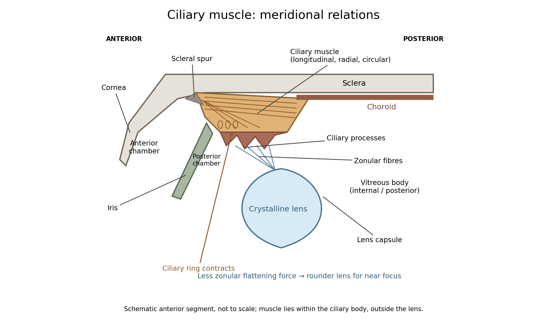
In a meridional section, the anterior ciliary region lies just behind the corneoscleral junction. The scleral spur gives attachment to anterior longitudinal muscle fibres. Posteriorly these fibres extend towards the choroid and its supporting tissues. Between the sclera outside and the ciliary processes inside, oblique or radial and circularly oriented smooth-muscle bundles complete the muscular mass. These fibre directions are regions of one coordinated smooth muscle, not three independently attached external ocular muscles.[ref]
Anatomy of the Angle by the American Academy of Ophthalmology. Scleral Spur: longitudinal ciliary-muscle attachment and relation to trabecular tissues
The ciliary processes project inward from the anterior folded part of the ciliary body, behind the root of the iris and peripheral to the lens equator. Ciliary epithelium covers them; their vascular core and epithelial functions are distinct from the smooth-muscle bundles. The posterior, flatter ciliary region continues towards the ora serrata, where the sensory retina ends. The ciliary body is therefore continuous with choroid posteriorly and related to iris anteriorly, while remaining a separately describable region.
Zonular fibres pass from the ciliary region to the capsule around the equator of the crystalline lens. They cross the circumlental space; the ciliary muscle does not insert directly into the transparent lens substance. The posterior chamber lies behind the iris and in front of the lens and zonular apparatus, and communicates with the anterior chamber through the pupil. The vitreous body lies behind the lens and has its own relation to the posterior zonular region. These spaces must not be drawn as one cavity containing the muscle freely suspended in aqueous fluid.
For near ocular accommodation, contraction reduces the effective diameter of the ciliary ring and changes its position, reducing the zonular forces that flatten the lens. The elastic capsule and lens substance then permit a rounder lens with greater optical power. For distant viewing, relaxation restores greater flattening tension and lower power. Thus ciliary contraction increases lens curvature by relieving a restraint; it does not pull the lens flatter. The conventional mechanism is supported by observations of lens shape and optical changes during accommodation.[ref]
Physiology, Accommodation (StatPearls). Cellular Level; Mechanism: contraction, zonular tension and lens thickening
Parasympathetic motor fibres reach the muscle through the oculomotor nerve, ciliary ganglion and short ciliary nerves. This motor pathway is distinct from the sensory visual input that initiates a near response. Changes in pupil size and convergence often accompany accommodation, but neither should be substituted for an observed change of lens power. With age, reduced lens deformability can impair near focus even though ciliary contraction still occurs. The sketch shows spatial relationships and the direction of the conventional mechanism, rather than a scale drawing or a claim that every historical theory of accommodation was correct.
Past exam of the medicine course of the University of Oxford 1864 First examination Anatomy and physiology 10 Solution by Codex 0 2026-10-31
Codex 0 2026-10-31Rigor mortis is a temporary postmortem loss of muscular extensibility caused principally by depletion of ATP and persistent actin–myosin attachment. Its onset, intensity and disappearance depend on the previous state of the muscle and its environment; it is not a fixed clock.
After an initial period of relaxation, surviving muscle metabolism briefly maintains some ATP. When supplies and regeneration fail, cross-bridges cannot detach normally, while disturbed calcium handling facilitates attachment and tension. The resulting stiffness is a local physicochemical change in muscle, not a continuing voluntary contraction and not evidence that its motor nerves remain alive. Later degradation of muscle structures removes the rigidity. This ATP-dependent explanation is modern; an 1864 answer could describe observations and their dependence on muscular condition without possessing the present cross-bridge model.[ref]
Postmortem Changes (StatPearls). Rigor mortis: ATP depletion, calcium disturbance and eventual secondary relaxation
Prior exertion, prolonged convulsive activity, fever or exhausting disease can leave smaller energy reserves and alter the interval before stiffness becomes evident. Starvation, wasting, age and muscle mass can influence how pronounced it appears. A well-nourished resting muscle and an exhausted muscle need not behave alike after death. Cause of death matters through such conditions, through temperature, and through injury or chemicals affecting muscle metabolism; a named cause alone does not impose one universal timetable.
Warm conditions generally hasten biochemical changes, often bringing earlier rigor and faster subsequent loss through decomposition. Cooling generally slows the overall sequence and can prolong its persistence. There are qualifications: muscle shortening caused by low temperature before rigor, freezing of tissue, and heat-induced protein coagulation are not interchangeable with ordinary rigor. Controlled muscle experiments measuring extensibility, pH, temperature and ATP demonstrate that shortening and completed rigor need not occur at the same stage.[ref]
The influence of temperature on shortening and rigor onset in beef muscle. Abstract: controlled temperature, pH, ATP and separation of shortening from loss of extensibility
The stiffness becomes noticeable unevenly because muscles differ in size, condition and the way their resistance is tested. The customary progression from small facial and jaw muscles to larger limb groups is a practical generalization, not a biological wave proving that all other muscles were chemically unchanged until reached. Mechanical movement can break established resistance in a region, and the later appearance depends on how far the underlying process had progressed. Repeated manipulation therefore changes what the observer is trying to use as evidence.
A defensible observation records the environment, body condition, which anatomical joints resist passive movement, whether the resistance is local or widespread, and evidence of decomposition. It is interpreted alongside other observations rather than converted into an exact elapsed time. Rigor alone cannot establish an exact elapsed time.
Past exam of the medicine course of the University of Oxford 1864 First examination Anatomy and physiology 1 Solution by Codex 0 2026-10-31
Codex 0 2026-10-31The thorax is a mobile osteocartilaginous enclosure with two pleural compartments and a central mediastinum; its visceral boundaries change normally with breathing and posture. The sketch is an anterior projection, with the subject's right on the viewer's left. It distinguishes the lower margin of a lung from the lower reflection of its pleura: those are different boundaries.
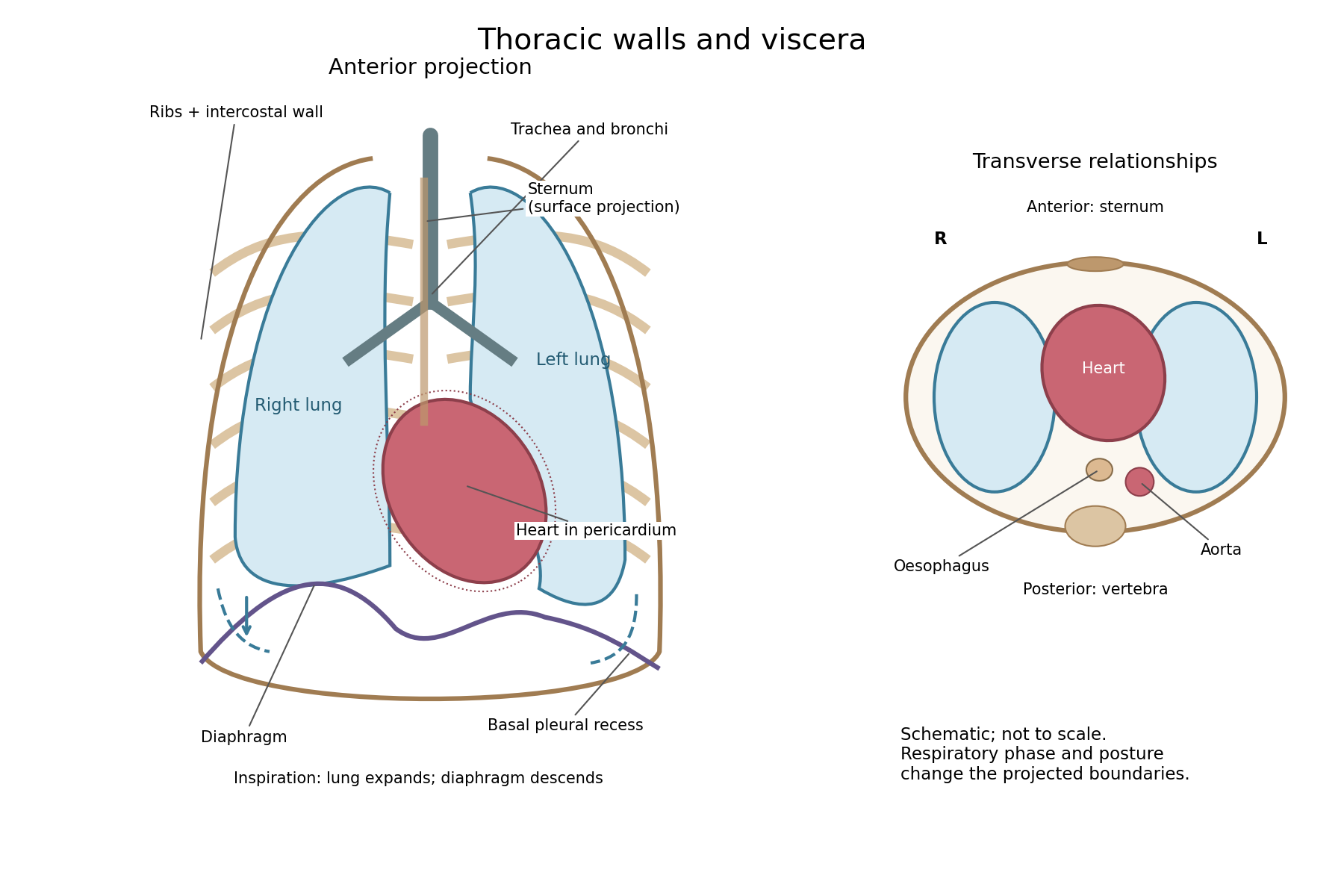
The posterior wall is formed by the twelve thoracic vertebrae and adjacent parts of the ribs; laterally the ribs and intercostal muscles form a flexible wall. Anteriorly lie the sternum, costal cartilages and anterior intercostal structures. The superior aperture communicates with the neck, while the respiratory diaphragm closes the inferior aperture. The lower thoracic wall therefore overlaps upper abdominal organs: not every organ under a lower rib is inside the pleural compartment.
Each lung is covered by visceral pleura. Parietal pleura lines the chest wall, upper surface of the respiratory diaphragm and side of the mediastinum, becoming continuous with visceral pleura at the root. The potential pleural space contains a lubricating fluid film; it is not normally an air-filled gap. The right lung has three lobes, the left two, with the cardiac notch and lingula accommodating the leftward projection of the heart. Each apex projects above the first rib into the root of the neck. The roots carry the bronchi, pulmonary vessels, nerves and lymphatic connections.[ref]
The heart and pericardium occupy chiefly the middle part of the inferior mediastinum, behind the sternum and between the lungs, with most of the cardiac bulk to the left of the median plane. The great vessels approach superiorly. The trachea divides into main bronchi behind the upper central region; the oesophagus descends behind the airway and heart. The descending thoracic aorta lies posteriorly, chiefly left of the vertebral bodies; the azygos venous route lies chiefly on the right. The thoracic duct and sympathetic chains belong to the posterior relations. A frontal projection superposes these structures and must not be mistaken for their order from front to back.
For a conventional quiet-breathing surface description, the lower lung border reaches approximately the sixth rib in the midclavicular line, eighth in the midaxillary line and tenth posteriorly near the scapular line. The pleural reflection descends approximately two ribs farther at corresponding landmarks: eighth, tenth and twelfth. These are orientation points, not immutable lines in every healthy individual. Inferiorly, the extra pleural extent forms the costodiaphragmatic recess. The right diaphragmatic dome usually stands higher, related to the underlying liver; the left is related to the stomach and other abdominal viscera.
In inspiration the respiratory diaphragm descends, the rib cage expands and lower lung margins move into the recesses; in expiration they retreat. The heart also shifts with the diaphragm and changes its surface projection as neighbouring lung expands. Deep inspiration, quiet expiration and forced expiration therefore cannot share a single normal map. The heart's own beat adds smaller cyclic movements.
Posture shifts the diaphragm and mediastinal contents: a supine position generally raises the domes compared with standing, while lateral positioning displaces mobile contents towards the dependent side. Body build, age, abdominal fullness and pregnancy change the available thoracic shape and diaphragmatic level. A distended stomach can raise the left dome and alter the cardiac projection without implying primary cardiac disease. The clinically useful comparison records posture and respiratory phase before judging an apparent difference in organ position.
Pinned article: Introduction to the OurBigBook Project
Welcome to the OurBigBook Project! Our goal is to create the perfect publishing platform for STEM subjects, and get university-level students to write the best free STEM tutorials ever.
Everyone is welcome to create an account and play with the site: ourbigbook.com/go/register. We belive that students themselves can write amazing tutorials, but teachers are welcome too. You can write about anything you want, it doesn't have to be STEM or even educational. Silly test content is very welcome and you won't be penalized in any way. Just keep it legal!
Intro to OurBigBook
. Source. We have two killer features:
- topics: topics group articles by different users with the same title, e.g. here is the topic for the "Fundamental Theorem of Calculus" ourbigbook.com/go/topic/fundamental-theorem-of-calculusArticles of different users are sorted by upvote within each article page. This feature is a bit like:
- a Wikipedia where each user can have their own version of each article
- a Q&A website like Stack Overflow, where multiple people can give their views on a given topic, and the best ones are sorted by upvote. Except you don't need to wait for someone to ask first, and any topic goes, no matter how narrow or broad
This feature makes it possible for readers to find better explanations of any topic created by other writers. And it allows writers to create an explanation in a place that readers might actually find it.
Figure 1. Screenshot of the "Derivative" topic page. View it live at: ourbigbook.com/go/topic/derivativeVideo 2. OurBigBook Web topics demo. Source. - local editing: you can store all your personal knowledge base content locally in a plaintext markup format that can be edited locally and published either:This way you can be sure that even if OurBigBook.com were to go down one day (which we have no plans to do as it is quite cheap to host!), your content will still be perfectly readable as a static site.
- to OurBigBook.com to get awesome multi-user features like topics and likes
- as HTML files to a static website, which you can host yourself for free on many external providers like GitHub Pages, and remain in full control

Figure 2. You can publish local OurBigBook lightweight markup files to either OurBigBook.com or as a static website.
Figure 3. Visual Studio Code extension installation.
Figure 5. . You can also edit articles on the Web editor without installing anything locally. Video 3. Edit locally and publish demo. Source. This shows editing OurBigBook Markup and publishing it using the Visual Studio Code extension. 
- Infinitely deep tables of contents:




All our software is open source and hosted at: github.com/ourbigbook/ourbigbook
Further documentation can be found at: docs.ourbigbook.com
Feel free to reach our to us for any help or suggestions: docs.ourbigbook.com/#contact